Prediction of CD8
Epitopes in
braziliensis
Proteins Using EPIBOT:
In Silico
Search and
In Vivo
Validation
Angelo Duarte1☯, Artur T. L. Queiroz2☯, Rafael Tosta1, Augusto M. Carvalho2, Carlos
Henrique Barbosa3, Maria Bellio3, Camila I. de Oliveira2,4*, Manoel Barral-Netto2,4*
1Departmento de Tecnologia, Universidade Estadual de Feira de Santana, Av. Transnordestina, s/n, DTEC-Módulo 3, 44036–900, Feira de Santana, BA, Brazil,2CPqGM—FIOCRUZ, R. Waldemar Falcão, 121, 40296–710, Salvador, BA, Brazil,3Instituto de Microbiologia Paulo de Góes, Centro de Ciências da Saúde, Universidade Federal do Rio de Janeiro (UFRJ), Avenida Carlos Chagas Filho, 373 Bloco D, sala 35, Cidade Universitária, 21941–902, Rio de Janeiro, RJ, Brazil,4Instituto de Investigação em Imunologia, São Paulo, Brazil
☯These authors contributed equally to this work.
*[email protected](CIO);[email protected](MBN)
Abstract
Background
Leishmaniasis is caused by intracellularLeishmaniaparasites that induce a T-cell mediated
response associated with recognition of CD4+and CD8+T cell Line 1Lineepitopes.
Identifi-cation of CD8+antigenic determinants is crucial for vaccine and therapy development.
Herein, we developed an open-source software dedicated to search and compile data ob-tained from currently available on line prediction algorithms.
Methodology/Principal Findings
We developed a two-phase algorithm and implemented in an open source software called EPIBOT, that consolidates the results obtained with single prediction algorithms, generating a final output in which epitopes are ranked. EPIBOT was initially trained using a set of 831 known epitopes from 397 proteins from IEDB. We then screened 63Leishmania braziliensis
vaccine candidates with the EPIBOT trained tool to search for CD8+T cell epitopes. A proof-of-concept experiment was conducted with the top eight CD8+epitopes, elected by EPIBOT. To do this, the elected peptides were synthesized and validated for theirin vivo
cy-totoxicity. Among the tested epitopes, three were able to induce lysis of pulsed-target cells.
Conclusion
Our results show that EPIBOT can successfully search across existing prediction tools, gen-erating a compiled list of candidate CD8+epitopes. This software is fast and a simple search engine that can be customized to search over different MHC alleles or HLA haplotypes.
OPEN ACCESS
Citation:Duarte A, Queiroz ATL, Tosta R, Carvalho AM, Barbosa CH, Bellio M, et al. (2015) Prediction of CD8+Epitopes inLeishmania braziliensisProteins Using EPIBOT:In SilicoSearch andIn Vivo Validation. PLoS ONE 10(4): e0124786. doi:10.1371/ journal.pone.0124786
Academic Editor:Simona Stager, INRS - Institut Armand Frappier, CANADA
Received:September 27, 2014
Accepted:March 5, 2015
Published:April 23, 2015
Copyright:© 2015 Duarte et al. This is an open access article distributed under the terms of the Creative Commons Attribution License, which permits unrestricted use, distribution, and reproduction in any medium, provided the original author and source are credited.
Data Availability Statement:All relevant data are within the paper and its Supporting Information files.
Funding:This work was supported with a grant from FAPESB (Fundação de Amparo à Pesquisa da Bahia) (SUS0003/2009). The funders had no role in study design, data collection and analysis, decision to publish, or preparation of the manuscript.
Introduction
Leishmaniasis is an infectious disease with significant economic impact in several countries. Over three hundred million people are exposed to the parasites, with 12 million infected world-wide, predominantly in tropical and subtropical countries (World Health Organization page:
http://www.who.int/emc/diseases/leish/leisdis1.html). Leishmaniasis can be caused by different species ofLeishmania spp. protozoans that infect macrophages in the human host. The treat-ments available for all forms of leishmaniasis are toxic and drug resistance is on the rise, further increasing the need for vaccine development [1].
In Brazil, cutaneous leishmaniasis (CL) is caused mostly byLeishmania braziliensisand pres-ents as a skin ulcer associated with an intense inflammatory reaction with the presence of T cells [2]. The main immune mechanism for the control ofLeishmaniainfection is IFN-γproduction
and subsequent macrophage activation, enabling the elimination of intracellularLeishmania par-asites. It has been shown that CD4+T cells are the main source of IFN-γproduction, and,
hence, macrophage activation (rev. in [3]). CD8+T cells, on the other hand, are cytotoxic and contribute with pathogenesis of CL caused byL.braziliensis: CD8+T cells promote lysis of leish-mania-infected cells [4,5]. Moreover, the frequency of CD8+T cells expressing cytotoxic media-tors such as granzyme is directly correlated with the intensity of inflammatory reaction CL lesions [6]. The activation of CD8+T cells is dependent on the recognition of antigenic peptide, presented by major histocompatibility complex molecules at the surface of infected cells [7]. Given the association of CD8+T cell with pathogenesis of CL, it has become important to identi-fy putativeL.braziliensisCD8+peptides associated with the cytotoxic response.
Experimental identification of MHC-binding peptides requires an assay for each peptide, a time consuming and costly process [8].In silicoepitope and MHC-peptide binding prediction, on the other hand, allow optimization of epitope discovery in vaccine design studies, therefore reducing the experimental workload [9]. A variety of algorithms have been developed and used in the field of epitope prediction and these algorithms range from Simplest Sequence Motifs and position-specific scoring matrices (PSSM) [10] to more complex machine-learning proba-bilistic approaches, such as Hidden Markov Models (HMM) [11], Artificial Neural Networks (ANN) [12] and Support Vector Machines (SVM) [13]. However, epitope discovery using dis-tinct algorithms results in contrasting outputs, rendering candidate selection a cumbersome task. Since epitope mapping is important for the screening of cellular immunity in protected individuals, for example, an algorithm than combines different search methodologies and gen-erates a unique list of candidates becomes a useful tool.
Here, we developed a two-phase algorithm that merges the results generated by individual prediction algorithms generating a unified final rank of elected epitopes. The algorithm was implemented in EPIBOT, a free software for non-commercial use developed in JAVA language. The zip source is available at:https://sites.google.com/a/ecomp.uefs.br/angeloduarte/epibot? pli=1. To run the friendly-interface, the user needs to extract the EPIBOT.rar file and navigate to folder. To execute in Windows and Mac OS, user double clicks the. jar file. For Linux OS ex-ecute the shell command java—jar Epibot.jar. More information is available in the software manual. EPIBOT was tested with 63Leishmania braziliensisproteins and the top predicted eight epitopes were validatedin vivofor their potential to induce CD8+T cell cytotoxicity.
Materials and Methods
Software development
proteins containing known CD8+epitopes: we used a set of 4251 H2-Kdepitopes, available in IEDB. However, among these only 831 proteins (S1 Table) with CD8+epitopes were recovered from NCBI. Entries without reports at duplicates and proteins with more than 1 epitope were also removed from the dataset leaving 397 proteins (S2 Table) with identified CD8+epitopes. These were used to assess the accuracy of the different prediction algorithms. Each calibration protein was then individually submitted to the following epitope prediction algorithm: BIMAS [14], SYFPEITHII [10], netMHC [15], SVMHC [16] and IEDB In every case, we searched for peptides presenting high affinity with mouse MHC class I molecules of BALB/c mice (H2- Kd). Each search resulted in an epitope rank, where the best ranked epitope is expected to present a top score (rank = 1). This list was ordered according to the peptide score and the top epitopes ranked equal or close to 1. Therefore, each epitope rank (its compiled list position) is the algo-rithm´s prediction quality indicator. A perfect algorithm would classify known CD8 epitopes at the top. Thus, a Specific Prediction Score (SPS) was defined: this scores the algorithm’s qual-ity for a specific calibration protein (and its calibration epitope). The SPS is the inverse of the Rank of the Calibration Epitope (RCE), from the list of epitopes yielded by the algorithm, pre-sented inEq 1:
SPS¼1=RCE ð1Þ
To assess overall algorithm quality, a General Prediction Score (GPS) was determined for each prediction algorithm, using all proteins SPS averages measured from the calibration set (defined inEq 2). The Number of Calibration Proteins (NCP) was applied. The factor 10 was used to normalize GPS range to 0–10 interval. Good prediction algorithms are expected to have a GPS near 10.SPS
GPS¼ 10
NCP XNCP
i¼1 GPSi ð2Þ
After calibration phase, the software determines the GPS value for each prediction algo-rithm. Thus, the EPIBOT algorithm is trained and ready to perform epitope screening in the query protein set.
EPIBOT Unified prediction
After EPIBOT calibration, the GPS values are used to predict epitopes in a query dataset. Thereby, the two-step consolidation algorithm submits each query protein individually to the prediction algorithms (netMHC, SYFPEITHII, BIMAS, SVMHC and IEDB) and then mea-sures the Unified Epitope Score (UES). First, the epitope-predicted list generated by each algo-rithm was normalized, where normalized scores (NS) range from [0–1] where 1 is the top epitope score and 0 is the bottom epitope score. Second, for a given Number of Prediction Al-gorithms (NPA), the UES is calculated usingEq 3, by summing epitope normalized scores, from each prediction algorithm, multiplied by the general prediction score from each different algorithm.
UES¼XNPAi 1 NSiGPSi ð3Þ
Thefinal EPIBOT output is an epitope-predicted list descending by UES order, based on
EPIBOT prediction using
L
.
braziliensis
query proteins
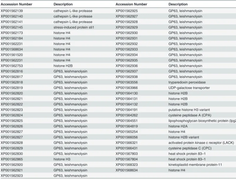
Sixty-threeLeishmania braziliensisproteins were screened with EPIBOT for the discovery of potential 9-mer epitopes presented by H2-Kd(Table 1). These epitopes were then validated in vivo, using a cytotoxicity assay, as described below. The full sequences of the proteins were ex-tracted from NCBI. The selected proteins have been previously defined as candidate antigens [17–20] and were tested with the trained EPIBOT algorithm, using the five prediction tools mentioned above. The prediction output was evaluated and sorted by UES. The top epitopes were compared to mouse proteins using BLAST [11].Epitopes with query coverage of 100% and identity superior to 90% with self-murine proteins from ref_seq database were discarded. The predicted peptides were also compared to known epitopes from IEDB database to avoid known-epitopes re-evaluation.
Table 1. L.braziliensisprotein query set.
Accession Number Description Accession Number Description
XP001562139 cathepsin L-like protease XP001562925 GP63, leishmanolysin
XP001562140 cathepsin L-like protease XP001562927 GP63, leishmanolysin
XP001562141 cathepsin L-like protease XP001562928 GP63, leishmanolysin
XP001562145 stress-induced protein sti1 XP001562929 GP63, leishmanolysin
XP001562173 histone H4 XP001562930 GP63, leishmanolysin
XP001562184 histone H4 XP001562931 GP63, leishmanolysin
XP001562231 histone H4 XP001562932 GP63, leishmanolysin
XP001568634 histone H4 XP001562933 GP63, leishmanolysin
XP001561520 histone H4 XP001562934 GP63, leishmanolysin
XP001562231 histone H4 XP001562935 GP63, leishmanolysin
XP001562753 histone H2B XP001562936 GP63, leishmanolysin
XP001562816 GP63, leishmanolysin XP001562937 GP63, leishmanolysin
XP001562817 GP63, leishmanolysin XP001562938 GP63, leishmanolysin
XP001562818 GP63, leishmanolysin XP001563556 tryparedoxin peroxidase
XP001562819 GP63, leishmanolysin XP001563966 UDP-galactose transporter
XP001562820 GP63, leishmanolysin XP001564130 histone H2B
XP001562821 GP63, leishmanolysin XP001564131 histone H2B
XP001562822 GP63, leishmanolysin XP001564132 histone H2B
XP001562823 GP63, leishmanolysin XP001564191 putative histone H3 variant
XP001562824 GP63, leishmanolysin XP001564262 cysteine peptidase A (CPA)
XP001562825 GP63, leishmanolysin XP001564551 lipophosphoglycan biosynthetic protein (lpg2)
XP001562826 GP63, leishmanolysin XP001564819 histone H2A
XP001562827 GP63, leishmanolysin XP001565254 histone H4
XP001562827 GP63, leishmanolysin XP001566056 histone H2B variant
XP001562828 GP63, leishmanolysin XP001566321 activated protein kinase c receptor (LACK)
XP001562829 GP63, leishmanolysin XP001566431 cysteine peptidase C (CPC)
XP001562830 GP63, leishmanolysin XP001567803 heat shock protein 83–1
XP001562865 histone H3 XP001567804 heat shock protein 83–1
XP001562920 GP63, leishmanolysin XP001568323 kinetoplastid membrane protein-11
XP001562921 GP63, leishmanolysin XP001568634 histone H4
XP001562923 GP63, leishmanolysin
In vivo
validation of predicted epitopes
Ethics Statement. Female BALB/c mice, 6–8 weeks of age, 25–30 grams, were obtained from CPqGM/FIOCRUZ animal facility where they were maintained under pathogen-free conditions. Mice were housed in groups of five. Environmental conditions were a temperature of 21°C ±2°, humidity of 55% ±10%, and a 12:12 light:dark cycle with lights on at 0700 and off at 1900. Animals were housed in 422×230×203 mm cages (Domi Series, Alesco, Brazil) and given access to mouse maintenance food (Biobase Bio Tec, Brazil) and waterad libitum. Envi-ronmental enrichment included bedding (maravalhaPinus elliotti, Hemo In Produtos para Biotério, Brazil), one red tinted igloo (Alesco, Brazil). During housing, animals were monitored twice daily for health status. No adverse events were observed. In vivo validation was per-formed using 36 animals. Splenocytes to be pulsed with peptides were obtained from 12 naive mice. Pulsed and labeled splenocytes were injected into 24L.braziliensis-infected mice. All ani-mal work was conducted according to the Guidelines for Aniani-mal Experimentation of the Colé-gio Brasileiro de Experimentação Animal and of the Conselho Nacional de Controle de Experimentação Animal. The local Ethics Committee on Animal Care and Utilization (CEUA) approved all procedures involving animals (L-03/2011). All sections of this report adhere to the ARRIVE Guidelines for reporting animal research. A completed ARRIVE guidelines check-list is included inS1 ARRIVEChecklist. To obtain splenocytes and draining lymph node cells, mice were euthanized using compressed CO2under a flow rate of 1–3 liters per minute for a 10
liter (volume) chamber. CO2flow was maintained for a minimum of 1 minute until lack of
res-piration and faded eye color were observed.
Parasite culture, intradermal inoculation and lesion measurement. L.braziliensis pro-mastigotes strain MHOM/BR/01/BA788 were grown in Schneider medium (Sigma) supple-mented with 100U/ml of penicillin, 100ug/ml of streptomycin, 10% heat-inactivated fetal calf serum (all from Life Technologies). Stationary-phase promastigotes (105parasites in 10μl of sa-line) were inoculated into the right ear dermis of age-matched BALB/c mice using a 27.5-gauge needle. Lesion size was monitored weekly, for 5 weeks, using a digital calliper (Thomas Scientific).
In vivocytotoxicity assay. Splenocytes were obtained from naive BALB/c (H2-Kd). Cells
were divided into three groups and were labeled with the fluorogenic dye CFDA (Invitrogen) at final concentrations of 8μM (CFDAhigh), 2μM (CFDAintermediate) or 0.5μM (CFDAlow). CFDAhigh cells were previously pulsed for 40 minutes at 37°C with 5μM of H-2Kd-restrictedL.braziliensis peptides (AYLASCDFI, AYIDGHVTI, KYQHSTEML or TYQRVYATL. CFDAintermediatecells were pulsed in the same way with WYLATHSLI, SYMGYFQNI, IYVSYADLI or VYLSFGFRL). CFDAlowcells remained unpulsed. Subsequently, CFDAhighand CFDAintermediatecells were washed and mixed with equal numbers of CFDAlowcells before injecting intravenously (15–20 x 106 total cells) intoL.braziliensis-infected and into control non-infected mice. Draining lymph node and spleen cells of recipient mice were collected 20 hours after cell transfer, fixed and ana-lyzed by cytometry, using a FACSCalibur Cytometer (BD Biosciences). Percentage of CFDAlow (M1), CFDAintermediate(M2) and CFDAhigh(M3) cells were obtained using CellQuest software (BD Biosciences). Percentage of specific lysis was determined using the formula: 1–((M2 in-fected/M1 infected) / (M2 naïve/ M1 naïve)) x 100% or 1–((M3 infected/M1 infected) / (M3 naïve/ M1 naïve)) x 100% [21].
Statistical analysis
Results
In silico
analysis of
L
.
braziliensis
proteins for potential candidate
peptides
To identify potential CD8+T cell activating 9-mer epitopes presented by H2-Kdwe used the trained EPIBOT on a query dataset of 63L.braziliensisprotein candidates (Table 1). EPIBOT screening generated an output of 10950 UES-associated epitopes. The output list was sorted and the eight best-predicted epitopes with UES above 9.5 (Table 2) were chosen: AYLASCDFI, WYLATHSLI, AYIDGHVTI, SYMGYFQNI, GYVGIVVAL, KYQHSTEML, IYVSYADLI, VYLSFGFRL. These peptides were synthesized and validatedin vivo.
In vivo
cytotoxicity following inoculation of peptide-pulsed splenocytes
into
L
.
braziliensis
-infected mice
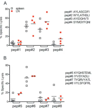
Following epitope prediction using EpiBot, we validated the top eight peptidesin vivo, using an assay that measures cytotoxicity of CD8+T cells following an encounter with peptide-pulsed cells. No expressive cytotoxicity activity was observed when cells were pulsed with
AYLATCDFI or SYMGYFQNI peptides (Fig 1A). On the other hand, the percentage of specific lysis of cells pulsed with WYLATHSLI was 42.2% in the spleen and 47.6% in the draining lymph node, while specific lysis for cells pulsed with AYIDGHVTI was 35.5%, in the spleen and and 52.1% % in the draining lymph node. Among the other four peptides tested, specific lysis was found only for peptide IYVSYADLI which displayed 52% and 56% of specif lysis in the spleen and draining lymph nodes, respectively (Fig 1B).
Discussion
The identification of MHC-associated epitopes, recognized by T cells, is essential to measure epitope-specific T cell responses. Several approaches have been developed to improve epitope discovery (rev. in [9]), however, the variability observed among existing prediction tools rises important questions concerning the approaches´ consensus. Herein, we developed an analysis tool, EPIBOT, capable of epitope prediction across different prediction algorithms currently
Table 2. TopL.braziliensisepitopes as predicted by EPIBOT.
EPITOPE UESa Accession Description % Lysis (mean of spleen and dLN values)
AYLASCDFI 13.296 XP001562821 GP63 leishmanolysin 5.75
AYLASCDFL 13.193 XP001562826 GP63 leishmanolysin NTb
AYLATCDFL 13.003 XP001562925 GP63 leishmanolysin NT
WYLATHSLI 12.534 XP001562139 cathepsin L-like protease(CPB) 44.88 AYIDGHVTI 12.063 XP001562141 cathepsin L-like protease(CPB) 43.82
SYMGYFQNI 11.183 XP001564262 cysteine peptidase A (CPA) 8.28
GYVGIVVAL 10.56 XP001564551 lipophosphoglycan biosynthetic protein (lpg2) NT
KYQHSTEML 10.505 XP001564191 histone H3 variant 12.02
IYVSYADLI 9.979 XP001562817 GP63 leishmanolysin 54.17
TYQRVYATL 9.947 XP001562141 cathepsin L-like protease(CPB) 7.15
VYLSFGFRL 9.601 XP001562184 histone H4 9.91
aUES, Uni
fied Epitope Score
bNT: not tested
available online. EPIBOT was tested on query set of 63L.braziliensisand the top eight epitopes were synthesized for in vivo validation.
Although the use of EPIBOT on 63 proteins from theL.braziliensisproteome yielded a large number of predicted epitopes, such result was lower than that obtained using different prediction tool (BIMAS, SYFPEITHII, netMHC, SVMHC and IEDB) individually.
After calibration, EPIBOT automatically submits each protein to the prediction tools select-ed by the user and all outputs are storselect-ed in an SQL database. The user only defines the protein queries set in a single initial submission. This solves all multi-submissions and high throughput issues. These features allow the user to test different prediction tools in large queries, contain-ing many different proteins, thereby improvcontain-ing epitope search. In this way, EPIBOT handles large data sets, generated following the search by each predictor, combining different method-ologies together with a rational output, yielding a unified score.
EPIBOT was able to handle the data groups generated by each predictor, combining differ-ent methodologies together with a rational output and generating a unique predictor score. The approach of combining results from several prediction tools is advantageous since integra-tion of many predicintegra-tion methods improves the overall predicintegra-tion performance [22]. Addition-ally, this helps solving a crucial issue regarding different epitope prediction algorithms: the different score metrics used by each algorithm. For example, BIMAS [14] uses HLA class I half time dissociation, while SYFPEITHI [10] uses log-based score. Moreover, EPIBOT enables the use of different predictors tools in a set of proteins, generating a predicted epitopes pool, allow-ing epitope comparison based on a sallow-ingle score. This poses advantages over the use of a sallow-ingle
Fig 1. Specific CD8+T cell-mediated immune response inL.braziliensis-infected BALB/c mice.Target
cells were pulsed withL.braziliensispeptides (A) AYLASCDFI, WYLATHSLI, AYIDGHVTI and SYMGYFQNI and (B) GYVGIVVAL, KYQHSTEML, IYVSYADLY and VYLSFGFRL. Target cells were then transferred toL.
braziliensisinfected mice. In vivo cytoxoxicity was analyzed in spleen (white squares) and in draining lymph
nodes (red squares) of recipient mice, by flow cytometry, as described in Materials and Methods. Data are shown individually.
prediction algorithm at a time so that the user can submit a query dataset at once instead of submitting each protein to each algorithm, one at a time. Besides, EPIBOT generates a simpler rational output in which the resulting epitopes are already ranked. Lastly, we compared the output of EPIBOT with that of some existing algorithms, using the same set of proteins tested in two published studies: Seyed et al. [23] described the results ofin-silicoprediction for sixL. majorproteins and indicated 18 epitopes for the HLA-0201 allele. Using the same set of pro-teins, EPIBOT identified 10 of the 18 epitopes in the top 10 positions (of the output list) and 15 of the 18 epitopes in the top 19 positions (of the output list). Agallou et al. [24] evaluated four leishmania proteins: 18 epitopes were predicted by SYFPEITHI and 24 epitopes predicted Bimas for the H2-Kdallele. Bimas did not predict certain epitopes predicted by SYFPEITHI and vice-versa; only 12 epitopes were predicted by both algorithms. Using this same set of pro-teins, EPIBOT yielded a result list in which seven of these 12 epitopes were in the top 10 posi-tions, one epitope was in the 59thposition and the remaining four epitopes above the position 200. These results confirmed that EPIBOT performs well and is capable of combining the re-sults of different prediction sites, yielding a more robust and meaningful prediction.
Another crucial issue generally observed across epitope prediction tools is the setting of out-put threshold. The choice of the threshold remains unclear and it is generally decided upon by the user: in one work, authors chose 20 as the threshold score for SYFPEITHI and 100 for BIMAS for the identification ofL.majorepitopes [23]. Elsewhere, the output threshold was de-fined by the top 5 highest scoring epitopes, among multiple predictors used [25]. Threshold cut-offs were also applied for epitope discovery inL.infantum. However, a score18 for SYF-PEITHI and binding affinity<500 nM for NetMCH were used for merging queries´ search [24]. EPIBOT, on the other hand, executes a rational comparison of the outputs from several prediction tools, which employ different prediction metrics.
Using EPIBOT with a query set of 63L.braziliensisproteins, the software identified 10,950 L.braziliensispeptides. We then opted to validate the top eight predicted epitopes,in vivoas targets for CD8+T cells. InT.cruziinfection, a similar assay detected>90% specific lyses in in-fected mice when cells were pulsed with theT.cruzipeptide VNHRFTLV [26]. The epitopes validatedin vivoderived fromL.braziliensiscathepsin L-like protein and Histone H3 [27]. Dif-ferent studies showed that CD8+T cells contribute with immunity to leishmania [28–31]. In the case ofL.braziliensisinfection, however, mice depleted of CD8+T cells develop smaller le-sions when compared with mice treated with isotype control antibody [5]. In addition, transfer of CD8+T cells to Rag–/–mice, that lack T cells, infected withL.braziliensisresults in the devel-opment of uncontrolled and metastatic lesions. Nonetheless, CD8+T cells producing IFN-γare
also detected inL.braziliensis-infected mice [32], indicating that these cells can contribute with macrophage activation and parasite elimination. Therefore, EPIBOT will contribute towards the identification of CD8 epitopes, enabling a better understanding of the role of CD8 cells dur-ing leishmania infection. Of note, compardur-ing EPIBOT, BIMAS, SYFPEITHII, IEDB, NetMHC and SVMHC, only BIMAS was able to identify, among the 63L.braziliensisproteins, the same three epitopes we herein validated in vivo. The other predictors, when used alone, identified ei-ther one or two epitopes only (data not shown).
Supporting Information
S1 Arrive Checklist.(DOCX)
S1 Table. List of previously described CD8+IEDB epitopes used to train EPIBOT.
(DOCX)
S2 Table. List of proteins with known CD8+epitopes used to train EPIBOT.
(DOCX)
Acknowledgments
MBN and CIO are senior investigators from CNPq.
Author Contributions
Conceived and designed the experiments: AD ATLQ MB CIO MBN. Performed the experi-ments: AD ATLQ RT AMC CHB. Analyzed the data: AD ATLQ RT AMC CHB CIO MBN. Contributed reagents/materials/analysis tools: AD ATLQ MB CIO. Wrote the paper: AD ATLQ AMC MB CIO MBN.
References
1. Costa CH, Peters NC, Maruyama SR, de Brito EC Jr, Santos IK. Vaccines for the leishmaniases: pro-posals for a research agenda. PLoS Negl Trop Dis. 2011; 5: e943. doi:10.1371/journal.pntd.0000943
PMID:21468307
2. Llanos Cuentas EA, Cuba CC, Barreto AC, Marsden PD. Clinical characteristics of human Leishmania braziliensis braziliensis infections. Trans R Soc Trop Med Hyg. 1984; 78: 845–846. PMID:6533860
3. Noben-Trauth N, Lira R, Nagase H, Paul WE, Sacks DL. The relative contribution of IL-4 receptor sig-naling and IL-10 to susceptibility to Leishmania major. J Immunol. 2003; 170: 5152–5158. PMID:
12734362
4. Brodskyn CI, Barral A, Boaventura V, Carvalho E, Barral-Netto M. Parasite-driven in vitro human lym-phocyte cytotoxicity against autologous infected macrophages from mucosal leishmaniasis. J Immunol. 1997; 159: 4467–4473. PMID:9379046
5. Novais FO, Carvalho LP, Graff JW, Beiting DP, Ruthel G, Roos DS et al. Cytotoxic T cells mediate pa-thology and metastasis in cutaneous leishmaniasis. PLoS Pathog. 2013; 9: e1003504. doi:10.1371/ journal.ppat.1003504PMID:23874205
6. Santos Cda S, Boaventura V, Ribeiro Cardoso C, Tavares N, Lordelo MJ, Noronha A et al. CD8(+) granzyme B(+)-mediated tissue injury vs. CD4(+)IFNgamma(+)-mediated parasite killing in human cu-taneous leishmaniasis. J Invest Dermatol. 2013; 133: 1533–1540. doi:10.1038/jid.2013.4PMID:
23321919
7. Harty JT, Badovinac VP. Influence of effector molecules on the CD8(+) T cell response to infection. Curr Opin Immunol. 2002; 14: 360–365. PMID:11973135
8. Lin HH RS, Tongchusak S, Reinherz EL, Brusic V. Evaluation of MHC class I peptide binding prediction servers: applications for vaccine research. BMC Immunol. 2008; 16:8
9. Davies MN, Flower DR. Harnessing bioinformatics to discover new vaccines. Drug Discov Today. 2007; 12: 389–395. PMID:17467575
10. Rammensee H, Bachmann J, Emmerich NP, Bachor OA, Stevanovic S.SYFPEITHI: database for MHC ligands and peptide motifs. Immunogenetics. 1999; 50: 213–219. PMID:10602881
11. Hughey R, Krogh A. Hidden Markov models for sequence analysis: extension and analysis of the basic method. Comput Appl Biosci. 1996; 12: 95–107. PMID:8744772
12. Presnell SR, Cohen FE. Artificial neural network for pattern recognition in biochemical sequences. Ann Rev Biophys Biomol Struct. 1993; 22:283–298. PMID:8347992
14. Parker KC, Bednarek MA, Coligan JE. Scheme for ranking potential HLA-A2 binding peptides based on independent binding of individual peptide side-chains. J Immunol. 1994; 152: 163–175. PMID:
8254189
15. Lundegaard C, Lund O, Nielsen M. Accurate approximation method for prediction of class I MHC affini-ties for peptides of length 8, 10 and 11 using prediction tools trained on 9mers. Bioinformatics. 2008, 24: 1397–1398. doi:10.1093/bioinformatics/btn128PMID:18413329
16. Donnes P, Kohlbacher O. SVMHC: a server for prediction of MHC-binding peptides. Nucleic Acids Res. 2006; 34: W194–197. PMID:16844990
17. Salay G, Dorta ML, Santos NM, Mortara RA, Brodskyn C, Oliveira CI et al. Testing of four Leishmania vaccine candidates in a mouse model of infection with Leishmania (Viannia) braziliensis, the main causative agent of cutaneous leishmaniasis in the New World. Clin Vaccine Immunol. 2007; 14: 1173– 1181. PMID:17626159
18. Herrera-Najera C, Pina-Aguilar R, Xacur-Garcia F, Ramirez-Sierra MJ, Dumonteil E. Mining the Leish-mania genome for novel antigens and vaccine candidates. Proteomics. 2009; 9: 1293–1301. doi:10. 1002/pmic.200800533PMID:19206109
19. Okwor I, Uzonna J. Vaccines and vaccination strategies against human cutaneous leishmaniasis. Hum Vaccin. 2009; 5:291–301. PMID:19221514
20. Alvar J, Croft SL, Kaye P, Khamesipour A, Sundar S, Reed SG. (2013) Case study for a vaccine against leishmaniasis. Vaccine. 2013; 18:B244–249. doi:10.1016/j.vaccine.2012.11.080PMID:23598489
21. Barber DL, Wherry EJ, Ahmed R. Cutting edge: rapid in vivo killing by memory CD8 T cells. J Immunol. 2003; 171: 27–31. PMID:12816979
22. Vita R, Zarebski L, Greenbaum JA, Emami H, Hoof I, Salimi N et al. The immune epitope database 2.0. Nucleic Acids Res. 2010; 38: D854–862. doi:10.1093/nar/gkp1004PMID:19906713
23. Seyed N, Zahedifard F, Safaiyan S, Gholami E, Doustdari F, Azadmanesh K et al. In silico analysis of six known Leishmania major antigens and in vitro evaluation of specific epitopes eliciting HLA-A2 re-stricted CD8 T cell response. PLoS Negl Trop Dis. 2011; 5: e1295. doi:10.1371/journal.pntd.0001295
PMID:21909442
24. Agallou M, Athanasiou E, Koutsoni O, Dotsika E, Karagouni E. Experimental Validation of Multi-Epitope Peptides Including Promising MHC Class I- and II-Restricted Epitopes of Four Known Leishmania in-fantum Proteins. Front Immunol. 2014; 5: 268. doi:10.3389/fimmu.2014.00268PMID:24959167
25. Guerfali FZ, Ben-Abdallah H, Sghaier RM, Ben-Aissa K, Mkannez G, Attia H et al. An in silico immuno-logical approach for prediction of CD8+ T cell epitopes of Leishmania major proteins in susceptible BALB/c and resistant C57BL/6 murine models of infection. Infect Genet Evol. 2009; 9: 344–350. doi:
10.1016/j.meegid.2008.02.011PMID:18420466
26. Tzelepis F, de Alencar BC, Penido ML, Claser C, Machado AV, Bruna-Romero O et al. Infection with Trypanosoma cruzi restricts the repertoire of parasite-specific CD8+ T cells leading to immunodomi-nance. J Immunol. 2008; 180: 1737–1748. PMID:18209071
27. Peacock CS, Seeger K, Harris D, Murphy L, Ruiz JC, Quail MA et al. Comparative genomic analysis of three Leishmania species that cause diverse human disease. Nat Genet. 2007; 39: 839–847. PMID:
17572675
28. Gurunathan S, Sacks DL, Brown DR, Reiner SL, Charest H, Glaichenhaus N et al. Vaccination with DNA encoding the immunodominant LACK parasite antigen confers protective immunity to mice in-fected with Leishmania major. J Exp Med. 1997; 186: 1137–1147. PMID:9314562
29. Jayakumar A, Castilho TM, Park E, Goldsmith-Pestana K, Blackwell JM, McMahon-Pratt D. TLR1/2 ac-tivation during heterologous prime-boost vaccination (DNA-MVA) enhances CD8+ T Cell responses providing protection against Leishmania (Viannia). PLoS Negl. Trop. Dis. 2011; 5: e1204. doi:10.1371/ journal.pntd.0001204PMID:21695103
30. Kronenberg K, Brosch S, Butsch F, Tada Y, Shibagaki N, Udey MC et al. Vaccination with TAT-antigen fusion protein induces protective, CD8(+) T cell-mediated immunity against Leishmania major. J. In-vest. Dermatol. 2010; 130: 2602–2610. doi:10.1038/jid.2010.171PMID:20574442
31. Maroof A, Brown N, Smith B, Hodgkinson MR, Maxwell A, Losch FO et al. Therapeutic vaccination with recombinant adenovirus reduces splenic parasite burden in experimental visceral leishmaniasis. J. In-fect Dis. 2012; 205: 853–863. doi:10.1093/infdis/jir842PMID:22301630
32. de Moura TR, Novais FO, Oliveira F, Clarencio J, Noronha A, Barral A et al. Toward a novel experimen-tal model of infection to study American cutaneous leishmaniasis caused by Leishmania braziliensis. Infect Immun. 2005; 73: 5827–5834. PMID:16113301