Filopoda Formation, Migration and Invasion Abilities in
Lung Cancer Cells
Wen-Lung Wang1, Tse-Ming Hong2,3, Yih-Leong Chang4, Chen-Tu Wu4, Szu-Hua Pan2,5., Pan-Chyr
Yang2,6,7*.
1Graduate Institute of Life Sciences, National Defense Medical Center, Taipei, Taiwan,2Institute of Biomedical Sciences, Academia Sinica, Taipei, Taiwan,3Graduate Institute of Clinical Medicine, National Cheng Kong University, Tainan, Taiwan, 4Department of Pathology and Graduate Institute of Pathology, National Taiwan University, Taipei, Taiwan,5Graduate Institute of Clinical Genomics, National Taiwan University College of Medicine, Taipei, Taiwan,6Department of Internal Medicine, National Taiwan University College of Medicine, Taipei, Taiwan,7NTU Center of Genomic Medicine, National Taiwan University, Taipei, Taiwan
Abstract
LCRMP-1, a novel isoform of CRMP-1, can promote cancer cell migration, invasion and associate with poor clinical outcome in patients with non-small-cell lung cancer (NSCLC). However, the underlying regulatory mechanisms of LCRMP-1 in cancer cell invasiveness still remain obscure. Here, we report that GSK3b can phosphorylate LCRMP-1 at Thr-628 in consensus sequences and this phosphorylation is crucial for function of LCRMP-1 to promote filopodia formation, migration and invasion in cancer cells. Impediment of Thr-628 phosphorylation attenuates the stimulatory effects of LCRMP-1 on filopodia forming, migration and invasion abilities in cancer cells; simultaneously, kinase-dead GSK3b diminishes regulation of LCRMP-1 on cancer cell invasion. Furthermore, we also found that patients with low-level Ser-9-phosphorylated GSK3b
expression and high-level LCRMP-1 expression have worse overall survival than those with high-level inactive GSK3b
expressions and low-level LCRMP-1 expressions (P,0.0001). Collectively, these results demonstrate that GSK3b-dependent phosphorylation of LCRMP-1 provides an important mechanism for regulation of LCRMP-1 on cancer cell invasiveness and clinical outcome.
Citation:Wang W-L, Hong T-M, Chang Y-L, Wu C-T, Pan S-H, et al. (2012) Phosphorylation of LCRMP-1 by GSK3bPromotes Filopoda Formation, Migration and Invasion Abilities in Lung Cancer Cells. PLoS ONE 7(2): e31689. doi:10.1371/journal.pone.0031689
Editor:Adam I. Marcus, Emory University, United States of America
ReceivedDecember 15, 2011;AcceptedJanuary 11, 2012;PublishedFebruary 21, 2012
Copyright:ß2012 Wang et al. This is an open-access article distributed under the terms of the Creative Commons Attribution License, which permits unrestricted use, distribution, and reproduction in any medium, provided the original author and source are credited.
Funding:This work was supported by grants from the National Science Council (NSC 98-2628-B-002-086-MY3, NSC100-3112-B-006-005, and NSC100-2321-B-002-071). The funders had no role in study design, data collection and analysis, decision to publish, or preparation of the manuscript.
Competing Interests:The authors have declared that no competing interests exist.
* E-mail: pcyang@ntu.edu.tw
.These authors contributed equally to this work.
Introduction
Metastasis contributes to treatment failure and death in the majority of cancer patients [1]. The capacity of cancer cells to progressive metastasis is controlled by complicated cellular pro-cesses, involving microenvironmental changes, increasing ability of cell migration or invasion, multiple genetic events and regulatory factors. Until now, many master inducers and suppressors of cancer metastasis has been identified to be involved in these processes, and thus unraveling upstream regulatory pathways of these proteins may facilitate depicting detailed molecular mech-anisms for cancer metastasis [2].
Glycogen synthase kinase-3b (GSK3b) is known as a multi-tasking serine/threonine kinase that control numerous cellular processes including glycogen metabolism, cell differentiation, apoptosis, cytoskeleton rearrangement, cell cycle regulation, and cell proliferation [3,4]. GSK3bregulates a broad range of substrates through phosphorylation at optimal consensus motifs (Ser/Thr-X-X-X-Ser/Thr, where X is representative of any amino acid) [3,5]. Usually, most common substrates of GSK3bneed a specific priming kinase to increase the efficiency of first phosphorylation at serine or threonine residues that near to the four residues of GSK3b
phosphorylation site in the carboxyl terminus. For example, casein kinase 1 prior primesb-catenin to GSK3bphosphorylation [6], and casein kinase 2 is a priming kinase of glycogen synthase [7].
Collapsin response mediator protein-1 (CRMP-1) suppresses neuronal growth cone extension during development, and is also known as a cancer invasion suppressor [8,9]. Recently, we identified a novel isoform of CRMP-1, the long form CRMP-1 (LCRMP-1) [10]. LCRMP-1 can promote filopodia formation, cancer cell migration, invasion through functionally against CRMP-1, and its expression correlates with poor clinical outcome in non-small-cell lung cancer (NSCLC) patients. LCRMP-1 and CRMP-1 harbors identical C-terminus sequences from exon-2 to exon-14; however, N-terminal exon-1 sequence of LCRMP-1 is distinct with that of CRMP-1 [11]. Among human CRMP family, amino acid sequence of CRMP-2 is 78% and 76% identity with CRMP-1 and CRMP-3, respectively [12]. Previously, CRMP-2 has been reported to be phosphorylated by GSK3b at Thr-514, and associated with impairing neuronal polarization [13]. Notably,
CRMP-1 and CRMP-3 showed highly similar GSK3b
phos-phorylation consensus motifs with CRMP-2 [14]. Consistent with CRMP-1, LCRMP-1 also contain same motif for GSK3b
Since LCRMP-1 and CRMP-1 have opposite function on cancer migration and invasion, whether the function of LCRMP-1 may be regulated by GSK3b should be further studied. In the present report, we investigate possible regulation of GSK3b on LCRMP-1. Here, we demonstrated that GSK3b can phosphor-ylate LCRMP-1 and modulate filopodia formation, cancer cell migration and invasion. We further confirm the GSK3b -phosphorylated site in LCRMP-1, investigate its function for cell invasiveness and evaluate its clinical significant in NSCLC patients.
Results
GSK3bcan phosphorylate LCRMP-1 at Thr-628
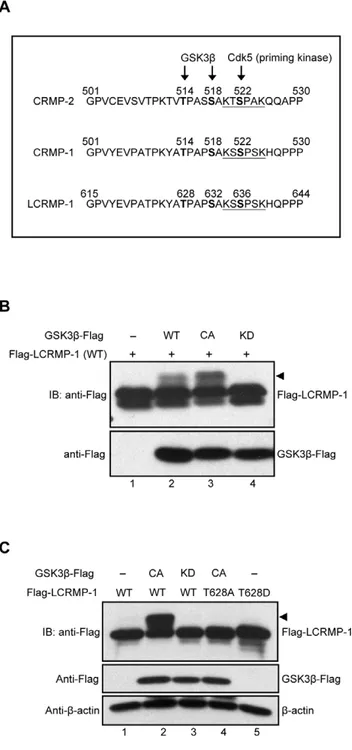
To predict whether the classic GSK3b phosphorylation consensus sequences are existed in LCRMP-1, we first aligned the protein sequences among CRMP-2, CRMP-1, and LCRMP-1 (Fig. 1A). Previous study showed that Cdk5 is a priming kinase that phosphorylates CRMP-2 at Ser-522, following with phosphoryla-tion of CRMP-2 at Thr-514 by GSK3band resulting in functional regulation of neuronal polarization [13]. Therefore, we found that protein sequences of LCRMP-1 contained highly consistent with Cdk5 and GSK3bphosphorylation motif, thus we speculated that a major potential phosphorylation site of LCRMP-1 is located at Thr-628 (Fig. 1A). To examine whether LCRMP-1 can be phosphorylated by GSK3b, HEK293T cells were cotransfected with wild-type Flag-tagged LCRMP-1 (WT) in the presence of empty vector, wild-type GSK3b (WT), constitutively active GSK3b(CA), or kinase-dead GSK3b(KD). Expression of GSK3b
(CA) was more obviously detected mobility shifts (arrowheads) of LCRMP-1 (WT) than GSK3b (WT) (Fig. 1B, lane 2 and 3). However, slow-migration upper bands were completely disap-peared in cells expressing kinase-deficient form of GSK3b (KD) (Fig. 1B, lane 4). These results suggest that LCRMP-1 is a substrate of GSK3band slowly migrating bands were caused by its phosphorylationin vivo.
Next, to determine whether GSK3bphosphorylates LCRMP-1 at Thr-628in vivo, a nonphosphorylated LCRMP-1 mutant was
generated by replacing Thr-628 to Ala (T628A). CL1-0 cells were cotransfected with LCRMP-1 (WT) or a LCRMP-1 (T628A) nonphosphorylated mutant in the presence of either empty vector, GSK3b (CA), or GSK3b (KD). Consistent with previous obser-vations, GSK3b (CA) and GSK3b (KD) were proved to display band shift and non-band shift (arrowheads) of LCRMP-1, respectively (Fig. 1C, lane 2 and 3). Notably, LCRMP-1 (T628A) was resistant to GSK3b (CA) activity and the shifted bands were almost abolished (Fig. 1C, lane 4). This result was similar to the conditions of LCRMP-1 (WT) plus GSK3b (KD) (Fig. 1C, lane 3 and 4), and further confirming that GSK3bindeed phosphorylated LCRMP-1 at Thr-628 residues. Collectively, all these results indicated that LCRMP-1 was specifically phosphor-ylated at Thr-628 by GSK3bin vivo.
Thr-628 phosphorylation of LCRMP-1 is required for cancer cell invasion, migration and filopodia formation
In our current reports, we have found that wild-type LCRMP-1 enhances filopodia formation, and promotes cell migration and invasion in noninvasive human lung cancer cell lines [10]. Since LCRMP-1 can be phosphorylated at Thr-628 by GSK3b, we next questioned whether the function of LCRMP-1 could be regulated by this phosphorylation. To address this, we generated a series of lentivirus that express GFP (control), non-tagged LCRMP-1 (WT), LCRMP-1 (T628A), or LCRMP-1 (T628D), and transduced them into low-invasive CL1-0 lung cancer cells which express low level
Figure 1. LCRMP-1 is a substrate of and phosphorylated by GSK3bat Thr-628.(A) Protein sequence analysis showed the potential consensus site of LCRMP-1 for the phosphorylation by GSK3b. Protein sequences are aligned among CRMP2, CRMP-1, and LCRMP-1. The numbers represent amino acid sites and underline shows a potential phosphorylation site for Cdk5. (B) GSK3bphosphorylates LCRMP-1 (WT) in vivo. HEK293T cells were co-transfected with Flag-tagged LCRMP-1 and distinct activity of Flag-tagged GSK3b(WT, CA and KD form). Equal amount of plasmids were transfected into every condition by using empty vectors. Cells were lysed 30 hr post-transfection and protein extracts were analyzed by immunoblotting with anti-Flag antibodies. (C) GSK3bphosphorylates LCRMP-1 (WT) at Thr-628in vivo. CL1-0 cells were co-transfected either Flag-tagged LCRMP-1(WT) or LCRMP-1 (T628A, T628D) mutants in the presence or absence of distinct activity of Flag-tagged GSK3b (CA and KD form). Empty vectors were used for supplement to equal amount of plasmids in transfection assay. Cell lysates were harvested 48 hr post-transfection and analyzed by immunoblotting with anti-Flag and anti-b-actin antibodies.
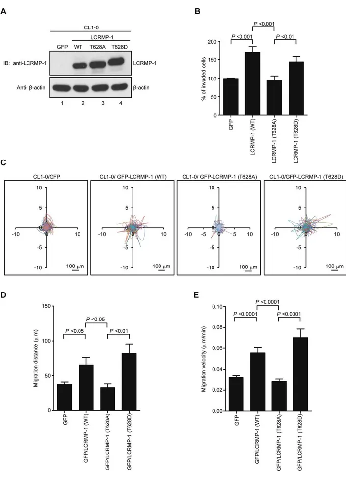
of endogenous LCRMP-1 [11]. Protein expression of wild-type or mutant LCRMP-1 was confirmed by immunoblotting analysis using anti-LCRMP-1 antibodies (Fig. 2A). These cells were then used to examine the ability of cell invasion. As expected, LCRMP-1 (WT) overexpression contributed to an increased cell invasive-ness compared with GFP control (Fig. 2B). However, T628A nonphosphorylated mutant of LCRMP-1 was greatly diminished cell invasiveness (Fig. 2B). Conversely, phospho-mimic LCRMP-1 (T628D), which was expected to mimic the phosphorylated form, displayed enhanced invasion ability similar to wild-type LCRMP-1 (WT). To further explore the effect of Thr-628 phosphorylation of LCRMP-1 expression on CL1-0 cell motility, we performed video time-lapse microscopy assay to monitor moving tracks of at least 10 individual cells over a 20-hour period. Lentivirus-transduced CL1-0 cells expressing GFP-LCRMP-1 (WT) or GFP-LCRMP-1 (T628D) increased both migration distance and migration velocity compared to GFP vector (Fig. 2C, D, and E). However, GFP-LCRMP-1 (T628A) showed marked compression of distance and velocity of migration (Fig. 2C, D, and E). These results suggested that phosphorylation of LCRMP-1 at Thr-628 is a prerequisite for cell invasiveness and cell migration.
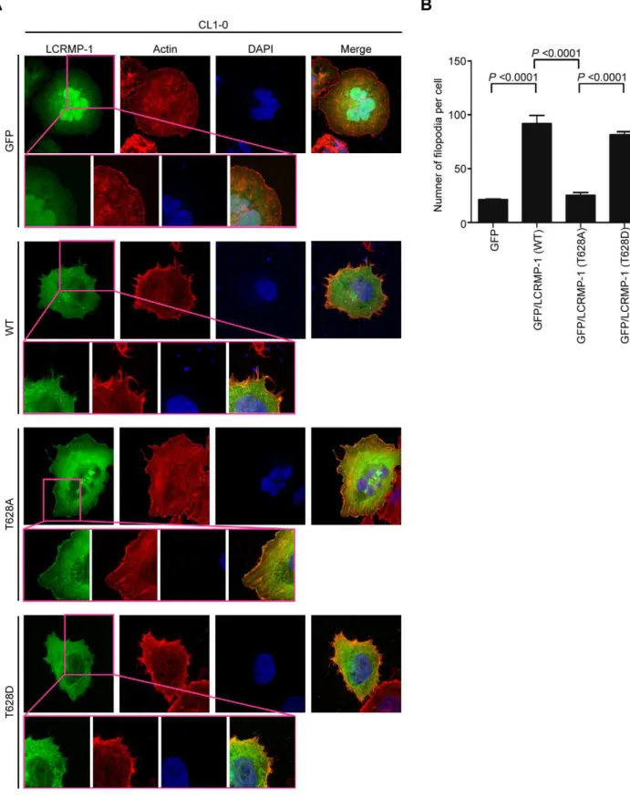
Next, we further examined the effects of GSK3bon LCRMP-1 induced filopodia formation. CL1-0 cells were transiently trans-fected with GFP control, GFP-LCRMP-1 (WT), GFP-LCRMP-1 (T628A), or GFP-LCRMP-1 (T628D) and following by staining with actin using rhodamine-conjugated phalloidin. Consistent with our current reports, immunofluorescence analysis revealed that numbers of filopodia that induced by ectopic expression GFP-LCRMP-1 (WT) in CL1-0 cells were more than that by GFP vector control. Conversely, numbers of filopodia formation were obviously attenuated in cells expressing nonphosphorylated mutant GFP-LCRMP-1 (T628A) (Fig. 3A; 3B, p,0.0001). In ad-dition, GFP-LCRMP-1 (T628D) was also induced more filopodia that similar to GFP-LCRMP-1 (WT), (Fig. 3A; 3B, p,0.0001). These data indicated that phosphorylation of LCRMP-1 at Thr-628 is crucial for filopodia formation.
GSK3bphosphorylates LCRMP-1 and modulates cancer cell invasion
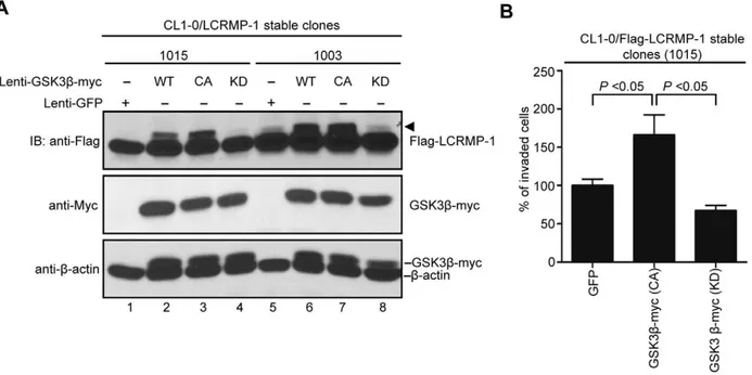
To further detect the effects of GSK3b on LCRMP-1(WT)-induced cancer cell invasion, lentivirus expressing GFP control, GSK3b(WT), GSK3b(CA), or GSK3b(KD) were infected in CL1-0/LCRMP-1 overexpression cells (lines 1015 and 1003) which have been previously shown to strongly induce cell invasiveness [10]. Consistent with our previous findings (Fig. 1B and C), immuno-blotting analysis showed that GSK3b(CA) induced LCRMP-1 with shifted band compared to GFP control (Fig. 4A, lane 1 and 3; lane 5 and 7), but a non-shifted band was observed in GSK3b (KD) (Fig. 4A, lane 4 and lane 8). Based on above conditions, the results of invasion assay were also shown that GSK3b(CA)-introduced CL1-0/LCRMP-1 cells (1015) could further promote cell invasion compared to control cells (Fig. 4B). However, GSK3b (KD)-introduced cells resulted in a decreased in invasion ability (Fig. 4B). Taken together, these results demonstrated that GSK3b could modulate LCRMP-1 activity through a phosphorylation-dependent manner to control cancer cell invasion.
Low expression of inactive GSK3band high expression of LCRMP-1 correlate with poor overall survival in NSCLC patients
Although our results consistently suggested that function of LCRMP-1 could be regulated by GSK3b phosphorylation, such studies do not fully reflect clinical malignancy. Accordingly, we
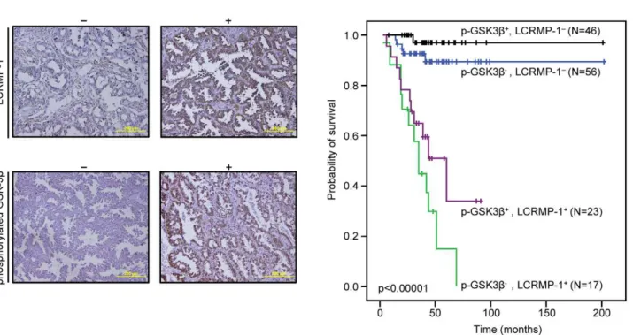
extended our analysis by examining inactive form GSK3b and LCRMP-1 protein expression levels in tumor specimens from 142 NSCLC patients. The clinical characteristics of these patients are summarized in Table 1. Serial sections of each specimen were stained with antibodies against LCRMP-1 and Ser-9-phosphory-lated GSK3bthat indicated the status of inactive form GSK3bdue to the Akt-mediated activation which results in suppression of GSK3b activity through phosphorylation at Ser-9 [15]. Our results showed typical staining of LCRMP-1 and Ser-9-phosphor-ylated GSK3bin patient’s specimen (Fig. 5A). Consistent with our previous reports, high-level LCRMP-1 had significantly poor overall survival compared with low-level LCRMP-1 in patients with NSCLC [10,11]. Notably, analysis of the combined effect of both proteins on patients’ prognoses revealed that patients with low-level expression of inactive form GSK3b and high-level expression of LCRMP-1 had poorer overall survival than those with high-level inactive form GSK3b expression and low-level LCRMP-1 expression (Fig. 5B, p,0.00001). Multivariable Cox proportional-hazards regression analyses, with a stepwise selection model, were present to evaluate the associations of various independent prognostic factors with patient survival (Table 2). These results suggest that high activity GSK3b and high-level LCRMP-1, possibly mimicking the phosphorylated status of LCRMP-1, are associated with increasing cancer invasiveness and poorer overall survival.
Discussion
Our study primarily investigated the regulatory mechanism of post-translation modification associated with the cancer cell migration and invasiveness of LCRMP-1. Here, we showed that GSK3b-dependent phosphorylation of LCRMP-1 positively regulates filopodia formation, migration and cancer cell invasion. On the basis of GSK3b-phosphorylated consensus motifs, Thr-628 amino acid residue of LCRMP-1 is the master phosphorylation site for GSK3b (Fig. 1). A substitution of Thr-628 for Ala in LCRMP-1 led to impair filopodia formation, migration and cancer cell invasion, whereas a replacement of Thr-628 to Asp greatly restored its function (Fig. 2 and 3). Consistent with these observations, ectopic expression of kinase-dead GSK3b dimin-ished LCRMP-1-induced invasive ability (Fig. 4). Moreover, clinical NSCLC patients with low-level of inactive GSK3b and high-level LCRMP-1 protein expression is associated with poor overall survival than those with high-level inactive form GSK3b
expression and low-level LCRMP-1 expression (Fig. 5B). Thus, our results provide evidence to support the crucial mechanism of GSK3b-dependent phosphorylation to control the LCRMP-1-mediated filopodia formation, migration and invasive abilities in cancer cells.
Sequence analysis indicated that there has a Cdk5 (priming kinase) phosphorylation site in both LCRMP-1 and CRMP-1 (Fig. 1A). After phosphorylation at Ser-636 by Cdk5, GSK3bin turn phosphorylates Ser-632 and Thr-628 sequentially. There-fore, GSK3b may induce slower migrating bands in LCRMP-1 including both Ser-632 and Thr-628 phosphorylation in cells, and the bands induced by constitutively active GSK3bwere more active than that of wild-type GSK3b (Fig. 1B). In detailed analysis, we found that LCRMP-1 mutant, T628A, could block most of GSK3b phosphorylation induced migrating bands (Fig. 1C). This may indicate that Thr-628 of LCRMP-1 may be the dominant and important phosphorylation site for GSK3b
The process of tumor invasion is primarily through alternations of the extracellular matrix including actin polymerization and filopodia formation [16]. Our findings reveal that blockage of GSK3b -mediated phosphorylation of LCRMP-1 at Thr-628 leads to a regression of filopodia formation. A previous report was described that GSK-3 phosphorylation for Paxillin is necessary for cytoskeletal rearrangement [17]. Thus, we speculate that GSK3bmay positively promote regulation of actin cytoskeleton and tumor invasion. In contrast to positively role, GSK3b is also reported to perform its suppressive roles for its substrates [18]. GSK3bphosphorylation for some nuclear transcription factors, such as b-catenin and Snail, trigger proteasomal degradation, following with suppression on epithelial–mesenchymal transition (EMT) and tumor invasion [6,19–21]. GSK3b simultaneously localizes in cytoplasm and nucleus [22], as consistent with our results of immunohistochemical staining (Fig. 5A), and LCRMP-1 is a stable cytosolic protein [10]. Therefore, GSK3bgovern cell invasion is possibly dependent on the characterization of its protein substrates. High-level LCRMP-1 expression is associated with poor overall and disease-free survival compared to low expression group in NSCLC patients [10,11]. Thus, LCRMP-1 potentially serves as a best candidate to identify high-risk patients. In this study, we showed a very interesting finding that patients with low-level phosphorylated GSK3band high-level LCRMP-1 expressions had worse overall survival than the other catalog groups. Focusing on the low-level LCRMP-1 expression or the high-level LCRMP-1 expression group, we could also found that high-level of phosphorylated GSK3b expression may discriminate better outcome from low-level phosphorylated GSK3bexpression, respectively. The combined effects of inactive form GSK3b and LCRMP-1 protein expression may have important clinical implica-tions to indicate the high-risk subset of NSCLC patients as candidates for additional effective adjuvant therapy. The results suggest that GSK3bphosphorylation of LCRMP-1 is associated with poor clinical outcome. However, there still have some limitations in our experiments. Although the reference indicated that Ser-9-phosphorylated GSK3b can indicate the status of inactive form GSK3b due to the Akt-mediated activation which results in suppression of GSK3b activity through phosphorylation at Ser-9 [15], whether Ser-9 phosphorylation of GSK3binhibits its ability to phosphorylate LCRMP-1 is still not clear. To solve this problem, generating a specific anti-phospho-Thr-628 LCRMP-1 antibody for immunohistochemistry should be the most important issues in the future. This may confirm the clinical significance of GSK3binduced phosphorylation of LCRMP-1 in NSCLC patients.
In addition, we also found that patients with low-levels (n = 73) or high-levels (n = 69) of Ser-9-phosphorylated GSK3bexpression were almost equal in distribution. Cells exist at different actual levels of active GSK3b unless distinct signaling pathways for inhibiting GSK3bare triggered concurrently, such as MAPK and phosphatidylinositol-3-OH kinase/Akt pathways [20]. Thus, we speculate that distinct activated extent of signaling pathways to induce an inhibition of GSK3b activity may lead to different outcome for patients with LCRMP-1 expression. Therefore, further investigating upstream signaling pathway for regulation of GSK3b may provide a better diagnosis for NSCLC patients with low-levels or high-levels of LCRMP-1 expression.
In conclusion, we show a new regulatory mechanism for GSK3b to phosphorylate an invasion enhancer LCRMP-1 and thus could further fine-tune cancer cell invasion abilities. Additionally, LCRMP-1 expression and Ser-9-phosphorylated GSK3b levels may have clinical implications in the outcome prediction of patients with NSCLC.
Materials and Methods
Ethics statement
This investigation was approved by the Institutional Review Board of the National Taiwan University Hospital and obtained informed written consent statement from all participant patients involved in our study.
Patients and Tumor Specimens
Lung tumor tissue specimens were obtained from patients (n = 142) with histologically confirmed NSCLC who had under-gone complete surgical resections at the National Taiwan University Hospital (Taipei, Taiwan) between December 28, 1995, and December 26, 2005. This investigation was approved by the Institutional Review Board of the National Taiwan Uni-versity Hospital. The enrolled patients had not been treated with neoadjuvant chemotherapy or irradiation therapy. All specimens were formalin fixed, sectioned, stained with hematoxylin and eosin, and examined by microscopy. Pathological staging was performed by Dr. Yih-Leong Chang (Department of Pathology and Graduate Institute of Pathology, National Taiwan University) according to the international staging system for lung cancer [23].
Immunohistochemical analysis
Immunohistochemical staining of tumor tissue samples from patients with NSCLC was carried out as previously described [11]. In brief, the sections for analysis of LCRMP-1 or phosphorylated GSK3b protein expression were first autoclaved in Trilogy Solution (Cell Marque Corp., Rocklin, CA.) or Antigen Retrieval Citra Solution (Biogenex, San Ramon, CA) at 121uC for 10 minutes. The samples were subsequently made a treatment of 3% H2O2-methanol, incubation with DakoCytomation Dual EndogenousEnzyme Block (DakoCytomation, Inc., Carpinteria, CA) for 10 minutes, Ultra V Block (Lab Vision Corporation, Fremont, CA) for 10 minutes, antibody-dilution buffer (Ventana Medical Systems, Inc., Tucson, AZ) for 10 minutes, and finally with a phosphorylated GSK3b (Cell signaling, Danvers, MA) antibody for 6 hours at room temperature or a polyclonal anti– LCRMP-1 antibody (C2; 1:300 dilution) overnight at 4uC. Detection of the immunostaining was determined using Super Sensitive Non-Biotin Polymer HRP Detection System (BioGenex, San Ramon, CA) according to the manufacturer’s protocol.
Modified Boyden chamber invasion assay
Modified Boyden chambers with polycarbonate-membrane inserts (pore size 8mm; Falcon, Becton Dickinson) coated with
30mg Matrigel (BD) were performed cell invasion assays. 2.56104 cells suspended in RPMI medium containing 10% NuSerum assessing with immunoblotting using anti-LCRMP-1 antibody and anti-b-actin antibodies. (B) Nonphosphorylated LCRMP-1 (T628A) mutant lowers activity of cell invasion. The invasive capacity of these cells was determined with the modified Boyden chambers invasion assayin vitro. Percentage of invasive ability was normalized to GFP control. Data were shown as means6SEM for three-independent experiments (n = 3). (C) Nonphosphorylated LCRMP-1 (T628A) mutant greatly suppressed cell migration tracks. Tract plots showed CL1-0 cells expressing GFP, GFP-LCRMP-1 (WT), GFP-LCRMP-1 (T628A), GFP-LCRMP-1 (T628D), respectively. Moving tracks of at least 10 representative cells at the start point all set to ‘0,0’ over a 20-hour period (different lines) showed the representative motility of cells. Scale bar, 100mm. (D, E) Total migration distance (D) and cell migration velocity (E) were quantified from cell tracking assay for 20 hours. Data were presented as means6SEM.
Figure 4. GSK3bmodulates ability of LCRMP-1-induced cancer cell invasion.(A) Lentivirus expressed GFP control, myc-tagged GSK3b(WT), GSK3b(CA), or GSK3b(KD) in CL1-0/LCRMP-1 (WT) overexpression cells (1015 and 1003). After 48 hours postinfection, these cells were lysed and subjected to immunoblotting analysis with using anti-Flag, anti-Myc, and anti-b-actin antibodies. (B) GSK3bactivity affects LCRMP-1-induced cancer cell invasion. CL1-0/LCRMP-1 overexpression cells (1015) were infected with lentivirus expressing GFP control, myc-tagged GSK3b(CA) or GSK3b(KD). After 48 hours postinfection, these cells were subjected to the modified Boyden chambers invasion assayin vitro. Normalization to GFP control served as percentage of invasive ability.
doi:10.1371/journal.pone.0031689.g004
Table 1.LCRMP-1 and phosphorylated GSK3bexpression in relation to clinical parameters and pathological characteristics*.
LCRMP-1 p-GSK3b
Category Subcategory Number #50% (%) .50% (%) P #70% (%) .70% (%) P
Total patients 142 102 (71.8) 40 (28.2) 73 (51.4) 69 (48.6)
Sex Female 78 55 (53.9) 23 (57.5) 0.7 44 (60.3) 34 (49.3) 0.188
Male 64 47 (46.1) 17 (42.5) 29 (39.7) 35 (50.7)
Histological type{ Adenocarcinoma 123 86 (87.8) 37 (94.9) 0.215 60 (85.7) 63 (94.0) 0.108
Squamous cell carcinoma 14 12 (12.2) 2 (5.1) 10 (14.3) 4 (6.0)
Tumor size, cm .3 74 57 (55.9) 17 (42.5) 0.151 41 (56.2) 33 (47.8) 0.32
#3 68 45 (44.1) 23 (57.5) 32 (43.8) 36 (52.2)
Vascular invasion Positive 25 19 (18.6) 6 (15.0) 0.61 14 (19.2) 11 (15.9) 0.613
Negative 117 83 (81.4) 34 (85.0) 59 (80.8) 58 (84.1)
Lymph node metastasis Positive 32 18 (17.7) 14 (35.0) 0.026 18 (24.7) 14 (20.3) 0.534
Negative 110 84 (82.4) 26 (65.0) 55 (75.3) 55 (79.7)
Extranodal extension Positive 20 10 (9.8) 10 (25.0) 0.019 11 (15.1) 9 (13.0) 0.729
Negative 122 92 (90.2) 30 (75.0) 62 (84.9) 60 (87.0)
Tumor stage Stage I 107 82 (80.4) 25 (62.5) 53 (72.6) 54 (78.3)
Stage II 17 13 (12.8) 4 (10.0) 11 (15.1) 6 (8.7)
Stage III–IV 18 7 (6.9) 11 (27.5) 0.004 9 (12.3) 9 (13.0) 0.505
LCRMP-1 expression, % .50 102 – – – 56 (76.7) 46 (66.7) 0.184
#50 40 – – 17 (23.3) 23 (33.3)
p-GSK3bexpression, % .70 69 46 (45.1) 23 (57.5) 0.184 – – –
#70 73 56 (54.9) 17 (42.5) – –
*Pvalues were calculated using a two-sided chi-squared test. Abbreviations: LCRMP-1, long-form collapsin response mediator protein-1; p-GSK3b, phosphorylated Glycogen synthase kinase-3b.
(Invitrogen, Eugene, OR) were plated in the upper chambers, and 1 ml medium was added to cover the lower chambers. After 24 hours incubation at 37uC, cells were fixed with methanol at room temperature for 10 minutes. After fixation, samples were stained with a 50mg/ml solution of propidium iodide (Sigma, St. Louis, MO) at room temperature for 30 minutes. Each membrane was photographed and counted the number of cells under a
microscope at a magnification of 650, using the Analytical Imaging Station software package (Imaging Research Inc., St. Catharines, ON, Canada). Each experiment was assayed in triplicate.
Immunofluorescence staining for observation of filopodia formation
Transfected or lentivirus-infected cells were fixed with 3.7% cold paraformaldehyde, washed with PBS, following by permea-bilizing with 0.1% Triton X-100. The cells were then stained with rhodamine-conjugated phalloidin (red, Molecular Probes, Eugene, OR). The cells were mounted onto microscope slides with ProLongH Gold antifade reagent with DAPI (Molecular Probes) and then examined and photographed using LSM 700 laser scanning confocal microscope from Carl Zeiss.
Cell migration analysis
Moving tracks of migrating cells were performed by video time-lapse microscopy as previously described [24]. In brief, cells were maintained in growth medium at 37uC/5% CO2and time-lapse images were observed under a AF 6000 LX microscope (Meyer Instruments,Inc.) for the time period of 20 hours. Images were taken with a CoolSNAP HQ CCD camera (Roper Scientific, NJ) at 5-minute intervals and processed by MetaMorph 5.0 software (Universal Imaging, Downingtown, PA).
Cell culture and transfection
The human lung adenocarcinoma cell lines (CL1-0 cells) were isolated from a 64-year-old male patient with a poorly differen-Table 2.Hazard ratios for death (from any cause) among
patients with NSCLC, according to multivariable Cox regression analysis*.
Variable Hazard ratio (95% C.I.) P
LCRMP-1 26.22 (7.56 to 90.94) ,.0001
p-GSK3b 0.48 (0.2 to 1.16) 0.102
Sex 2.48 (0.97 to 6.37) 0.058
Histological type 0.15 (0.04 to 0.52) 0.003
Tumor stage 0.47 (0.3 to 0.73) 0.001
*Stepwise selection was used to choose the optimal multivariable Cox proportional hazard regression model. LCRMP-1 and phosphorylated GSK3b expression was designated as ‘high’ or ‘low’ using 50% and 70% cell positivity as the cut-off point respectively, and were adjusted by histological type (squamous cell carcinoma as the referent vs. adenocarcinoma), and stage (stage I as the referent vs. stage II vs. stage III).Pvalues (two-sided) were calculated using a chi-square test. Abbreviations, LCRMP-1, long-form collapsin response mediator protein-1; p-GSK3b, phosphorylated Glycogen synthase kinase-3b; CI, confidence interval.
doi:10.1371/journal.pone.0031689.t002
Figure 5. Kaplan-Meier survival plots for NSCLC patients grouped by phosphorylated GSK3band LCRMP-1 protein expression levels.(A) Typical protein expression patterns of phosphorylated GSK3band LCRMP-1 were detected by immunohistochemistry using anti-phospho-GSK3b(Ser9) and anti-LCRMP-1 antibodies (C2) in serial dissections of primary tumor specimens from 142 NSCLC patients who underwent surgical resections. Results are shown+and2denotes tumors with and without over-expression with indicated protein respectively. Scale bars, 100mm. p-GSK3bwas represented to phosphorylated GSK3b. (B) Kaplan–Meier analysis of overall survival for 142 NSCLC patients with p-GSK3b2-LCRMP-12, p-GSK3b2-LCRMP-1+, p-GSK3b+-LCRMP-12, and p-GSK3b+-LCRMP-1+.Pvalues were performed by 2-sided log-rank tests.
tiated adenocarcinoma and selected in our laboratory by in vitro Transwell invasion to get 5 sublines with progressive invasiveness, with similar genotypic background (designated CL1-1, CL1-2, CL1-3, CL1-4, and CL1-5) as previously described [25]. HEK293T cell lines were purchased from American Type Culture Collection (ATCC, USA). The CL1-0 and HEK293T cells were grown in RPMI and DMEM medium containing 10% FBS and 2 mM L-glutamine (all from Invitrogen, Eugene, OR) at 37uC in a humidified atmosphere of 5% CO2-95% air, respectively. All cell lines in this study were tested with mycoplasma-free condition. All transfection experiments were carried out using Lipofectamine or Lipofectamine 2000 reagents (Invitrogen) according to the manufacturer’s instructions.
Protein sequences alignment and Plasmids
Amino acid sequences alignment of CRMP-2, CRMP-1, and LCRMP-1 are based on their Gene bank accession number NP_001377, NP_001304, and NP_001014809, respectively. The LCRMP-1 expression plasmid pCMV-Tag2A-LCRMP-1(WT) and pEGFP-LCRMP-1(WT) were described as previously [10]. Amino-acid substitution mutants of LCRMP-1 were generated by PCR-based site-directed mutagenesis with a QuikChange kit (Stratagene, Santa Clara, CA) and verified by DNA sequencing.
FLAG-tagged GSK3b (WT, CA and KD form) expression
plasmids were subcloned from pCMV-5A-GSK3b (WT, CA and KD form, a gift from M.-C. Hung) into pFLAG -CMV-5a vector (Sigma).
Antibodies
Primary antibodies for immunoblotting were as follows: monoclonal anti-Flag (M2; Sigma), anti-Myc (9E11; Millipore, Billerica, MA), anti-b-actin (Sigma) and polyclonal anti-LCRMP-1 antibody. The HRP conjugated goat mouse and goat anti-rabbit secondary antibodies were purchased from (Amersham Biosciences, Pittsburgh, PA).
Lentivirus production and transduction
GFP-tagged, untagged LCRMP-1 (WT, T628A and T628D),
and myc-tagged GSK3b (WT, CA and KD form) were
constructed by cloning their cDNA into pTYEF lentiviral vector. Briefly, HEK293T cells were contransfected with the indicated lentiviral vector and three helper plasmids pHP-dl-N/A, pHEF-VSVG, and pCEP4-Tat by using Lipofectamine 2000 reagents according to manufacturer’s protocols. Virus-containing medium was collected at 24, 48, 72 hours post-transfection, centrifuged,
and filtered through 0.45mm-pore-size filters. The percentage of pTYEF-GFP-infected cells by flow cytometry were determined the relative lentivirus titers. Cells were infected with GFP or the indicated lentivirus in media containing polybrene (8mg/ml). After twenty-four hours post-infection, cells were treated with fresh medium for 24–48 hours and then used for all experiments.
Cell lysate preparation and immunoblotting
All experiments were performed according to standard protocols. Briefly, preparation of whole-cell lysates for immuno-blotting and immunoprecipitation were using IP lysis buffer (20 mM Tris, pH 7.5, 150 mM NaCl, 0.5% Nonidet P-40, 100mM Na3VO4, 50 mM NaF, 30 mM sodium pyrophosphate) containing protease inhibitors (protease inhibitor cocktail; Roche Diagnostics, Basel, Switzerland). After brief sonication and centrifugation, protein samples were resolved by SDS-PAGE gels, transferred into PVDF membranes (Millipore), blotted with the indicated antibodies and finally detected chemiluminescent signals using X-ray films.
Statistical analysis
Data are shown as Mean6SEM. and statistical analyses were performed by Student’s t-test or Pearson’s x2 test. The overall
survivals for patient groups with different expression signatures were determined using SPSS software (v10.0; SPSS, Inc., Chicago, IL) by the Kaplan–Meier method and two-sided log-rank tests. Immunoreactivity in more than 50% and 70% of the tumor specimens was defined to high level of LCRMP-1 and phosphor-ylated GSK3b expression, respectively. P values,0.05 were considered to be statistically significant.
Acknowledgments
We thank H. K. Sytwu (Graduate Institute of Medical Sciences, National Defense Medical Center, Taipei, Taiwan) for providing the lentiviral plasmids for the lentivirus infection system, and Mien-Chie Hung (Department of Molecular and Cellular Oncology, Breast Cancer Basic Research Program, The University of Texas M. D. Anderson Cancer Center, Houston, Texas) for providing the pCMV-5A-GSK3b(WT, CA and KD form) plasmids, and Pei-Fang Hung, Shih-Han Kao, and Shuenn-Chen Yang for technical assistance.
Author Contributions
Conceived and designed the experiments: W-LW T-MH S-HP P-CY. Performed the experiments: W-LW S-HP. Analyzed the data: W-LW T-MH Y-LC C-TW S-HP. Wrote the paper: W-LW S-HP P-CY.
References
1. Steeg PS (2006) Tumor metastasis: mechanistic insights and clinical challenges. Nat Med 12: 895–904.
2. Steeg PS (2003) Metastasis suppressors alter the signal transduction of cancer cells. Nat Rev Cancer 3: 55–63.
3. Frame S, Cohen P (2001) GSK3 takes centre stage more than 20 years after its discovery. Biochem J 359: 1–16.
4. Doble BW, Woodgett JR (2003) GSK-3: tricks of the trade for a multi-tasking kinase. J Cell Sci 116: 1175–1186.
5. Eldar-Finkelman H (2002) Glycogen synthase kinase 3: an emerging therapeutic target. Trends Mol Med 8: 126–132.
6. Liu C, Li Y, Semenov M, Han C, Baeg GH, et al. (2002) Control of beta-catenin phosphorylation/degradation by a dual-kinase mechanism. Cell 108: 837–847. 7. Fiol CJ, Mahrenholz AM, Wang Y, Roeske RW, Roach PJ (1987) Formation of protein kinase recognition sites by covalent modification of the substrate. Molecular mechanism for the synergistic action of casein kinase II and glycogen synthase kinase 3. J Biol Chem 262: 14042–14048.
8. Shih JY, Yang SC, Hong TM, Yuan A, Chen JJ, et al. (2001) Collapsin response mediator protein-1 and the invasion and metastasis of cancer cells. J Natl Cancer Inst 93: 1392–1400.
9. Shih JY, Lee YC, Yang SC, Hong TM, Huang CY, et al. (2003) Collapsin response mediator protein-1: a novel invasion-suppressor gene. Clin Exp Metastasis 20: 69–76.
10. Pan SH, Chao YC, Hung PF, Chen HY, Yang SC, et al. (2011) The ability of LCRMP-1 to promote cancer invasion by enhancing filopodia formation is antagonized by CRMP-1. J Clin Invest 121: 3189–3205.
11. Pan SH, Chao YC, Chen HY, Hung PF, Lin PY, et al. (2010) Long form collapsin response mediator protein-1 (LCRMP-1) expression is associated with clinical outcome and lymph node metastasis in non-small cell lung cancer patients. Lung Cancer 67: 93–100.
12. Wang LH, Strittmatter SM (1996) A family of rat CRMP genes is differentially expressed in the nervous system. J Neurosci 16: 6197–6207.
13. Yoshimura T, Kawano Y, Arimura N, Kawabata S, Kikuchi A, et al. (2005) GSK-3beta regulates phosphorylation of CRMP-2 and neuronal polarity. Cell 120: 137–149.
15. Cross DA, Alessi DR, Cohen P, Andjelkovich M, Hemmings BA (1995) Inhibition of glycogen synthase kinase-3 by insulin mediated by protein kinase B. Nature 378: 785–789.
16. Pollard TD, Borisy GG (2003) Cellular motility driven by assembly and disassembly of actin filaments. Cell 112: 453–465.
17. Cai X, Li M, Vrana J, Schaller MD (2006) Glycogen synthase kinase 3- and extracellular signal-regulated kinase-dependent phosphorylation of paxillin regulates cytoskeletal rearrangement. Mol Cell Biol 26: 2857–2868. 18. Luo J (2009) Glycogen synthase kinase 3beta (GSK3beta) in tumorigenesis and
cancer chemotherapy. Cancer Lett 273: 194–200.
19. Brembeck FH, Schwarz-Romond T, Bakkers J, Wilhelm S, Hammerschmidt M, et al. (2004) Essential role of BCL9-2 in the switch between beta-catenin’s adhesive and transcriptional functions. Genes Dev 18: 2225–2230.
20. Zhou BP, Deng J, Xia W, Xu J, Li YM, et al. (2004) Dual regulation of Snail by GSK-3beta-mediated phosphorylation in control of epithelial-mesenchymal transition. Nat Cell Biol 6: 931–940.
21. Conacci-Sorrell M, Simcha I, Ben-Yedidia T, Blechman J, Savagner P, et al. (2003) Autoregulation of E-cadherin expression by cadherin-cadherin interac-tions: the roles of beta-catenin signaling, Slug, and MAPK. J Cell Biol 163: 847–857.
22. Bijur GN, Jope RS (2003) Glycogen synthase kinase-3 beta is highly activated in nuclei and mitochondria. Neuroreport 14: 2415–2419.
23. Sobin L, Wittekind C (2002) TNM Classification of Malignant Tumours. 6th ed. New Jersey, USA: John Wiley & Sons.
24. Chao YC, Pan SH, Yang SC, Yu SL, Che TF, et al. (2009) Claudin-1 is a metastasis suppressor and correlates with clinical outcome in lung adenocarci-noma. Am J Respir Crit Care Med 179: 123–133.