Evaluation of Intestinal Phosphate Binding to
Improve the Safety Profile of Oral Sodium
Phosphate Bowel Cleansing
Stef Robijn, Benjamin A. Vervaet, Patrick C. D’Haese*, Anja Verhulst
Laboratory of Pathophysiology, University of Antwerp, Antwerp, Belgium
Abstract
Prior to colonoscopy, bowel cleansing is performed for which frequently oral sodium phos-phate (OSP) is used. OSP results in significant hyperphosphos-phatemia and cases of acute kidney injury (AKI) referred to as acute phosphate nephropathy (APN; characterized by nephrocalcinosis) are reported after OSP use, which led to a US-FDA warning. To improve the safety profile of OSP, it was evaluated whether the side-effects of OSP could be pre-vented with intestinal phosphate binders. Hereto a Wistar rat model of APN was developed. OSP administration (2 times 1.2 g phosphate by gavage) with a 12h time interval induced bowel cleansing (severe diarrhea) and significant hyperphosphatemia (21.79±5.07 mg/dl 6h after the second OSP dose versus 8.44±0.97 mg/dl at baseline). Concomitantly, serum PTH levels increased fivefold and FGF-23 levels showed a threefold increase, while serum calcium levels significantly decreased from 11.29±0.53 mg/dl at baseline to 8.68±0.79 mg/dl after OSP. OSP administration induced weaker NaPi-2a staining along the apical proximal tubular membrane. APN was induced: serum creatinine increased (1.5 times baseline) and nephrocalcinosis developed (increased renal calcium and phosphate content and calcium phosphate deposits on Von Kossa stained kidney sections). Intestinal phos-phate binding (lanthanum carbonate or aluminum hydroxide) was not able to attenuate the OSP induced side-effects. In conclusion, a clinically relevant rat model of APN was devel-oped. Animals showed increased serum phosphate levels similar to those reported in hu-mans and developed APN. No evidence was found for an improved safety profile of OSP by using intestinal phosphate binders.
Introduction
Colonoscopy is the standard procedure for diagnostic evaluation of the colon, with more than 14 million examinations performed annually in the United States alone. [1] As its efficacy largely depends on the degree to which fecal material is removed from the colon, bowel cleansing is car-ried out for which frequently oral sodium phosphate (OSP) solutions or tablets are used. OSP increases the osmolality of the intestinal lumen, thereby retaining fluid and inducing peristalsis
OPEN ACCESS
Citation:Robijn S, Vervaet BA, D’Haese PC, Verhulst A (2015) Evaluation of Intestinal Phosphate Binding to Improve the Safety Profile of Oral Sodium Phosphate Bowel Cleansing. PLoS ONE 10(3): e0116590. doi:10.1371/journal.pone.0116590
Academic Editor:Utpal Sen, University of Louisville, UNITED STATES
Received:September 19, 2014
Accepted:December 11, 2014
Published:March 19, 2015
Copyright:© 2015 Robijn et al. This is an open access article distributed under the terms of the Creative Commons Attribution License, which permits unrestricted use, distribution, and reproduction in any medium, provided the original author and source are credited.
Data Availability Statement:All relevant data are within the paper and its Supporting Information files.
Funding:SR was funded by a PhD grant of the Agency for Innovation by Science and Technology in Flanders (http://www.iwt.be/). AV is a postdoctoral fellow of the Fund for Scientific Research Flanders (http://www.fwo.be/). This research was funded by FWO research grant G.0583.09.
and colonic evacuation. [2,3] The major advantage of OSP is the relatively low volume required for efficient bowel cleansing, generally resulting in a higher patient compliance and better effica-cy compared to other agents such as polyethylene glycol (PEG). [4–11] For years, a standard OSP procedure consisted of two 45 ml (or 20 tablets) doses which had to be taken with a time in-terval of 10–12 hours (usually the evening before and the morning of the colonoscopy). As each dose contained a mixture of NaH2PO4and Na2HPO4equivalent to*6 g phosphorus (*18 g
phosphate), it far exceeded the normal daily intake of*1 g phosphorus. [12]
Since the case report of Desmeuleset al. [13] in 2003, an increasing number of cases [14–26]
are described in which patients develop (in some cases irreversible) acute kidney injury (AKI) after the use of OSP. Renal biopsy findings include calcium phosphate (CaP) deposits in the distal tu-bules and collecting ducts, often accompanied by tubular atrophy and mild to moderate interstitial fibrosis, which led to the term“acute phosphate nephropathy”(APN). [12,14,27] This condition can be attributed to the acute OSP load which leads to significantly increased serum phosphate lev-els that stimulate PTH secretion which, in turn, leads to endocytosis of the NaPi-2a/c co-transport-ers from the brush border of the proximal tubules. This endocytosis inhibits renal phosphate reabsorption and, hence, results in significant urinary phosphate wasting. [28–30] After OSP ad-ministration, serum phosphate levels double compared to baseline and urinary phosphate excre-tion increases eightfold, which may result in intratubular CaP crystal formaexcre-tion and retenexcre-tion. [12,29,31,32]
Following its initial use, the recommended OSP dose was reduced (a 45 ml or 20 tablets dose followed by a 30 ml or 12 tablets dose), providing equally effective bowel cleansing with less pro-nounced, however still significant, hyperphosphatemia. [10,33] In 2008, however, the US Food and Drug Administration issued a safety alert stating that OSP for bowel cleansing should only be avail-able by prescription which eventually caused a discontinuation of many (over-the-counter) OSP products. Currently, OSP remains available in tablet form by prescription only. [3,12,27,34–36]
As OSP generally results in a higher patient compliance and better efficacy compared to PEG, an important therapeutic approach could consist of the prevention of phosphate absorp-tion to improve the safety profile of OSP. [10,27] In this context, the potential use of lantha-num carbonate, an efficient non-calcium containing phosphate binder with very low systemic absorption, used to treat hyperphosphatemia in dialysis patients is worthy of consideration. [37] Aluminum-based phosphate binders are also highly effective. Despite their association with cognitive disturbances, osteomalacia and anemia due to aluminum accumulation over time in patients with end-stage renal disease (aluminum is excreted exclusively by the kidney), they still remain in use either in combination with other phosphate binding agents or for acute control of hyperphosphatemia where risks of accumulation are minimal. [38,39] Its elimina-tion pathway implies that there is no concern about aluminum accumulaelimina-tion in subjects with normal renal function.
In this study, a model of APN was developed in the Wistar rat. As well as the known acute effects of OSP (hyperphosphatemia, hypocalcemia and APN), the effects of OSP on serum PTH and FGF-23 levels and on the expression of NaPi co-transporter proteins in the kidney (NaPi-2a) were investigated. Finally, it was determined whether OSP induced side-effects could be prevented by intestinal phosphate binding using either lanthanum carbonate or aluminum hydroxide.
Materials and Methods
Optimization of the composition of the OSP dose
In a pilot experiment, 20 male Wistar rats (*300 g; Charles River) were randomly assigned to
(iii) 0.5 g and (iv) 0.55 g phosphorus (corresponding to 1.2 g; 1.35 g; 1.5 g and 1.65 g phosphate, respectively) by gavage. At different time points (baseline, 1 hour, 3 hours, 6 hours and 24 hours after the OSP dose) blood was drawn from the tail vein in restrained, conscious animals for the determination of serum phosphate. In a second phase, the optimized OSP dose (corre-sponding to 0.4 g phosphorus; 1.2 g phosphate) was administered to 6 male Wistar rats (*360
g; Charles River) by gavage based on the standard OSP dosing regimen (2 OSP doses with a time interval of 12 hours). Animals were fasted during the study period, but had access to tap waterad libitum. At different time points (baseline, 12 hours after the first OSP dose and 3
hours, 6 hours, 24 hours and 48 hours after the second dose) blood was drawn from the tail vein for the determination of serum phosphate.
The composition of the OSP dose was based on the therapeutically used Fleet Phospho-soda [2,12,40] and contained Na2HPO4. 2 H2O (Analar) and NaH2PO4. H2O (Merck) in a weight ratio of*1:2.3.
Evaluation of the rat model of APN and effect of intestinal phosphate
binding
30 male Wistar rats (*380 g; Charles River) were randomly assigned to five experimental
groups (n = 6 per group): (i) a group receiving 1000 mg lanthanum carbonate (suspended in 0.1% carboxymethylcellulose; CMC) followed by OSP corresponding to 0.4 g phosphorus (1.2 g phosphate; 0.3x molar lanthanum/phosphate ratio), (ii) a group receiving 289.5 mg aluminum hydroxide (suspended in 0.1% CMC) followed by the same OSP dose (0.3x molar aluminum/phosphate ratio), (iii) a group receiving 1000 mg aluminum hydroxide (suspended in 0.1% CMC) followed by the same OSP dose (1x molar aluminum/phosphate ratio), (iv) a group receiving vehicle (0.1% CMC) followed by the same OSP dose and (v) a control group receiving vehicle followed by water by gavage. Based on the standard OSP dosing regimen, two OSP doses (together with lanthanum carbonate, aluminum hydroxide or vehicle) were admin-istered with a time interval of 12 hours. Animals were fasted during the study period, but had access to tap waterad libitum.
Lanthanum carbonate, aluminum hydroxide (or vehicle) and OSP (or water) were adminis-tered through separate gavages to avoid phosphate binding prior to arrival in the intestine which would not reflect actual intestinal complexation.
At different time points (baseline, 12 hours after the first OSP dose and 3 hours after the sec-ond OSP dose) blood was drawn from the tail vein in restrained, conscious animals for the de-termination of a series of serum parameters. At sacrifice (6 hours after the second OSP dose), animals were anaesthetized with 30 mg/kg sodium pentobarbital (Nembutal; Ceva Santé Ani-male) via intravenous injection, opened through a midline incision and exsanguinated through the aorta abdominalis. The left kidney and intestinal tract were removed and slices of renal tis-sue were fixed in neutral buffered formalin (FNB) for 4 hours, rinsed with 70% isopropanol and embedded in paraffin for (immuno-) histology or digested in 65% nitric acid for chemical analysis. The content of the entire colon and terminal 5 cm of the ileum was collected to exam-ine bowel cleansing.
Ethics statement
Serum biochemistry
Serum phosphate (mg/dl) was measured using the Ecoline Phosphate FS assay (DiaSys). Serum calcium (mg/dl) was determined by flame atomic absorption spectrometry (FAAS; Perkin-Elmer). Serum creatinine (mg/dl) was measured using the kinetic alkaline picrate (Jaffe) reaction. Serum intact PTH (Immutopics) (pg/ml) and intact FGF-23 (Kainos Laboratories) (pg/ml) were both de-termined using ELISA kits.
Bowel cleansing
Besides visual observation of diarrhea, bowel cleansing was examined by collecting the bowel content of the entire colon as well as the terminal 5 cm of the ileum in a 15 ml centrifuge tube during sacrifice. Water was added up to a volume of 14 ml and content was vigorously mixed. Subsequently, the tubes were centrifuged at 3000 rpm (1620 g) for 45 minutes and the percent-age of visible insoluble bowel content (pellet; 14 ml equals 100%) was used as a measure of bowel cleansing. [40]
Renal CaP calcifications
Renal calcifications were visualized by Von Kossa staining. Deparaffinized 4μm tissue sections
were incubated in 5% silver nitrate for 45 minutes. Slides were rinsed in water, incubated in 1% pyrogallol for 3 minutes, rinsed with water, fixed in 5% sodium thiosulfate for 1 minute and counterstained with hematoxylin/eosin. In each section CaP deposits were quantified histo-morphometrically using Axiovision 4.5 software (Carl Zeiss). Renal calcification is expressed as area% and was calculated as the ratio of Von Kossa positive (pixel²) on renal tissue
(pixel²) area.
Renal crystal content was also quantitatively assessed by measuring calcium and phosphate content in renal tissue digestion solutions. Briefly, transversal slices of renal tissue were weighed after removal, digested in 65% nitric acid at 60°C overnight and diluted in 0.1% lanthanum ni-trate to eliminate chemical interference during subsequent spectrometric analysis. In this solu-tion total calcium content was measured by FAAS (Perkin-Elmer) and total phosphate content using the Ecoline Phosphate FS assay (DiaSys).
Immunohistochemistry
Immunohistochemical staining was performed on FNB-fixed, deparaffinized renal sections for NaPi-2a. Heat-induced epitope retrieval was performed by microwaving the sections (2x 5 min) in 0.1 M citrate buffer (pH 6.0). The tissue sections were then blocked with normal goat serum (20% in TSB with 1% Triton X-100) for 20 min and incubated overnight with polyclonal rabbit anti-human NaPi-2a (1:40; Novus Biologicals; NBP2-13328). Biotinylated goat anti-rabbit (Vector Laboratories) was used as secondary antibody. Avidin/biotinylated peroxidase complex (VEC-TASTAIN ABC KIT, Vector Laboratories) was added as signal amplifier and 3-Amino-9-ethyl-carbazole (AEC, Sigma-Aldrich) was used as substrate. The sections were counterstained with hematoxylin. Sections in which the primary antibody was omitted were used as negative controls.
Statistical analysis
calcium and phosphate content and the histomorphometrical calcification score and between the serum parameters (FGF-23, PTH, calcium and phosphate). Statistics were performed with SPSS Statistics 20.0.
Results
Optimization of the composition of the OSP dose
Administration of a single OSP dose resulted in significantly increased serum phosphate levels for all tested doses (0.4 g, 0.45 g, 0.5 g and 0.55 g phosphorus) and induced diarrhea in all ani-mals, reflecting efficient bowel cleansing. However, because of the mortality in the groups re-ceiving 0.5 g and 0.55 g phosphorus (20% and 80% respectively), it was decided to use the lowest dose (0.4 g phosphorus) in the second phase of the optimization study.
Two OSP doses (corresponding each to 0.4 g phosphorus) with a 12 hours’time interval in-duced diarrhea in all animals and resulted in significantly increased serum phosphate concentra-tions with the highest levels (nearly two times baseline and thus resembling the clinical situation in humans receiving OSP) [31,32] occurring 6 hours after the second dose (14.8 ± 3.1 vs 9.2 ± 0.6 mg/dl at baseline) and without mortality. This OSP dosing regimen was further evaluated.
Evaluation of the rat model of APN
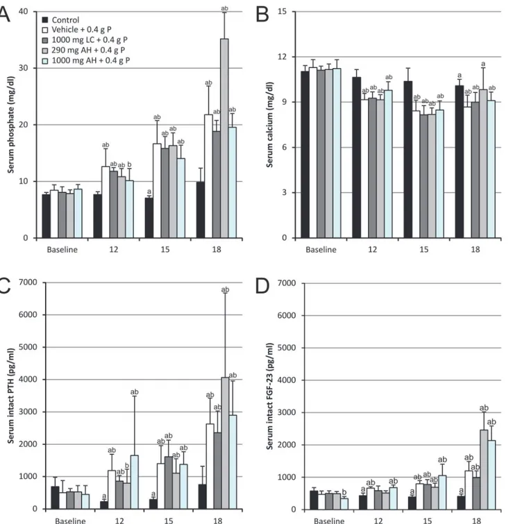
Acute serum effects of OSP. Administration of two OSP doses (corresponding each to 0.4 g phosphorus or 1.2 g phosphate) with a 12 hours’time interval resulted in transiently increased serum phosphate levels compared to control animals (Fig. 1A). Serum phosphate levels peaked at 6 hours after the administration of the second OSP dose and increased up to more than twofold, i.e. 21.79 ± 5.07 mg/dl versus 8.44 ± 0.97 mg/dl at baseline. Concomitantly, serum calcium signifi-cantly decreased from 11.29 ± 0.53 mg/dl at baseline to 8.68 ± 0.79 mg/dl after OSP (Fig. 1B). Serum phosphate and calcium negatively correlated with each other (r = -0.75, p<0.01;Table 1).
Together with the acute changes in serum phosphate and calcium levels, serum (intact) PTH (Fig. 1C) and (intact) FGF-23 (Fig. 1D) levels also significantly increased already at 12 hours after the first OSP dose. The increase in serum PTH concentration was more pronounced than that of FGF-23. While for PTH a fivefold increase versus baseline was seen, FGF-23 levels showed a threefold increase after OSP administration. Both serum FGF-23 and serum PTH strongly corre-lated with serum phosphate (r = 0.85 and r = 0.80 respectively, p<0.01;Table 1), whereas for
serum calcium levels there was a negative correlation with PTH as well as FGF-23 (r = -0.55 and r = -0.69 respectively, p<0.01;Table 1).
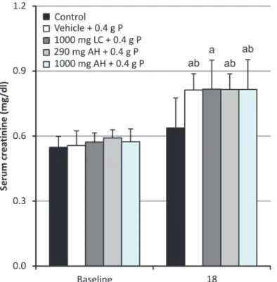
Acute phosphate nephropathy. Serum creatinine concentrations (Fig. 2) significantly in-creased to 1.5 times baseline after OSP administration, indicating the development of AKI (KDIGO). [41]
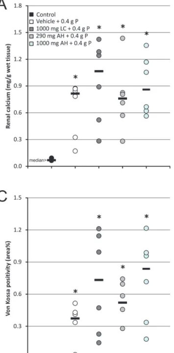
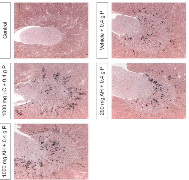
Renal calcium content (Fig. 3A) was significantly higher in animals receiving OSP as com-pared to control animals with a median of 0.815 mg/g wet renal tissue (range: 0.170–0.868 mg/g) compared to 0.068 mg/g wet renal tissue (range: 0.064–0.092 mg/g). A similar pattern was ob-served for the renal phosphate content (Fig. 3B) with a median of 2.64 mg/g wet renal tissue (range: 2.21–2.84 mg/g) in animals receiving OSP compared to 1.96 mg/g wet renal tissue (range: 1.79–2.20 mg/g) in controls. Histomorphometric quantification of Von Kossa stained kidney sections (Fig. 3C) confirmed these results. Renal calcium and phosphate concentrations strongly correlated with each other (r = 0.80, p<0.01) as well as with the area% Von Kossa
posi-tivity (r = 0.82 and r = 0.74 respectively, p<0.01). The calcifications were mainly confined to the
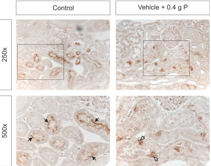
Expression of renal NaPi co-transporters. NaPi-2a was clearly localized to the apical membrane of proximal tubules in control animals. In animals receiving OSP, NaPi-2a staining was weaker along the apical membrane and appeared more intracellular (Fig. 5).
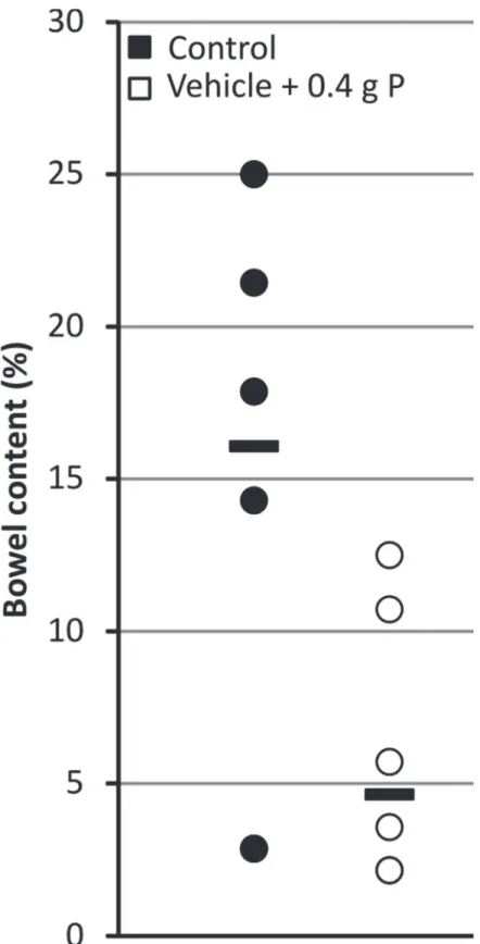
Bowel cleansing. OSP administration induced diarrhea in all animals. The amount of insoluble bowel content of the entire colon and terminal 5 cm of the ileum (used as a measure
Fig 1. Serum (A) phosphate, (B) calcium, (C) intact PTH and (D) intact FGF-23 levels.Data are presented as mean±standard deviation. (aP<0.05 vs baseline;bP<0.05 vs control) (12: 12 hours after the first OSP dose; 15: 3 hours after the second OSP dose and 18: 6 hours after the second OSP dose; n = 6 per group; statistical tests: Mann–Whitney-U and Wilcoxon-signed-rank).
of bowel cleansing) decreased from a median of 16.1% (range: 2.9–25.0%) in control animals to 4.6% (range: 2.1–12.5%) in animals receiving OSP (Fig. 6).
Effect of intestinal phosphate binding
Neither phosphate binder treatment was able to attenuate the serological effects of OSP (serum phosphate, calcium, PTH and FGF-23 levels;Fig. 1) and as a result did not have any effect on the development of APN (Figs.2,3and4).
Discussion
It is presumed that the incidence of APN is highly underestimated because many cases would remain undiagnosed (clinically silent) when the amount of affected renal tubules is limited. [24,29] As OSP generally results in a higher patient compliance and better efficacy compared
Table 1. Correlations between serum parameters in animals receiving OSP.
Spearman correlation coefficient (r) Serum phosphate Serum calcium Serum PTH Serum FGF-23
Serum phosphate X -0.75** 0.80** 0.85**
Serum calcium -0.75** X -0.55** -0.69**
Serum PTH 0.80** -0.55** X 0.79**
Serum FGF-23 0.85** -0.69** 0.79** X
**p<0.01
doi:10.1371/journal.pone.0116590.t001
Fig 2. Serum creatinine levels.Data are presented as mean±standard deviation. (aP<0.05 vs baseline;b P<0.05 vs control; 18: 6 hours after the second OSP dose; n = 6 per group; statistical tests: Mann–Whitney-U
and Wilcoxon-signed-rank).
to PEG, an important therapeutic approach could consist of the prevention of phosphate ab-sorption to improve its safety profile, [10,27] as adequate hydration only may not be able to prevent hyperphosphatemia and precipitation of CaP crystals. [42,43] Therefore, in this study, it was investigated whether the acute side-effects of OSP administration (hyperphosphatemia, hypocalcemia and APN) could be prevented by inhibition of intestinal phosphate absorption with two therapeutically used phosphate binding agents, i.e. lanthanum carbonate or
aluminum hydroxide. Moreover, the effects of OSP on serum PTH and FGF-23 levels and on the expression of NaPi co-transporter proteins in the kidney (NaPi-2a) were examined.
The optimized OSP doses used in this study each contained 0.4 g phosphorus and, as such, correspond to approximately four times the normal daily phosphorus intake in rats (based on a diet containing 0.8% phosphorus and daily food intake of 25 g). Both OSP doses not only caused diarrhea, resulting from its osmotic effect and thus presenting evidence of efficient bowel cleansing, but also resulted in a nearly twofold increase in serum phosphate levels com-pared to baseline, which is comparable to the range reported in patients. [31,32] Moreover, nephrocalcinosis was induced which was accompanied by an increase in serum creatinine
Fig 4. Von Kossa stained renal sections, representative of each treatment group.Magnification x31.25.
of 1.5 times baseline, indicating the development of AKI (KDIGO). [41] Taken together, these data present a relevant rat model of APN.
The phosphate binder doses used in this study, i.e. a 0.3x molar lanthanum/phosphate ratio and a 0.3x and 1x molar aluminum/phosphate ratio, did not attenuate the acute side-effects of OSP administration. In patients undergoing bowel cleansing, each OSP dose is equivalent to*6 g phosphorus (*18 g phosphate). [12] When the molar ratios of the phosphate binders
in this study are extrapolated, this would correspond to a dose of 15 g lanthanum carbonate, and a dose of 4.3 g and 15 g aluminum hydroxide, illustrating that the use of even higher phos-phate binder doses to attenuate the acute effects of OSP is not feasible and that further studies, investigating other strategies, are required to prevent phosphate absorption after OSP use to improve its safety profile.
Interestingly, both serum PTH and FGF-23 levels increased after OSP administration. Al-though well known for PTH and in the setting of hyperphosphatemia in chronic kidney disease (CKD), only scarce data are available on the effect of acute hyperphosphatemia on FGF-23 levels. While in patients with chronic renal failure serum FGF-23 levels have been shown to already in-crease at the early stages of CKD in the absence of any significant change of serum phosphate or
Fig 5. Immunohistochemical analysis of the apical (closed arrows) and intracellular (open arrows) NaPi-2a expression in the kidney.
Fig 6. Effect of OSP on bowel content.Data are presented as individual values (circles) and median (horizontal bars). (n = 6 per group; statistical test: Mann–Whitney-U).
PTH, [44,45] in the present study the increase in serum PTH levels was more pronounced and preceded the rise in serum FGF-23 values. This latter observation is in line with findings of a re-cent phosphate enema case report, [46] in which it was shown that FGF-23 only peaks after cor-rection of hypocalcemia. This could be a mechanism to avoid reductions in calcitriol which would exacerbate hypocalcemia. [47]
Based on our rather unexpected findings showing no effect of both phosphate binders on the OSP-induced hyperphosphatemia, it was hypothesized that in untreated animals the first OSP dose would decrease NaPi-2b expression, resulting in less phosphate absorption from the second OSP dose as compared to animals receiving phosphate binder treatment. In these latter groups, an amount of phosphate of the first OSP dose would be bound in the intestine, result-ing in less suppression of the NaPi-2b transporter and consequently more phosphate absorp-tion from the second OSP dose. This hypothesis was examined by administraabsorp-tion of the phosphate binder only with the second OSP dose, so that the first reaction of the intestine to-wards an increased phosphate load would be the same as in animals only receiving OSP. This strategy, however, also resulted in a twofold increase in serum phosphate levels as compared to baseline (data not shown).
It was observed by others that in the duodenum an adaptive increase in NaPi-2b expression occurs in response to an acute switch from a low-to-high phosphate diet which is accompanied by decreased renal NaPi-2a expression and transient postprandial hyperphosphatemia. [48,49] NaPi-2b mediates the bulk (223C90%) of sodium-dependent intestinal phosphate absorption, [48,50] accounting for*40–45% of total intestinal phosphate absorption. This indicates that,
in contrast to the kidney, a significant component of intestinal phosphate transport is sodium-independent or occurs in a passive (paracellular) way. [48,51] One can hypothesize that this latter pathway may increase in importance following OSP intake, due to the very high phos-phate load and the resulting osmotic effect in the intestinal lumen, which may explain why no significant inhibition of phosphate absorption was observed.
A possible limitation of this study consists in the fact that no urinary parameters were mea-sured. Urine sampling, however, was not feasible as these samples were contaminated with feces (diarrhea) during the housing of the animals in metabolic cages.
In conclusion, a clinically relevant rat model of APN was developed. Animals showed in-creased serum phosphate and dein-creased serum calcium levels similar to those reported in hu-mans and developed APN. Serum PTH and FGF-23 levels significantly increased. NaPi-2a expression in the apical membrane of proximal tubules was reduced, confirming the particular involvement of NaPi also in acute phosphate exposure. No evidence was found for an im-proved safety profile of OSP by using phosphate binders.
Acknowledgments
We are very grateful for the excellent technical assistance of Geert Dams, Simonne Dauwe, Hilde Geryl, Ludwig Lamberts and Rita Marynissen. Dirk De Weerdt is thanked for his graphical assistance.
Author Contributions
References
1. Seeff LC, Richards TB, Shapiro JA, Nadel MR, Manninen DL, et al. (2004) How many endoscopies are performed for colorectal cancer screening? Results from CDC's survey of endoscopic capacity. Gastro-enterology 127: 1670–1677. PMID:15578503
2. Heher EC, Thier SO, Rennke H, Humphreys BD (2008) Adverse renal and metabolic effects associated with oral sodium phosphate bowel preparation. Clin J Am Soc Nephrol 3: 1494–1503. doi:10.2215/
CJN.02040408PMID:18596115
3. Landreneau SW, Di Palma JA (2013) Colon cleansing for colonoscopy 2013: current status. Curr Gas-troenterol Rep 15: 341. doi:10.1007/s11894-013-0341-5PMID:23852571
4. Belsey J, Epstein O, Heresbach D (2007) Systematic review: oral bowel preparation for colonoscopy. Aliment Pharmacol Ther 25: 373–384. PMID:17269992
5. Vanner SJ, MacDonald PH, Paterson WG, Prentice RS, Da Costa LR, et al. (1990) A randomized pro-spective trial comparing oral sodium phosphate with standard polyethylene glycol-based lavage solu-tion (Golytely) in the preparasolu-tion of patients for colonoscopy. Am J Gastroenterol 85: 422–427. PMID:
2183591
6. Rostom A, Jolicoeur E, Dube C, Gregoire S, Patel D, et al. (2006) A randomized prospective trial com-paring different regimens of oral sodium phosphate and polyethylene glycol-based lavage solution in the preparation of patients for colonoscopy. Gastrointest Endosc 64: 544–552. PMID:16996347 7. Lichtenstein GR, Grandhi N, Schmalz M, Lottes SR, Forbes WP, et al. (2007) Clinical trial: sodium
phosphate tablets are preferred and better tolerated by patients compared to polyethylene glycol solu-tion plus bisacodyl tablets for bowel preparasolu-tion. Aliment Pharmacol Ther 26: 1361–1370. PMID:
17983368
8. Tan JJ, Tjandra JJ (2006) Which is the optimal bowel preparation for colonoscopy—a meta-analysis. Colorectal Dis 8: 247–258. PMID:16630226
9. Johanson JF, Popp JW Jr, Cohen LB, Lottes SR, Forbes WP, et al. (2007) A randomized, multicenter study comparing the safety and efficacy of sodium phosphate tablets with 2L polyethylene glycol solu-tion plus bisacodyl tablets for colon cleansing. Am J Gastroenterol 102: 2238–2246. PMID:17573796 10. Rex DK (2007) Dosing considerations in the use of sodium phosphate bowel preparations for
colonos-copy. Ann Pharmacother 41: 1466–1475. PMID:17652123
11. Korsten MA, Spungen AM, Rosman AR, Ancha HR, Post JB, et al. (2010) A prospective assessment of renal impairment after preparation for colonoscopy: oral sodium phosphate appears to be safe in well-hydrated subjects with normal renal status. Dig Dis Sci 55: 2021–2029. doi:
10.1007/s10620-009-1013-zPMID:19834806
12. Markowitz GS, Perazella MA (2009) Acute phosphate nephropathy. Kidney Int 76: 1027–1034. doi:10.
1038/ki.2009.308PMID:19675530
13. Desmeules S, Bergeron MJ, Isenring P (2003) Acute phosphate nephropathy and renal failure. N Engl J Med 349: 1006–1007. PMID:12954755
14. Aasebo W, Scott H, Ganss R (2007) Kidney biopsies taken before and after oral sodium phosphate bowel cleansing. Nephrol Dial Transplant 22: 920–922. PMID:17138571
15. Bourquin V, Ponte B, Zellweger M, Levy M, Hadengue A, et al. (2011) Phosphate nephropathy: how to avoid it? Rev Med Suisse 7: 2227–2228. PMID:22400350
16. Gonlusen G, Akgun H, Ertan A, Olivero J, Truong LD (2006) Renal failure and nephrocalcinosis associ-ated with oral sodium phosphate bowel cleansing: clinical patterns and renal biopsy findings. Arch Pathol Lab Med 130: 101–106. PMID:16390223
17. Han SH, Lee JE, Lee SH, Li JJ, Hong SW, et al. (2010) A case of acute phosphate nephropathy in a pa-tient with nephrotic syndrome and decreased serum fetuin-A. Clin Nephrol 74: 159–163. PMID:
20630138
18. Lochy S, Jacobs R, Honore PM, De Waele E, Joannes-Boyau O, et al. (2013) Phosphate induced crys-tal acute kidney injury—an under-recognized cause of acute kidney injury potentially leading to chronic kidney disease: case report and review of the literature. Int J Nephrol Renovasc Dis 6: 61–64. doi:10.
2147/IJNRD.S41428PMID:23662071
19. Mackey AC, Green L, Amand KS, Avigan M (2009) Sodium phosphate tablets and acute phosphate ne-phropathy. Am J Gastroenterol 104: 1903–1906. doi:10.1038/ajg.2009.342PMID:19661931 20. Manfro RC, Pedroso JA, Pegas KL, Goncalves LF (2009) Acute phosphate nephropathy in a kidney
transplant recipient with delayed graft function. Transplantation 87: 618–619. doi:10.1097/TP.
21. Markowitz GS, Nasr SH, Klein P, Anderson H, Stack JI, et al. (2004) Renal failure due to acute nephro-calcinosis following oral sodium phosphate bowel cleansing. Hum Pathol 35: 675–684. PMID:
15188133
22. Markowitz GS, Whelan J, D'Agati VD (2005) Renal failure following bowel cleansing with a sodium phosphate purgative. Nephrol Dial Transplant 20: 850–851. PMID:15772276
23. Markowitz GS, Stokes MB, Radhakrishnan J, D'Agati VD (2005) Acute phosphate nephropathy follow-ing oral sodium phosphate bowel purgative: an underrecognized cause of chronic renal failure. J Am Soc Nephrol 16: 3389–3396. PMID:16192415
24. Palmadottir VK, Gudmundsson H, Hardarson S, Arnadottir M, Magnusson T, et al. (2010) Incidence and outcome of acute phosphate nephropathy in iceland. PLoS One 5: e13484. doi:10.1371/journal. pone.0013484PMID:20976065
25. Rocuts AK, Waikar SS, Alexander MP, Rennke HG, Singh AK (2009) Acute phosphate nephropathy. Kidney Int 75: 987–991. doi:10.1038/ki.2008.293PMID:18580858
26. Slee TM, Vleming LJ, Valentijn RM (2008) Renal failure due to acute phosphate nephropathy. Neth J Med 66: 438–441. PMID:19011271
27. Hurst FP, Abbott KC (2009) Acute phosphate nephropathy. Curr Opin Nephrol Hypertens 18: 513– 518. doi:10.1097/MNH.0b013e32833096afPMID:19636249
28. Bacic D, Lehir M, Biber J, Kaissling B, Murer H, et al. (2006) The renal Na+/phosphate cotransporter NaPi-IIa is internalized via the receptor-mediated endocytic route in response to parathyroid hormone. Kidney Int 69: 495–503. PMID:16514432
29. Lien YH (2008) Is bowel preparation before colonoscopy a risky business for the kidney? Nat Clin Pract Nephrol 4: 606–614. doi:10.1038/ncpneph0939PMID:18797448
30. Segawa H, Yamanaka S, Onitsuka A, Tomoe Y, Kuwahata M, et al. (2007) Parathyroid hormone-de-pendent endocytosis of renal type IIc Na-Pi cotransporter. Am J Physiol Renal Physiol 292: F395– F403. PMID:16985216
31. Caswell M, Thompson WO, Kanapka JA, Galt DJ (2007) The time course and effect on serum electro-lytes of oral sodium phosphates solution in healthy male and female volunteers. Can J Clin Pharmacol 14: e260–e274. PMID:18000319
32. DiPalma JA, Buckley SE, Warner BA, Culpepper RM (1996) Biochemical effects of oral sodium phos-phate. Dig Dis Sci 41: 749–753. PMID:8674396
33. Rex DK, Schwartz H, Goldstein M, Popp J, Katz S, et al. (2006) Safety and colon-cleansing efficacy of a new residue-free formulation of sodium phosphate tablets. Am J Gastroenterol 101: 2594–2604. PMID:17029618
34. Rex DK, Vanner SJ (2009) Colon cleansing before colonoscopy: does oral sodium phosphate solution still make sense? Can J Gastroenterol 23: 210–214. PMID:19319385
35. Hassan C, Bretthauer M, Kaminski MF, Polkowski M, Rembacken B, et al. (2013) Bowel preparation for colonoscopy: European Society of Gastrointestinal Endoscopy (ESGE) guideline. Endoscopy 45: 142–150. doi:10.1055/s-0032-1326186PMID:23335011
36. Connor A, Tolan D, Hughes S, Carr N, Tomson C (2012) Consensus guidelines for the safe prescription and administration of oral bowel-cleansing agents. Gut 61: 1525–1532. PMID:22842619
37. Behets GJ, Verberckmoes SC, D'Haese PC, De Broe ME (2004) Lanthanum carbonate: a new phos-phate binder. Curr Opin Nephrol Hypertens 13: 403–409. PMID:15199290
38. Hutchison AJ (2009) Oral phosphate binders. Kidney Int 75: 906–914. doi:10.1038/ki.2009.60PMID:
19279554
39. Becaria A, Campbell A, Bondy SC (2002) Aluminum as a toxicant. Toxicol Ind Health 18: 309–320. PMID:15068131
40. Patel V, Nicar M, Emmett M, Asplin J, Maguire JA, et al. (2009) Intestinal and renal effects of low-vol-ume phosphate and sulfate cathartic solutions designed for cleansing the colon: pathophysiological studies in five normal subjects. Am J Gastroenterol 104: 953–965. doi:10.1038/ajg.2008.124PMID:
19240703
41. Kidney Disease: Improving Global Outcomes (KDIGO) Acute Kidney Injury Work Group (2012) KDIGO Clinical Practice Guideline for Acute Kidney Injury. Kidney Int Suppl 2: 1–138.
42. Pelham RW, Cleveland MV, DiPalma JA (2010) Sodium phosphate tablets and acute phosphate ne-phropathy: how much hydration is "adequate"? Am J Gastroenterol 105: 476–477. doi:10.1038/ajg.
2009.571PMID:20139888
44. Isakova T, Wolf MS (2010) FGF23 or PTH: which comes first in CKD? Kidney Int 78: 947–949. doi:10.
1038/ki.2010.281PMID:21030968
45. Isakova T, Wahl P, Vargas GS, Gutierrez OM, Scialla J, et al. (2011) Fibroblast growth factor 23 is ele-vated before parathyroid hormone and phosphate in chronic kidney disease. Kidney Int 79: 1370– 1378. doi:10.1038/ki.2011.47PMID:21389978
46. Gracia-Iguacel C, Gonzalez-Parra E, Rodriguez-Osorio L, Sanz AB, Almaden Y, et al. (2013) Correc-tion of hypocalcemia allows optimal recruitment of FGF-23-dependent phosphaturic mechanisms in acute hyperphosphatemia post-phosphate enema. J Bone Miner Metab 31: 703–707. doi:10.1007/
s00774-013-0435-zPMID:23677707
47. Rodriguez-Ortiz ME, Lopez I, Munoz-Castaneda JR, Martinez-Moreno JM, Ramirez AP, et al. (2012) Calcium deficiency reduces circulating levels of FGF23. J Am Soc Nephrol 23: 1190–1197. doi:10.
1681/ASN.2011101006PMID:22581996
48. Sabbagh Y, O'Brien SP, Song W, Boulanger JH, Stockmann A, et al. (2009) Intestinal Npt2b Plays a Major Role in Phosphate Absorption and Homeostasis. J Am Soc Nephrol 20: 2348–2358. doi:10.
1681/ASN.2009050559PMID:19729436
49. Giral H, Caldas Y, Sutherland E, Wilson P, Breusegem S, et al. (2009) Regulation of rat intestinal Na-dependent phosphate transporters by dietary phosphate. Am J Physiol Renal Physiol 297: F1466– F1475. doi:10.1152/ajprenal.00279.2009PMID:19675183
50. Eto N, Tomita M, Hayashi M (2006) NaPi-mediated transcellular permeation is the dominant route in in-testinal inorganic phosphate absorption in rats. Drug Metab Pharmacokinet 21: 217–221. PMID:
16858125
51. Marks J, Debnam ES, Unwin RJ (2013) The role of the gastrointestinal tract in phosphate homeostasis in health and chronic kidney disease. Curr Opin Nephrol Hypertens 22: 481–487. doi:10.1097/MNH.