Features of Two New Proteins with
OmpA-Like Domains Identified in the Genome
Sequences of
Leptospira interrogans
Aline F. Teixeira1,2, Zenaide M. de Morais3, Karin Kirchgatter4, Eliete C. Romero5, Silvio A. Vasconcellos3, Ana Lucia T. O. Nascimento1,2*
1Centro de Biotecnologia, Instituto Butantan, Sao Paulo, SP, Brazil,2Programa de Pós-Graduação Interunidades em Biotecnologia,Instituto de Ciencias Biomedicas, Universidade de Sao Paulo, Sao Paulo, SP, Brazil,3Laboratório de Zoonoses Bacterianas, Faculdade de Medicina Veterinária e Zootecnia, Universidade de Sao Paulo, Sao Paulo, SP, Brazil,4Nucleo de Estudos em Malária, Superintendência de Controle de Endemias - Instituto de Medicina Tropical, Universidade de Sao Paulo, Sao Paulo, SP, Brazil, 5Centro de Bacteriologia, Instituto Adolfo Lutz, Sao Paulo, Brazil
Abstract
Leptospirosis is an acute febrile disease caused by pathogenic spirochetes of the genus
Leptospira. It is considered an important re-emerging infectious disease that affects humans worldwide. The knowledge about the mechanisms by which pathogenic leptospires invade and colonize the host remains limited since very few virulence factors contributing to the pathogenesis of the disease have been identified. Here, we report the identification and char-acterization of two new leptospiral proteins with OmpA-like domains. The recombinant pro-teins, which exhibit extracellular matrix-binding properties, are called Lsa46 - LIC13479 and Lsa77 - LIC10050 (Leptospiral surface adhesins of 46 and 77 kDa, respectively). Attachment of Lsa46 and Lsa77 to laminin was specific, dose dependent and saturable, with KDvalues
of 24.3±17.0 and 53.0±17.5 nM, respectively. Lsa46 and Lsa77 also bind plasma fibronec-tin, and both adhesins are plasminogen (PLG)-interacting proteins, capable of generating plasmin (PLA) and as such, increase the proteolytic ability of leptospires. The proteins corre-sponding to Lsa46 and Lsa77 are present in virulentL.interrogansL1-130 and in saprophyte
L.biflexaPatoc 1 strains, as detected by immunofluorescence. The adhesins are recognized by human leptospirosis serum samples at the onset and convalescent phases of the dis-ease, suggesting that they are expressed during infection. Taken together, our data could offer valuable information to the understanding of leptospiral pathogenesis.
Introduction
Leptospirosis, a worldwide zoonotic infection, is an important human and veterinary health problem. The etiologic agent of the disease is pathogenicLeptospira. Leptospirosis has greater incidence in tropical and subtropical regions [1,2]. The transmission occurs by exposure of in-dividuals in close proximity to wild or farm animals [3]. Recently, the disease became prevalent
OPEN ACCESS
Citation:Teixeira AF, de Morais ZM, Kirchgatter K, Romero EC, Vasconcellos SA, Nascimento ALTO (2015) Features of Two New Proteins with OmpA-Like Domains Identified in the Genome Sequences of
Leptospira interrogans. PLoS ONE 10(4): e0122762. doi:10.1371/journal.pone.0122762
Academic Editor:Odir A Dellagostin, Federal University of Pelotas, BRAZIL
Received:October 17, 2014
Accepted:February 13, 2015
Published:April 7, 2015
Copyright:© 2015 Teixeira et al. This is an open access article distributed under the terms of the Creative Commons Attribution License, which permits unrestricted use, distribution, and reproduction in any medium, provided the original author and source are credited.
Data Availability Statement:All relevant data are within the paper.
Funding:This work was supported by the Fundacao de Amparo a Pesquisa do Estado de Sao Paulo (FAPESP) grant number 2012/23913-9 and Conselho Nacional de Pesquisa e Desenvolvimento (CNPq) grant number 302758/2013-5. The funders had no role in study design, data collection and analysis, decision to publish, or preparation of the manuscript.
in cities with sanitation problems and a large population of urban rodent reservoirs, which con-taminate the environment through their urine [4]. In the host, leptospirosis has a biphasic clin-ical presentation beginning with a septicemic followed by an immune phase with antibody production and urinary excretion of leptospires. Because of the broad spectrum of symptoms, the disease remains mostly underdiagnosed and if not treated in a proper time, the patients may develop renal damage, liver failure, and in some cases, death may occur [3,5,6]. The most severe form of leptospirosis, known as Weil’s syndrome, seen in 5 to 15% of patients, is a multi-system febrile illness, chiefly with hepatic, renal and pulmonary involvement and a mortality rate of 5 to 40% [4]. Leptospirosis presents a great economic impact since the disease affects livestock resulting in abortions, stillbirths, infertility, reduced milk production and death [3,4].
Whole-genome sequencing analysis ofL.interrogansallowed identification of an array of putative leptospiral surface proteins categorized as hypothetical of unknown function [7,8]. In addition to acting as targets for the host´s immune system, it is possible that these proteins par-ticipate in initial adhesion to host cells. Indeed, many leptospiral adhesins have been identified [9]. Moreover, some adhesins are PLG-binding proteins able to generating PLA that could fa-cilitateLeptospirain the host-penetration process [10,11].
In the present study, we describe the functional and immunological evaluation of two novel proteins, LIC13479 and LIC10050, identified in the genome sequences ofL.interrogans serovar Copenhageni [7]. We show that these proteins are extracellular matrix (ECM) and PLG-binding proteins, which are probably expressed during infection and may participate in leptospiral pathogenesis.
Methods
ECM and biological components
Laminin, collagen, plasma and cellular fibronectin, elastin, vitronectin, and the control proteins fetuin and BSA were purchased from Sigma—Aldrich. (St. Louis, Mo., USA). Laminin-1 and collagen type IV were derived from the basement membrane of Engelbreth-Holm-Swarm mouse sarcoma; cellular fibronectin was derived from human foreskin fibroblasts; plasma fi-bronectin, vitronectin, and human complement serum were isolated from human plasma; elas-tin was derived from human aorta and collagen type I was isolated from rat tail. Native PLG, purified from plasma human, and factor H were purchased from EMD Chemicals, Inc. (San Diego, CA, USA). C4BP, isolated from normal human serum, was purchased from Comple-ment Technology, INC. (Tyler, TX, USA).
Leptospira
strains
Djasiman, Grippotyphosa, Hardjo, Hebdomadis, Icterohaemorrhagiae, Javanica, Panama, Patoc, Pomona, Pyrogenes, Sejroe, Shermani, Tarassovi and Wolffi.
In silico
sequence analysis
Predicted coding sequence (CDSs) LIC13479 and LIC10050 were indentified onL.interrogans serovar Copenhageni databasehttp://bioinfo03.ibi.unicamp.br/leptospira/[7]. CDSs selection was based on predicted cellular localization by PSORT and CELLO web servers,http://psort.hgc. jp/form.html[13] andhttp://cello.life.nctu.edu.tw/[14], respectively. The SMART,http://smart. embl-heidelbergde/[15], PFAM,http://www.sanger.ac.uk/Software/Pfam[16]and LipoP,http:// www.cbs.dtu.dk/services/LipoP/[17] web servers were used to search for predicted functional and structural domains. Conservation analyses of the coding sequences were assessed using Clus-tal Omega multiple-sequence alignment,http://www.ebi.ac.uk/Tools/msa/clustalo/[18].
Cloning, expression and purification of LIC13479 and LIC10050
The amplification of LIC13479 and LIC10050 was performed by PCR withL.interrogans sero-var Copenhageni strain FIOCRUZ L1-130 genomic DNA using specific primers (Table 1). The gene sequence was amplified without the signal sequence. The PCR fragments of 1176bp (LIC13479) and 2004 bp (LIC10050) were ligated into theE.coliexpression vector pAE [19] at the restriction sites presented inTable 1. Sequences were confirmed by DNA sequencing with an ABI 3100 automatic sequencer (PE Applied Biosystems, Foster city, CA). Then, plasmids pAE-LIC13479 and pAE-LIC10050 were used to transformE.coliBL21 (DE3) Star pLysS. Re-combinant proteins were expressed upon addition of 1mM IPTG for 3 h under constant agita-tion at 37°C in the presence of 50μg/mL ampicillin and 34μg/mL chloramphenicol. The cells
were harvested by centrifugation, and the resulting bacterial pellet was resuspended in lysis buffer (20mM Tris/HCL- pH8.0, 200mM NaCl, 200mg/mL lysozyme, 2 mM PMSF and 1% Triton-X114). The bacteria cells were lysed on ice with the aid of a sonication apparatus (ultra-sonic processor; GE Healthcare Bio-Sciences). The insoluble fraction was recovered and resus-pended in a buffer containing 20mM Tris/HCL-pH8.0, 500mM NaCl and 8M urea. The proteins were then purified through Ni+2- charged chelating chromatography in a Sepharose fast flow columns and dialyzed against buffer containing 500 mM NaCl and 20 mM Tris/HCL-pH 8.0 for 72 h. The efficiency of the purification and protein loss were evaluated after dialysis by 12% SDS-PAGE. Protein concentrations were estimated by comparing with predetermined concentrations of albumin (BSA—Bovine Serum Albumin).
Circular dicrhoism (CD) spectroscopy
Purified recombinant proteins were dialyzed against sodium phosphate buffer pH 7.4 and CD spectroscopy measurements were performed at 20°C using a Jasco J-810 spectropolarimeter
Table 1. Gene locus, given names, NCBI reference sequence number, sequence of the primers used for DNA amplification and molecular mass of expressed recombinant proteins.
Gene locus1 Given name NCBI reference sequence number2 Primer sequence (restriction site bolded) Molecular mass (kDa)
LIC13479 Lsa46 YP_0033801 F: 5´-CTCGAGAGTATAAATCAAAATCCT 3´ - Xho I 46.34
R: 5´ -AAGCTTCTAACGACTGATAATCTG 3´ - Hind III
LIC10050 Lsa77 YP_0000501 F: 5´ -CTCGAGTCTCAACCTCTACCG 3´ - Xho I
R: 5´ -AAGCTTTCAGAGCTTTCTAAAAC 3´ - Hind III 76.67
1http://aeg.ibi.ic.unicamp.br/world/lic/; LIC:Leptospira interrogansCopenhageni; 2http://www.ncbi.nlm.nih.gov/protein/
(Japan Spectroscopic, Tokyo) equipped with a Peltier unit for temperature control. Far-UV CD spectra were measured using a 1 mm—path—length cell at 0.5 nm intervals. The spectra were presented as an average of five scans recorded from 180 to 260 nm. The residual molar elliptici-ty was expressed in degree cm2dmol-1. Spectrum data were evaluated with CAPITO software (http://capito.nmr.fli-leibniz.de/) that calculates the secondary structure content from the ellip-ticity experimental data [20].
Antiserum production against Lsa46 and Lsa77
BALB/c mice (4–6 weeks old) were immunized subcutaneously with 10μg of the recombinant
proteins mixed with 10% (v/v) Alhydrogel (2% Al(OH)3,BrenntagBiosector) as an adjuvant.
Negative control mice were injected with PBS mixed with adjuvant. Two weeks after each im-munization, the mice were bled from the retro-orbital plexus, and the resulting pooled sera analysed by ELISA for the determination of antibody titres and concentration.
Lymphoproliferation assay and cytokine production
At the end of the immunization protocols, BALB/c mice were sacrificed, their spleens were aseptically removed and cells were cultured for lymphoproliferation assay and cytokine pro-duction, essentially as described in [21].
Immunoblotting assay
The purified recombinant proteins were loaded into 12% SDS-PAGE and transferred to nitrocel-lulose membranes (Hybond ECL; GE Healthcare) in a semidry equipment. Membranes were blocked with 10% non-fat dried milk in PBS containing 0.05% Tween 20 (PBS-T) and then incu-bated with anti-Lsa66 (1:800), Lsa77 (1: 1,500) or anti-OmpL1 (1:800) mouse polyclonal serum for 2h at room temperature. The membranes were incubated with HRP-conjugated anti-mouse IgG (1:3,000, Sigma) for 1h. Monoclonal HRP-conjugated anti-his tag antibodies (1:10,000, Sigma) were also used. The protein reactivity was revealed by a ECL reagent kit (GE Healthcare).
Identification of LIC13479 and LIC10050 CDSs among leptospiral
strains
Bacterial cultures ofLeptospiraspp. were harvested by centrifugation and washed with PBS containing 5mM MgCl2.After centrifugation cells were resuspended in PBS, lysed by
sonica-tion, and the resulting protein extracts were loaded into 12% SDS-PAGE and transferred to ni-trocellulose membranes (Hybond ECL; GE Healthcare) in semidry equipment. Membranes were blocked with 10% non-fat dried milk in PBS containing 0.05% Tween 20 (PBS-T) and then incubated with anti-Lsa46 or Lsa77 (1: 100) mouse polyclonal serum for 2h at room tem-perature. Next, the membranes were incubated with HRP-conjugated anti-mouse IgG (1:3,000, Sigma). The protein reactivity was revealed by ECL reagent kit (GE Healthcare).
Immunofluorescence assay (IFA)
The localization of LIC13479 and LIC10050 CDSs proteins by IFA was performed as follows: L.interrogans(FIOCRUZ L1-130) andL.biflexa(Patoc1), suspensions containing approxi-mately 109cells/mL of live leptospires were harvested at 3,800 times g for 15 min, washed twice with PBS (with 50mM NaCl), resuspended in 200μl of PBS with 2% paraformaldehyde for 40
washed (PBS containing 1% BSA) and incubated with anti-mouse IgG antibodies conjugated to fluorescein isothiocyante (FITC, Sigma) at a dilution 1:50 for 50 min. Leptospires were then washed and resupended in 50μl of PBS containing 0.03μg propidium iodide (Sigma- Aldrich),
50μl anti-fading solution (ProLong Gold, Molecular Probes) for total volume of 100μl. The
immunofluorescence—labeled leptospires were examined using a confocal LSM 510 META immunofluorescence microscope (Zeiss, Germany).
Microscopic agglutination test (MAT)
The microscopic agglutination test was performed according to Faine et al [4]. In brief, an array of serovars ofLeptospiraspp. as antigens were employed, as previously described. A laboratory-confirmed case of leptospirosis was defined by demonstration of a four-fold microagglutination titer rise between paired serum samples. The serovar was considered to be the one with the high-est dilution that could cause 50% of agglutination. MAT was considered negative when the titer was below 100.
Reactivity of recombinant proteins with serum samples of human
leptospirosis and of unrelated febrile diseases
Human IgG antibodies against Lsa46 and Lsa77 were evaluated by ELISA. Serum samples of negative and positive MAT from confirmed leptospirosis patients and of febrile unrelated dis-eases, were diluted 1:100 and evaluated for total IgG using peroxidase-conjugated anti-human IgG antibodies (1:3,000, Sigma, USA). Commercial healthy human sera were used as control, and cutoff values were set at three standard deviations above the mean OD492of sera from
con-trol (healthy human sera).
Binding of recombinant proteins to ECM and serum components
Protein attachment to individual macromolecules of ECM and serum components was ana-lyzed according to previously reported procedures [23] with some modifications. In brief, ELISA plates (Costar High Binding; Corning) were coated with 1μg each component or the
negative controls BSA and fetuin in 100μl PBS for 16h at 4°C. At the next day, plates were
blocked with 10% non-fat dried milk in PBS-T for 2h; thereafter 1μg of each recombinant
pro-tein was added per well allowing binding to the different components for 2h at 37°C. After washing with PBS-T, bound proteins were detected by addition of an appropriate dilution of mouse antiserum that resulted in an A492value of 1 in previous titrations in 100μl PBS (1:800
for Lsa46 and 1:1,500 for Lsa77). Incubation proceeded for 1h at 37°C and after 3 washes with PBS-T, 100μl of a 1:3,000 dilution of HRP-conjugated goat anti-mouse IgG in PBS was added
per well, followed by 1 h incubation at 37°C. The reactivity was detected with OPD substrate (1mg/ml) in citrate phosphate buffer (pH5.0) plus 1μl/mL H2O2in 100μl per well. The
reac-tion proceeded for 10 min and was interrupted by the addireac-tion of 50μl of 4N H2SO4. The
ab-sorbance at 492nm was determined in a microplate reader (TP- reader, Thermo). Binding was also confirmed by using HRP-conjugated anti-His mAbs previously titrated against the recom-binant protein and used at a dilution that generates an A492value of approximately 1.
Dose-response curves and K
Dvalues
ELISA plates were coated overnight with 1μg laminin, plasma fibronectin or PLG. Plates were
mice against each protein followed by HRP- conjugated anti-mouse IgG. The ELISA data, when reactions reached a saturation point, were used to calculate the equilibrium dissociation constant (KD), according to a method described elsewhere [24], following the equation KD=
(Amax[protein])/A)-[protein], where A is the absorbance at a given protein concentration, Amaxis the maximum absorbance for the ELISA plate reader (equilibrium), [protein] is the
pro-tein concentration and KDis the equilibrium dissociation constant for a given protein
concen-tration (ELISA data point).
Binding characterization of recombinant proteins to PLG and PLA
generation assay
To determine the role of lysine residues in PLG-recombinant protein interactions, the lysine analogue 6-aminocaproic acid (ACA) (Sigma), together with the recombinant protein at a final concentration of 2 or 20mM, was added to the PLG-coated wells. The detection of bound pro-tein was performed as described above. For accessing the PLA generation from PLG bound to the recombinant proteins, ELISA plates were coated overnight with 10μg/mL recombinant
pro-teins in PBS at 4°C. BSA was employed as negative control. Plates were washed with PBS-T and blocked (PBS-T 10% non-fat dry milk) for 2h at 37°C. The blocking solution was discarded and 10μg/mL human PLG was added, followed by incubation for 2h at 37°C. Wells were
washed and then 4ng/well of human uPA (Sigma-Aldrich) was added. Subsequently, 100μl/
well of plasmin—specific substrateD-valyl-leucyl-lysine-p-nitroanilide dihydrochloride
(Sigma-Aldrich) was added at a final concentration of 0.4 mM in PBS. Plates were incubated overnight at 37°C and substrate degradation was measured by taking readings at 405 nm.
Fibrinogen degradation assay
Lsa46 or Lsa77 (10μg/mL) was immobilized onto 96 wells plate for 16h. Plates were washed
three times with PBS-T and blocked for 2h at 37°C with 3% BSA diluted in PBS. The blocking solution was discarded and PLG (20μg/mL) was added and incubated for 1h at 37°C. Wells
were washed three times with PBS-T, in order to remove free PLG, and 1μg of human purified
fibrinogen (Sigma, USA) together with plasminogen activator uPA (3U) were added. Reaction mixtures were incubated for 16h at 37°C, separated by SDS-PAGE and transferred into nitro-cellulose membranes. The membranes were blocked by incubating overnight at 4°C with 10% non-fat dry milk. The fibrinogen detection was performed by incubations with goat anti-human fibrinogen antibodies (1:3,000) and rabbit anti-goat secondary antibodies conjugated with HRP (1:30,000). The membranes were developed with ECL (GE Healthcare).
Antibody inhibition assay
Ethics statements
All animal studies were approved by the Ethics Committee of the Instituto Butantan, Sao Paulo, SP, Brazil, under protocol number 890/12. The Committee in Animal Research in Instituto Butantan adopts the guidelines of the Brazilian College of Animal Experimentation. Confirmed-leptospirosis human serum samples were from Instituto Adolfo Lutz collection, Sao Paulo, Bra-zil, and were donated for research purposes. Serum samples from patients with other infectious diseases were obtained from the collections of the Laboratorio de Imunoepidemiologia, SUCEN, Sao Paulo, Brazil; Laboratorio de Protozoologia, IMT/USP, Sao Paulo, Brazil (sera from patients with Chagas’disease) Laboratoriode Virologia, IMT/USP, Sao Paulo, Brazil (sera from patients with human immune deficiency virus [HIV] infection and dengue); and Nucleo de Estudos em Malária, SUCEN/IMT/USP, Sao Paulo, Brazil (sera from patients with malaria). The Ethics Committee for Research with Human Beings of ICB/University of Sao Paulo has deliberated that this project is exempt of ethics approval because it does not involve human manipulation.
Statistical analysis
All results are expressed as the ±SD. Student’s pairedt-test was used to determine the signifi-cance of differences between means and p<0.05 was considered statistically significant. Three
or two independent experiments were performed, each one in triplicate.
Results
Bioinformatics analysis of the coding sequences
Expression and purification of recombinant proteins
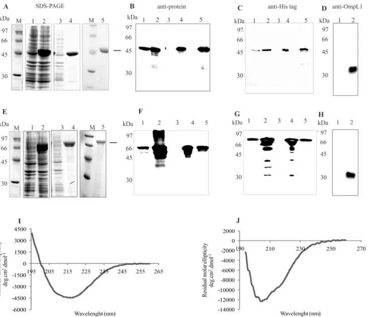
The selected coding sequences, without the signal peptide sequence were PCR amplified, cloned and expressed as His-tagged proteins inE.coli. Gene locus, given name, NCBI reference number, primer sequences with restriction cloning sites used for PCR amplifications and ex-pected molecular mass of the recombinant proteins are depicted inTable 1. The recombinant proteins were purified by nickel affinity chromatography, and an aliquot of each protein was analyzed by SDS-PAGE and shown in Fig2Aand2E, for LIC13479 (Lsa46) and LIC10050 (Lsa77), respectively. The results show that both proteins are expressed in their insoluble forms, in bacterial cell pellets, as seen in lane 4 of each Coomassie blue stained figure. Purifica-tion was successfully achieved, as shown by the presence of protein major bands in lane 5 of
Fig 1. Scheme of proteins with their putative domains and sequence conservation amongLeptospira spp. by Clustal Omega alignments. (A)Depicted are PD40 (from the WD40-like Beta Propeller Repeat family protein) and OmpA-like (outer membrane protein A) predicted domains identified in LIC13479 and LIC10050 CDSs by the BLAST and PFAM programs. Also shown are the regions of secondary structures, alpha helices, beta strands, and random coil structures predicted by the CAPITO program.(B)Blast analysis was performed among sequences of amino acids available in GenBank database and leptospiral sequences were employed to perform Clustal Omega multiple sequence alignments. The resulting phylograms show the high level of sequence conservation for LIC13479 and for LIC10050 among pathogenic strains ofLeptospira. Intermediate and saprophyte strains show lower degree of sequence identity, and are organized in more distant branches.
the same figures. Western blotting analysis of Lsa46 (Fig2Band2C) and Lsa77 (Fig2Fand 2G) were performed and the proteins were probed with anti-Lsa46 (Fig 2B) and anti-Lsa77 (Fig 2F) polyclonal antibodies, whereas in Fig2Cand2G, the proteins Lsa46 and Lsa77 were detected with anti-His mAbs, respectively. In the case of Lsa46, western blotting probed with both sera detected Lsa46 as the only protein band, while for Lsa77 additional protein bands were detected. In the case of polyclonal serum, these protein bands are probably due to non-specific reaction, while for mAbs the reactivity are with lower mass protein bands, possibly caused by some Lsa77 degradation. In any event, after purification, only Lsa77 protein bands were detected (Fig2Fand2G, lane 5). The specificity of the antibodies raised against both pro-teins was assessed by including OmpL1, a leptospiral His-tagged recombinant protein [21]. No reactivity was observed when OmpL1 was probed either with anti-Lsa46 or anti-Lsa77, indicat-ing that these antibodies were not directed against His-tag (data not shown). As expected, anti-OmpL1 recognized anti-OmpL1 but not Lsa46 and Lsa77 proteins (Fig2Dand2H, lane 1). Struc-tural integrity of the purified proteins was assessed by circular dichroism (CD) spectroscopy, depicted inFig 2Ifor Lsa46 andFig 2Jfor Lsa77, and the spectral data per wavelength analyzed by the CAPITO software [20]. The results show 10, 36 and 51% of alpha helix, beta-strands and random for Lsa46, and 21, 0.1 and 70% of alpha helix, beta-strands and random secondary structures in the case of Lsa77. Although with different percentage, a combination of secondary structures was predicted by the capito program, including random structures for both proteins, 19 and 33% for Lsa46 and Lsa77, respectively. Random secondary structure (23%) has also been found for another adhesin, LipL53 [34], but whether this structure might affect the func-tion of these proteins is unknown and remains to be studied.
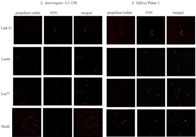
Presence of Lsa46 and Lsa77 orthologs among virulent and saprophyte
strains of
Leptospira
by IFA
In order to assess whether the chosen CDSs are located at the bacterial surface, we set out to an-alyze the protein location by using immunofluorescence microscopy. We also evaluated the
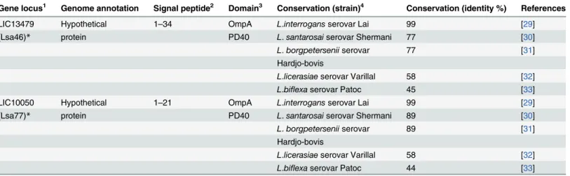
Table 2. Gene locus, features, predicted signal peptide, protein domain and sequence conservation.
Gene locus1 Genome annotation Signal peptide2 Domain3 Conservation (strain)4 Conservation (identity %) References
LIC13479 Hypothetical 1–34 OmpA L.interrogansserovar Lai 99 [29]
(Lsa46)* protein PD40 L.santarosaiserovar Shermani 77 [30]
L.borgpeterseniiserovar 77 [31] Hardjo-bovis
L.licerasiaeserovar Varillal 58 [32]
L.biflexaserovar Patoc 45 [33]
LIC10050 Hypothetical 1–21 OmpA L.interrogansserovar Lai 99 [29]
(Lsa77)* protein PD40 L.santarosaiserovar Shermani 89 [30]
L.borgpeterseniiserovar 89 [31] Hardjo-bovis
L.licerasiaeserovar Varillal 58 [32]
L.biflexaserovar Patoc 44 [33]
1http://aeg.ibi.ic.unicamp.br/world/lic/; LIC:Leptospira interrogansCopenhageni 2
http://www.cbs.dtu.dk/services/LipoP;
3
http://www.sanger.ac.uk/Software/Pfam;
4http://blast.ncbi.nlm.nih.gov/Blast.cgi/
*protein given name
presence of Lsa46 and Lsa77 orthologs inL.biflexasaprophyte strain. We have included LipL21, a leptospiral surface antigen, and DnaK, a cytoplasmic protein, as positive and negative controls, respectively. Leptospires were visualized by propidium iodide staining (Fig 3), fol-lowed by protein detection with the corresponding polyclonal antiserum, raised in mice against each protein, in the presence of anti-mouse IgG antibodies conjugated to FITC. Green fluores-cence could be observed for LipL21, Lsa46 and Lsa77 in both strains tested, but not with DnaK, used as a negative control. These assays also confirm the presence of both proteins in Leptos-pirastrains, most probably located at cell surface. The fluorescence observed with anti-Lsa46
Fig 2. Expression, purification and Western blotting of recombinant proteins.Expression and purification analysis of recombinant proteins Lsa46(A) and Lsa77(E)fromE.coliBL21 (DE3) Star pLysS were performed by SDS-PAGE. Lanes: molecular mass protein marker (M); non-induced total bacterial extract (1); induced total bacterial extract (2); soluble fraction (3); insoluble fraction (4); purified recombinant protein (5). Western blotting of Lsa46 or Lsa77 probed with the respective polyclonal antiserum(B)and(F)or with anti-His tag mAbs(C)and(G), respectively. Western blotting lanes 1 to 5 refer to the same sample condition as in SDS-PAGE. Lsa46 and Lsa77 in lane 1 of(D)and(H), respectively, and OmpL1 His-tag recombinant protein in lane 2 of(D)and(H) were probed with anti- OmpL1. Secondary structure evaluation obtained by circular dichroism spectra of the recombinant proteins: Lsa46(I)and Lsa77(J). The far-UV CD spectra are presented as an average of five scans.
and anti-Lsa77 seems be localized at the distal ends of the cells contrasting to the one observed with LipL21 that seems to be distributed along the bacteria. It is also possible that this visual pattern is due to low protein content of Lsa46 and Lsa77 orthologs as estimated by quantitative proteomics [28].
Immunological evaluation of Lsa46 and Lsa77
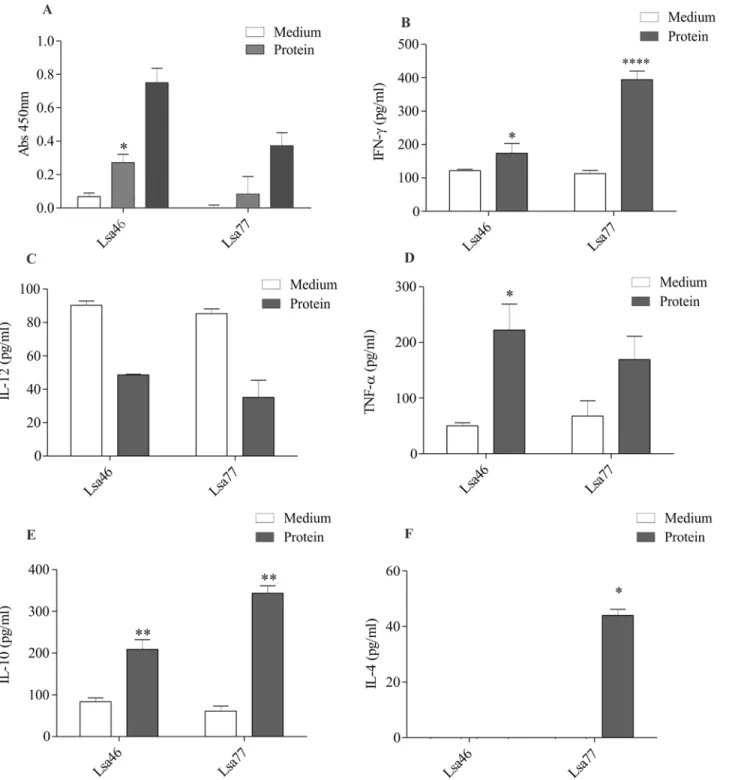
In order to characterize the humoral and cellular immune response of these proteins, mice were immunized with Lsa46 and Lsa77. After two boosters, antibodies were measure by ELISA and ti-tres of 20,000 and 100,000, were obtained for Lsa46 and Lsa77, respectively (data not shown). A high lymphoproliferation level was obtained when cells were treated with ConA, employed as a positive control (data not shown). The recombinant protein Lsa46 was capable to promote lym-phoproliferation on cultured cells of immunized animals, whereas for immunization with Lsa77 no statistically significant value was obtained, when compared to lymphocytes from animals that had not been primed with the recombinant protein (culture medium) (Fig 4A). Superna-tants of cultured spleen cells from Lsa46 and Lsa77 immunized mice were assessed for the pres-ence of the cytokines IL-10, IL-12, IL-4, IFN-γand TNF-α, selected to differentiate cellular Th1 (IFN-γ, TNF-α, and IL-12) and humoral Th2 (IL-10 and IL-4) immune responses. Lsa46 pro-moted an induction of IFN-γ, TNF-αand IL-10 cytokines (Fig4B,4Dand4E), with statistically significant values when compared to immunized but not stimulated animal cells. In addition to
Fig 3. Cellular localization of native proteins inLeptospira.VirulentL.interrogansserovar Copenhageni strain FIOCRUZ L1-130 (left panel) and saprophyteL.biflexaserovar Patoc strain Patoc1 (right panel) were fixed with paraformaldehyde and polyclonal anti-Lsa46 and anti-Lsa77 were used to identify surface-exposed protein; serum against LipL21 and DnaK were used as a marker for surface exposed and non-exposed, cytoplasmic protein, respectively. FITC-conjugated secondary antibodies were used to reveal the surface-bound antibodies. Leptospires were identified by propidium iodide staining of the DNA. Co-localization is shown in the merged images.
all cytokines listed for Lsa46, Lsa77 also promoted an enhancement of IL-4 (Fig4B,4Eand4F). Although both proteins elicited IFN-γand TNF-α, neither promoted an increase in IL-12 level.
Reactivity of Lsa46 and Lsa77 with human serum samples
To examine whether Lsa46 and Lsa77 are capable of inducing an immune response in infected host, we assessed the reactivity of the proteins measuring IgG antibodies present in paired serum samples at the onset (MAT-) and at the convalescent (MAT+) phase of leptospirosis. We performed an ELISA using 36- and 38-paired samples, half for each phase, for Lsa46 and Lsa77, respectively. The results depicted inFig 5show that both proteins are very reactive in both phases of the disease: 63 and 66% with MAT- and 84 and 55% with MAT+, for Lsa46 and Lsa77, respectively. The performances of proteins at the onset of leptospirosis, when MAT is still negative, are remarkable, suggesting that these proteins might be useful for diagnostic pur-poses. Due to the non-specific clinical symptoms of leptospirosis, we analyzed the reactivity of recombinant proteins Lsa46 and Lsa77 with serum samples from patients with unrelated infec-tious diseases that did not have a previous history of leptospirosis, including dengue (n = 12), malaria (n = 12), Chagas’disease (n = 12), and HIV infection (n = 12). The reactivity obtained with Lsa46 and Lsa77 and these serum samples was below the cut-off obtained from human healthy donors, except that Lsa46 showed reactivity with 2 and 1 serum samples of dengue and malaria, respectively (Fig 5). The specificity of Lsa77 and Lsa46 was calculated to be 100% for all unrelated diseases tested, except that Lsa46 with dengue and malaria dengue, the specificity was calculated to be 83.3 and 91.2%, respectively.
Binding of recombinant proteins to ECM components
Fig 4. Assessment of mice immune response elicited by Lsa46 and Lsa77. (A)Lymphocytes proliferation in response to mice immunization with recombinant proteins. Cells were cultured and stimulated with medium alone (negative control) or recombinant proteins (5μg / ml). ConA was used as a
positive control (not shown). The proliferative response was measured by a colorimetric Brdu-ELISA. Bars represent the mean absorbance at 450nm±the standard deviation of three replicates and are representative of two independent experiments. Spleen cells were isolated and cultured by 48h in presence of medium or recombinant proteins. Cell-free supernatants were collected and the level of cytokines IFN-γ(B), IL-12(C), TNF-α(D), IL-10(E)and IL-4(F)was assayed by ELISA. For statistical analyses, comparisons were made between cells from immunized animals that received stimuliin vitro(gray bars) and the ones treated with medium only (white bars) by the two-tailedt-test (*P<0.05,**P<0.01 and****P<0.0001).
and Lsa77 (Fig 6F). Binding saturation level was reached by Lsa46 and Lsa77 at protein concen-tration of 1,500 and 3,000 nM, respectively. The calculated dissociation equilibrium constants (KD) for the recombinant proteins Lsa46 and Lsa77 were: 24.3±17.0 nM and 53 ±17.5 nM,
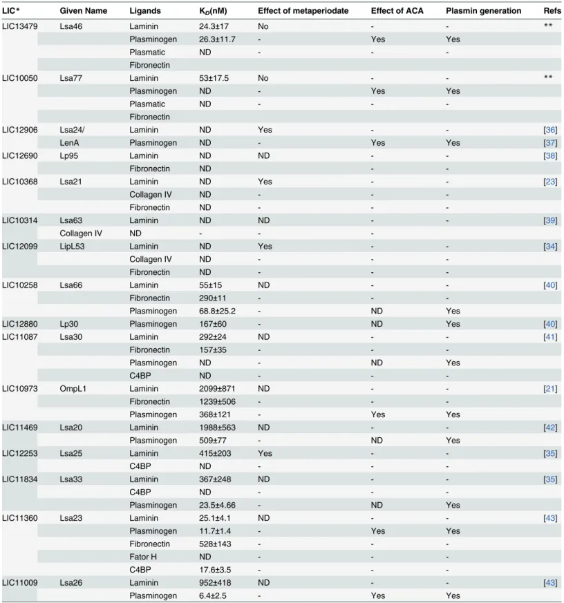
re-spectively.Table 3compares the affinities for the multiple ligands of the various proteins charac-terized in this laboratory.
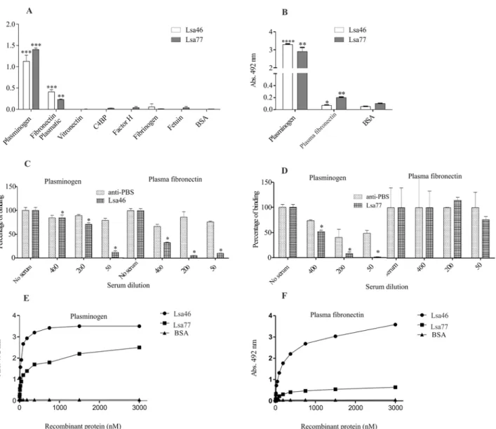
Binding of Lsa46 and Lsa77 to human plasma components
We have previously shown thatLeptospirabind PLG and that several proteins, including some adhesins, could act as binding proteins at the bacterial surface [10,46]. Hence, we decided to evaluate whether Lsa46 and Lsa77 were also capable of binding human PLGin vitro. We have also assayed other plasma components: plasma fibronectin, vitronectin, C4BP, factor H, and
Fig 5. Detection of antibodies against recombinant proteins in human leptospirosis and in unrelated febrile diseases serum samples.Reactivity of recombinant proteins, Lsa46 and Lsa77 with human leptospirosis paired serum samples at the onset (MAT-) and at the convalescent (MAT+) phase, and with human serum samples from patients diagnosed with unrelated febrile diseases. The cutoff values (dashed lines) are defined as the mean plus 3 standard deviations obtained with normal human sera.Leptospiraserum samples show 63 and 66% with MAT- and 84 and 55% with MAT+ serum samples, for Lsa46 and Lsa77, respectively.
Fig 6. Interaction of Lsa46 and Lsa77 with ECM components. (A)Wells were coated with 1μg of ECM macromolecules and the control proteins, BSA and
fetuin. Oneμg of Lsa46 and Lsa77 were added per well and attachments were measured by ELISA. Binding to laminin was confirmed by employing anti-His
mAbs(B). All data represent mean±the standard deviation from three independent experiments. For statistical analyses, the binding of recombinant proteins to the ECM components was compared to its binding to BSA by the two-tailedt-test (*P<0.05 and**P<0.01).(C)and(D)show the effect of mouse
polyclonal anti-Lsa46 and anti-Lsa77 sera upon the binding of the corresponding protein with laminin, as compared with the binding in the absence of antibodies (*P<0.05). Serum from mice immunized with PBS and adjuvant (Alhydrogel) was used as control. Effect of increasing protein concentration on
the binding to a constant laminin concentration: Lsa46(E)and Lsa77(F). Each point represents values determined in triplicate and data are expressed as the mean absorbance value at 492nm. BSA was used as a negative control. The dissociation constants (KD) were calculated based on ELISA data for the recombinant protein that reached equilibrium: 24.3±17.0 nM and 53±17.5 nM, for Lsa46 and Lsa77, respectively.
Table 3. Comparison of the binding affinities for the multiple ligands of the proteins characterized by this group.
LIC* Given Name Ligands KD(nM) Effect of metaperiodate Effect of ACA Plasmin generation Refs
LIC13479 Lsa46 Laminin 24.3±17 No - - **
Plasminogen 26.3±11.7 - Yes Yes
Plasmatic ND - -
-Fibronectin
LIC10050 Lsa77 Laminin 53±17.5 No - - **
Plasminogen ND - Yes Yes
Plasmatic ND - -
-Fibronectin
LIC12906 Lsa24/ Laminin ND Yes - - [36]
LenA Plasminogen ND - Yes Yes [37]
LIC12690 Lp95 Laminin ND ND - - [38]
Fibronectin ND -
-LIC10368 Lsa21 Laminin ND Yes - - [23]
Collagen IV ND - -
-Fibronectin ND - -
-LIC10314 Lsa63 Laminin ND ND - - [39]
Collagen IV ND - -
-LIC12099 LipL53 Laminin ND Yes - - [34]
Collagen IV ND - -
-Fibronectin ND - -
-LIC10258 Lsa66 Laminin 55±15 ND - - [40]
Fibronectin 290±11 - -
-Plasminogen 68.8±25.2 - ND Yes
LIC12880 Lp30 Plasminogen 167±60 - ND Yes [40]
LIC11087 Lsa30 Laminin 292±24 ND - - [41]
Fibronectin 157±35 - -
-Plasminogen ND - ND Yes
C4BP ND - -
-LIC10973 OmpL1 Laminin 2099±871 ND - - [21]
Fibronectin 1239±506 - -
-Plasminogen 368±121 - Yes Yes
LIC11469 Lsa20 Laminin 1988±563 ND - - [42]
Plasminogen 509±77 - ND Yes
LIC12253 Lsa25 Laminin 415±203 Yes - - [35]
C4BP ND - -
-LIC11834 Lsa33 Laminin 367±248 ND - - [35]
C4BP ND - -
-Plasminogen 23.5±4.66 - ND Yes
LIC11360 Lsa23 Laminin 25.1±4.1 ND - - [43]
Plasminogen 11.7±1.4 - Yes Yes
Fibronectin 528±143 - -
-Fator H ND - -
-C4BP 17.6±3.5 - -
-LIC11009 Lsa26 Laminin 952±418 ND - - [43]
Plasminogen 6.4±2.5 - Yes Yes
fibrinogen. Components were individually coated onto ELISA plates and allowed to interact with the recombinant proteins Lsa46 and Lsa77. The results show that Lsa46 and Lsa77 attach to PLG and to plasma fibronectin, when the reaction was probed with polyclonal antibodies against each protein (Fig 7A). The data were confirmed when the bindings were detected with anti-His mAbs (Fig 7B). No reactivity was detected with the other plasma components tested. We also investigate whether immune epitopes were involved in the binding of both proteins with PLG and plasma fibronectin, by pre-incubating the proteins with the respective antibody. The results demonstrate that immune epitopes are involved in the binding of both proteins with PLG (Fig7Cand7D) and of Lsa46 with plasma fibronectin (Fig 7C). Almost no inhibition on the binding of Lsa77 and plasma fibronectin was achieved when the protein was incubated with its antiserum, even at the lowest dilution employed (Fig 7D), suggesting that antibody binding regions do not participate in the interaction with this component.
The interactions between the recombinant proteins with PLG and plasma fibronectin were evaluated on a quantitative basis as depicted inFig 7EandFig 7F, respectively. Binding was dose-dependent when increasing concentrations of the recombinant protein Lsa46 and Lsa77 (0 to 3,000 nM) were added to constant amount of PLG (Fig 7E) and plasma fibronectin (Fig 7F). Binding saturation was reached only with Lsa46 and PLG at the protein concentration of 1,500 nM, with a dissociation equilibrium constant (KD) of 26.2± 11.7 nM (Table 3).
Binding of Lsa46 and Lsa77 with PLG occur via lysine residues and
generate PLA
PLG kringle domains frequently mediate interactions with lysine residues of the bacterial re-ceptors [47]. The involvement of these domains was shown to contribute in the binding of PLG withL.interrogansserovar Copenhageni strain FIOCRUZ L1–130, because ACA, an ana-logue of lysine, profusely inhibited the binding [46]. Based on these results, we decided to in-vestigate if lysine residues are involved in the binding of recombinant proteins with PLG, by the adding ACA to the reaction mixtures. The results strongly suggest that this is indeed the case for both Lsa46 and Lsa77 proteins, because the minimum ACA used in these assays nearly completely abolished the binding to PLG (Fig 8A).
Previous work of our group has reported that PLG bound to leptospiral binding proteins can be activated to PLA by activators [21,35,41,43,44]. To assess whether PLG attached to Lsa46 and Lsa77 proteins can also achieve proteolytic activity, as reported for several leptospiral proteins
Table 3. (Continued)
LIC* Given Name Ligands KD(nM) Effect of metaperiodate Effect of ACA Plasmin generation Refs
LIC11975 Lsa36 Laminin 120±97.6 ND - - [43]
Plasminogen 17.8±5.5 - Yes Yes
Fibronectin 34.8±10.4 - -
-LIC10645 Lsa44 Laminin 108±43 ND - - [44]
Plasminogen 53.5±18.4 - Yes Yes
LIC10731 Lsa45 Laminin 250.38 ND - - [44]
Plasminogen 36.8±20.3 - Yes Yes
LIC10829 Lsa32 Laminin ND ND - - [45]
Plasminogen 81.5±31.1 - Yes Yes
*Leptospira interrogansserovar Copenhageni L1-130 genome annotation;(http://aeg.lbi.ic.unicamp.br/world/lic/; Nacimento et al., 2004); **proteins in this study;
ND, not determined; KD, dissociation constant.
(seeTable 3), microplates individually coated with the recombinant proteins were incubated with PLG. The uPA-type PLG activator was added together with a plasmin-specific chromogen-ic substrate (described inmethodsection). The plasmin activity was indirectly evaluated by measuring the cleavage of the PLA-specific chromogenic substrate at 405 nm. The data show that only the complete system, Lsa46 or Lsa77, PLG, uPA and PLA substrate, can generate the expected PLA-derived product (Fig 8B). PLG bound to the proteins can be activated to PLA, via proteolytic cleavage through activators such as uPA, but Lsa77 seems to be more efficient, as seen by the higher amount of PLA generation when compared with Lsa46. BSA, which does not
Fig 7. Assessment of Lsa46 and Lsa77 binding with plasma components. (A)Wells were coated with 1μg of each plasma components and the control
proteins, BSA and fetuin. Oneμg of Lsa46 and Lsa77 were added per well and binding was measured by ELISA. Binding to plasminogen and plasma
fibronectin was confirmed by employing anti-His mAbs(B). All data represent mean±the standard deviation from three independent experiments. For statistical analyses, the binding of recombinant proteins to the plasma components was compared to its binding to BSA by using two-tailedt-test (*P<0.05,
**P<0.01 and****P<0.0001). Effect of mouse polyclonal anti-Lsa46(C)and anti-Lsa77(D)serum upon the binding of the corresponding protein with
plasminogen and plasma fibronectin was compared with the binding in the absence of antibodies (*P<0.05). Immunized mice serum with PBS in Alhydrogel
association was used as control. Dose-dependent binding of Lsa46(E)and Lsa77(F)to plasminogen and plasma fibronectin was performed. Each point represents values determined in triplicate and data are expressed as the mean absorbance value at 492nm. BSA was used as a negative control. The dissociation constant (KD) was calculated based on ELISA data for Lsa46 that reached equilibrium, as 26.2±11.7 nM.
bind PLG, was employed as negative control, and did not show any proteolytic activity. No cleavage of the chromogenic substrate was observed in controls omitting PLG, uPA or the chro-mogenic substrate, respectively. However, when PLG/PLA is bound to recombinant proteins no fibrinogen degradation products were detected (not shown).
Fig 8. The effect of lysine analogue (ACA) on the binding of Lsa46 and Lsa77 to PLG and evaluation of PLA generation in the presence of PLG activator. (A)Role of lysine residues in protein-PLG interaction was assessed by ELISA. BSA was used as a negative control. Bars represent the mean absorbance at 492nm±the standard deviation of three replicates and a representative of two independent experiments is depicted. For statistical analyses, the binding of the Lsa46 and Lsa77 in the presence of ACA was compared with the binding to PLG without ACA by the two-tailedt-test (*P<0.05 and**P<0.01).(B)PLA generation by PLG bound to Lsa46 and Lsa77 was measured indirectly by the cleavage of the specific PLA-specific substrate,D-valyl-leucyl-lysine-p-nitroanilide dihydrochloride, when the recombinant proteins were treated
with PLG+uPA+S or in the absence of one of the three components (PLG+uPA/ PLG+S/ uPA+S); BSA was employed as a control protein. Bars represent mean absorbance at 405nm as a measure of relative substrate cleavage±the standard deviation of three replicates; one representative, of two independent experiments, is shown. Statistically significant differences were observed relative to control BSA (**P<0.01 and
****P<0.0001).
Discussion
The characterization of leptospiral outer membrane proteins is critical to understanding lepto-spirosis pathogenicity. The OmpA-like domains (named after the description of C-terminal domain ofEscherichia coliOmpA protein) have been shown to be non-covalently associated with peptidoglycan [48]. Proteins having OmpA-like domains of important pathogens were described to be involved in different aspects of infection. OmpA outer membrane protein of Escherichia colihas been reported to act as adhesin/invasin and to participate in biofilm forma-tion [49]. Other OmpA-like proteins includeE.colilipoprotein PAL (Peptidoglycan-associated lipoprotein) [50], theNeisseria meningitidesRmp [51], and the peptidoglycan-associated lipo-protein (Pal) ofHaemophilus influenzathat is been considered a potential vaccine candidate against this bacteria [52]. The Loa22 was the first OmpA-like protein described inLeptospira and shown to be reactive with convalescent mouse sera. [53]. By mutagenesis experiments, this protein was reported to be essential for leptospiral virulence [54]. The OmpA-like proteins Omp52 and OmpA70 have been described forL.santarosaiserovar Shermani andL. interro-gansserovar Copenhageni, respectively [55,56]. The first protein is environmentally regulated and expressed in human confirmed leptospirosis patients, while the latter has been shown to be highly immunogenic in mice.
Although whole genome sequences of severalLeptospirahave created unquestionable contri-bution to understanding host-pathogen interactions, mechanisms driven by the bacteria during infection remain to be elucidated. We have been exploring the genome sequences ofL. interro-gansserovar Copenhageni searching for proteins annotated as hypothetical and surface-exposed. Through these criteria, we have identified and characterized several adhesins, including one with OmpA-like domain [9,40], proteins that interact with PLG [10,21,35,40,41,42,44,57], proteins that bind regulators of complement system [35,43] and proteins recognized by antibodies present in sera of human patients infected with leptospires [21,39,42,43,44,58].
In this work, we report the characterization of two novel hypothetical proteins having an OmpA-like domain at C-terminus, both of which are surface-exposed leptospiral adhesins, called Lsa46 and Lsa77. These proteins, encoded by the genes LIC13479 and LIC10050, were expressed inE.coli, as 46 and 77 kDa recombinant proteins, respectively.
The protein sequences are well preserved among pathogenic species ofLeptospira, whereas lower identities were found in intermediate and saprophyte strains. We confirm the expression of both proteins in low-passage virulent and saprophyte strains ofLeptospira. The expression of proteins was not detected by immunoblotting in saprophyte strains (data not shown), proba-bly due the lower sensitivity of the method when related to fluorescence. By comparing with the fluorescence of LipL21, a surface lipoprotein ofLeptospira[59], the proteins are most prob-ably surface exposed.
are localized at the cell surface as tested by immunofluorescence assay, their capacity to induce immune response in mice and their reactivity with confirmed leptospirosis serum samples.
The interaction between pathogens and the host fibrinolytic system has been shown for several pathogens including, invasive gram-positive, gram-negative bacteria, virus and para-sites [47,70,71,72,73]. Interactions with the fibrinolytic system byBorreliaspp. andTreponema denticolawere suggested to have an important role during infection [74,75]. Our group have reported for the first time thatLeptospiraspecies were also capable to bind PLG and generating PLA, in the presence of activator[46]. To date, several leptospiral proteins have been described as PLG-binding [57] and some of them also functioning as ECM-interacting proteins
[35,43,44]. We have previously reported an adhesin and PLG-binding protein with OmpA-like domain, named Lsa66 [40]. We have now identified Lsa46 and Lsa77, as novel PLG-binding proteins. The binding affinity was achieved only for Lsa46, and is of the same order of magni-tude of the values calculated for other recombinant proteins reported from our laboratory [10]. Bound PLG to both proteins could be converted to plasmin by the addition of PLG activator (uPA), with specific proteolytic activity. Although we have previously shown that PLG activat-ed to PLA on leptospiral surface is able to degrade laminin, fibronectin and fibrinogen [46,76], we did not detected fibrinogen degradation products when PLG/PLA is generated bound to re-combinant proteins. One possible explanation is the number of PLG binding proteins on lepto-spiral surface, 17 identified to this point [10] compared to one individual protein. Another possibility is that micro-environmental settings within the bacteria may provide better reaction conditions when compared to bacterial-free reaction medium.
The binding ability of Lsa46 and Lsa77 to host-derived molecules is different. Immunogenic epitopes seem to be involved on the interaction of Lsa46 with ECM and PLG, while with Lsa77 only the binding to PLG involved these sites. Interaction with laminin and this protein was par-tially prevented by anti-Lsa77. Though unexpectedly, the data suggest that with Lsa77 other non-immunogenic regions are involved on the interaction with laminin and plasma fibronec-tin. Similar data have been reported for the recombinant OmpL37 ofL.interrogans. The antise-rum against this protein did not exhibit any statistically significant effect on the binding of OmpL37 to fibronectin, fibrinogen and laminin [77].
Lsa46 and Lsa77 are immunogenic, capable of eliciting Th1 and Th2 immune responses in mice. These proteins have in common, with previously described Loa22 and Lsa66 adhesins having OmpA-like domains, positive reactivity with serum samples from patients diagnosed with leptospirosis and are probably expressed during the disease [39,40,53,64,77,78,79]. Most interestingly, both proteins have higher sensitivity to detect leptospirosis at the onset of the dis-ease than the standard reference test MAT [4], and could be further explored for early diagno-sis purposes. Moreover, both proteins showed high specificity among unrelated infections diseases, commonly found in tropical countries.
Acknowledgments
We are deeply indebted to Dr. Henrique Ronffato (Departamento de Parasitologia, Instituto Butantan, Sao Paulo, Brazil) for the use of confocal microscope facilities and helpful discussion. AFT has a fellowship from FAPESP (Brazil).
Author Contributions
Conceived and designed the experiments: AFT SAV ALTON. Performed the experiments: AFT ZMM ECR KK. Analyzed the data: AFT ECR KK ZMM SAV ALTON. Contributed reagents/materials/analysis tools: AFT ECR SAV KK ALTON. Wrote the paper: AFT ALTON.
References
1. Levett PN. Leptospirosis. Clin Microbiol Rev. 2001; 14: 296–326. PMID:11292640
2. Levett PN. Usefulness of serologic analysis as a predictor of the infecting serovar in patients with se-vere leptospirosis. Clin Infect Dis. 2003; 36: 447–452. PMID:12567302
3. Bharti AR, Nally JE, Ricaldi JN, Matthias MA, Diaz MM, Lovett MA, et al. Leptospirosis: a zoonotic dis-ease of global importance. Lancet Infect Dis. 2003; 3: 757–771. PMID:14652202
4. Faine S, Adler B, Bolin C, Perolat P, editors (1999) Leptospira and Leptospirosis. Melbourne, Australia.
5. Bajani MD, Ashford DA, Bragg SL, Woods CW, Aye T, Spiegel RA, et al. Evaluation of four commercial-ly available rapid serologic tests for diagnosis of leptospirosis. J Clin Microbiol. 2003; 41: 803–809.
PMID:12574287
6. Hull-Jackson C, Glass MB, Ari MD, Bragg SL, Branch SL, Whittington CU, et al. Evaluation of a com-mercial latex agglutination assay for serological diagnosis of leptospirosis. J Clin Microbiol. 2006; 44: 1853–1855. PMID:16672421
7. Nascimento AL, Verjovski-Almeida S, Van Sluys MA, Monteiro-Vitorello CB, Camargo LE, Digiampietri LA, et al. Genome features of Leptospira interrogans serovar Copenhageni. Braz J Med Biol Res. 2004; 37: 459–477. PMID:15064809
8. Nascimento AL, Ko AI, Martins EA, Monteiro-Vitorello CB, Ho PL, Haake DA, et al. Comparative geno-mics of two Leptospira interrogans serovars reveals novel insights into physiology and pathogenesis. J Bacteriol. 2004; 186: 2164–2172. PMID:15028702
9. Vieira ML, Fernandes LG, Domingos RF, Oliveira R, Siqueira GH, Souza NM, et al. Leptospiral extra-cellular matrix adhesins as mediators of pathogen-host interactions. FEMS Microbiol Lett. 2014; 352: 129–139. doi:10.1111/1574-6968.12349PMID:24289724
10. Vieira ML, Atzingen MV, Oliveira TR, Oliveira R, Andrade DM, Vasconcellos SA, et al. In vitro identifica-tion of novel plasminogen-binding receptors of the pathogen Leptospira interrogans. PLoS One. 2010; 5: e11259. doi:10.1371/journal.pone.0011259PMID:20582320
11. Vieira ML, de Morais ZM, Vasconcellos SA, Romero EC, Nascimento AL. In vitro evidence for immune evasion activity by human plasmin associated to pathogenic Leptospira interrogans. Microb Pathog. 2013; 51: 360–365.
12. Turner LH. Leptospirosis. 3. Maintenance, isolation and demonstration of leptospires. Trans R Soc Trop Med Hyg. 1970; 64: 623–646. PMID:4098633
13. Nakai K, Kanehisa M. Expert system for predicting protein localization sites in gram-negative bacteria. Proteins. 1991; 11: 95–110. PMID:1946347
14. Yu CS, Lin CJ, Hwang JK. Predicting subcellular localization of proteins for Gram-negative bacteria by support vector machines based on n-peptide compositions. Protein Sci. 2004; 13: 1402–1406. PMID:
15096640
15. Letunic I, Copley RR, Pils B, Pinkert S, Schultz J, Bork P. SMART 5: domains in the context of genomes and networks. Nucleic Acids Res. 2006; 34: D257–260. PMID:16381859
16. Finn RD, Mistry J, Schuster-Bockler B, Griffiths-Jones S, Hollich V, Lassmann T, et al. Pfam: clans, web tools and services. Nucleic Acids Res. 2006; 34: D247–251. PMID:16381856
17. Juncker AS, Willenbrock H, Von Heijne G, Brunak S, Nielsen H, Krogh A. Prediction of lipoprotein sig-nal peptides in Gram-negative bacteria. Protein Sci. 2003; 12: 1652–1662. PMID:12876315
19. Ramos CR, Abreu PA, Nascimento AL, Ho PL. A high-copy T7 Escherichia coli expression vector for the production of recombinant proteins with a minimal N-terminal His-tagged fusion peptide. Braz J Med Biol Res. 2004; 37: 1103–1109. PMID:15273812
20. Wiedemann C, Bellstedt P, Gorlach M. CAPITO—a web server-based analysis and plotting tool for
cir-cular dichroism data. Bioinformatics. 2013; 29: 1750–1757. doi:10.1093/bioinformatics/btt278PMID:
23681122
21. Fernandes LG, Vieira ML, Kirchgatter K, Alves IJ, de Morais ZM, Vasconcellos SA, et al. OmpL1 is an extracellular matrix- and plasminogen-interacting protein of Leptospira spp. Infect Immun. 2012; 80: 3679–3692. doi:10.1128/IAI.00474-12PMID:22802342
22. Atzingen MV, Rodriguez D, Siqueira GH, Leite LC, Nascimento AL. Induction of boosted immune re-sponse in mice by leptospiral surface proteins expressed in fusion with DnaK. Biomed Res Int. 2014; 2014: 564285. doi:10.1155/2014/564285PMID:25110682
23. Atzingen MV, Barbosa AS, De Brito T, Vasconcellos SA, de Morais ZM, Lima DM, et al. Lsa21, a novel leptospiral protein binding adhesive matrix molecules and present during human infection. BMC Micro-biol. 2008; 8: 70. doi:10.1186/1471-2180-8-70PMID:18445272
24. Lin YP, Lee DW, McDonough SP, Nicholson LK, Sharma Y, Chang YF. Repeated domains of leptos-pira immunoglobulin-like proteins interact with elastin and tropoelastin. J Biol Chem. 2009; 284: 19380–19391. doi:10.1074/jbc.M109.004531PMID:19473986
25. Adindla S, Inampudi KK, Guruprasad K, Guruprasad L. Identification and analysis of novel tandem re-peats in the cell surface proteins of archaeal and bacterial genomes using computational tools. Comp Funct Genomics. 2004; 5: 2–16. doi:10.1002/cfg.358PMID:18629042
26. Marchler-Bauer A, Anderson JB, Chitsaz F, Derbyshire MK, DeWeese-Scott C, Fong JH, et al. CDD: specific functional annotation with the Conserved Domain Database. Nucleic Acids Res. 2009; 37: D205–210. doi:10.1093/nar/gkn845PMID:18984618
27. Larkin MA, Blackshields G, Brown NP, Chenna R, McGettigan PA, McWilliam H, et al. Clustal W and Clustal X version 2.0. Bioinformatics. 2007; 23: 2947–2948. PMID:17846036
28. Malmstrom J, Beck M, Schmidt A, Lange V, Deutsch EW, Aebersold R. Proteome-wide cellular protein concentrations of the human pathogen Leptospira interrogans. Nature. 2009; 460: 762–765. doi:10.
1038/nature08184PMID:19606093
29. Ren SX, Fu G, Jiang XG, Zeng R, Miao YG, Xu H, et al. Unique physiological and pathogenic features of Leptospira interrogans revealed by whole-genome sequencing. Nature. 2003; 422: 888–893. PMID:
12712204
30. Chou LF, Chen YT, Lu CW, Ko YC, Tang CY, Pan MJ, et al. Sequence of Leptospira santarosai serovar Shermani genome and prediction of virulence-associated genes. Gene. 2012; 511: 364–370. doi:10.
1016/j.gene.2012.09.074PMID:23041083
31. Bulach DM, Zuerner RL, Wilson P, Seemann T, McGrath A, Cullen PA, et al. Genome reduction in Lep-tospira borgpetersenii reflects limited transmission potential. Proc Natl Acad Sci U S A. 2006; 103: 14560–14565. PMID:16973745
32. Ricaldi JN, Fouts DE, Selengut JD, Harkins DM, Patra KP, Moreno A, et al. Whole genome analysis of Leptospira licerasiae provides insight into leptospiral evolution and pathogenicity. PLoS Negl Trop Dis. 2012; 6: e1853. doi:10.1371/journal.pntd.0001853PMID:23145189
33. Picardeau M, Bulach DM, Bouchier C, Zuerner RL, Zidane N, Wilson PJ, et al. Genome sequence of the saprophyte Leptospira biflexa provides insights into the evolution of Leptospira and the pathogene-sis of leptospiropathogene-sis. PLoS One. 2008; 3: e1607. doi:10.1371/journal.pone.0001607PMID:18270594
34. Oliveira TR, Longhi MT, Goncales AP, de Morais ZM, Vasconcellos SA, Nascimento AL. LipL53, a tem-perature regulated protein from Leptospira interrogans that binds to extracellular matrix molecules. Mi-crobes Infect. 2010; 12: 207–217. doi:10.1016/j.micinf.2009.12.004PMID:20026283
35. Domingos RF, Vieira ML, Romero EC, Goncales AP, de Morais ZM, Vasconcellos SA, et al. Features of two proteins of Leptospira interrogans with potential role in host-pathogen interactions. BMC Micro-biol. 2012; 12: 50. doi:10.1186/1471-2180-12-50PMID:22463075
36. Barbosa AS, Abreu PA, Neves FO, Atzingen MV, Watanabe MM, Vieira ML, et al. A newly identified leptospiral adhesin mediates attachment to laminin. Infect Immun. 2006; 74: 6356–6364. PMID:
16954400
37. Verma A, Brissette CA, Bowman AA, Shah ST, Zipfel PF, Stevenson B. Leptospiral endostatin-like pro-tein A is a bacterial cell surface receptor for human plasminogen. Infect Immun. 2010; 78: 2053–2059.
doi:10.1128/IAI.01282-09PMID:20160016
39. Vieira ML, de Morais ZM, Goncales AP, Romero EC, Vasconcellos SA, Nascimento AL. Lsa63, a newly identified surface protein of Leptospira interrogans binds laminin and collagen IV. J Infect. 2010; 60: 52–64. doi:10.1016/j.jinf.2009.10.047PMID:19879894
40. Oliveira R, de Morais ZM, Goncales AP, Romero EC, Vasconcellos SA, Nascimento AL. Characteriza-tion of novel OmpA-like protein of Leptospira interrogans that binds extracellular matrix molecules and plasminogen. PLoS One. 2011; 6: e21962. doi:10.1371/journal.pone.0021962PMID:21755014
41. Souza NM, Vieira ML, Alves IJ, de Morais ZM, Vasconcellos SA, Nascimento AL. Lsa30, a novel adhe-sin of Leptospira interrogans binds human plasminogen and the complement regulator C4bp. Microb Pathog. 2012; 53: 125–134. doi:10.1016/j.micpath.2012.06.001PMID:22732096
42. Mendes RS, Von Atzingen M, de Morais ZM, Goncales AP, Serrano SM, Asega AF, et al. The novel leptospiral surface adhesin Lsa20 binds laminin and human plasminogen and is probably expressed during infection. Infect Immun. 2011; 79: 4657–4667. doi:10.1128/IAI.05583-11PMID:21844229
43. Siqueira GH, Atzingen MV, Alves IJ, de Morais ZM, Vasconcellos SA, Nascimento AL. Characterization of Three Novel Adhesins of Leptospira interrogans. Am J Trop Med Hyg. 2013;
44. Fernandes LG, Vieira ML, Alves IJ, de Morais ZM, Vasconcellos SA, Romero EC, et al. Functional and immunological evaluation of two novel proteins of Leptospira spp. Microbiology. 2014;
45. Domingos R, Fernandes L, Romero E, de Morais Z, Vasconcellos S, Nascimento AL. The novel Leptos-pira interrogans protein Lsa32 is expressed during infection and binds laminin and plasminogen. Micro-biology. 2015 Jan 27. pii: mic.0.000041. doi:10.1099/mic.0.000041
46. Vieira ML, Vasconcellos SA, Goncales AP, de Morais ZM, Nascimento AL. Plasminogen acquisition and activation at the surface of leptospira species lead to fibronectin degradation. Infect Immun. 2009; 77: 4092–4101. doi:10.1128/IAI.00353-09PMID:19581392
47. Lahteenmaki K, Kuusela P, Korhonen TK. Bacterial plasminogen activators and receptors. FEMS Microbiol Rev. 2001; 25: 531–552. PMID:11742690
48. Wang Y. The function of OmpA in Escherichia coli. Biochem Biophys Res Commun. 2002; 292: 396–401. PMID:11906175
49. Smith SG, Mahon V, Lambert MA, Fagan RP. A molecular Swiss army knife: OmpA structure, function and expression. FEMS Microbiol Lett. 2007; 273: 1–11. PMID:17559395
50. Cascales E, Bernadac A, Gavioli M, Lazzaroni JC, Lloubes R. Pal lipoprotein of Escherichia coli plays a major role in outer membrane integrity. J Bacteriol. 2002; 184: 754–759. PMID:11790745
51. Grizot S, Buchanan SK. Structure of the OmpA-like domain of RmpM from Neisseria meningitidis. Mol Microbiol. 2004; 51: 1027–1037. PMID:14763978
52. Parsons LM, Lin F, Orban J. Peptidoglycan recognition by Pal, an outer membrane lipoprotein. Bio-chemistry. 2006; 45: 2122–2128. PMID:16475801
53. Koizumi N, Watanabe H. Molecular cloning and characterization of a novel leptospiral lipoprotein with OmpA domain. FEMS Microbiol Lett. 2003; 226: 215–219. PMID:14553914
54. Ristow P, Bourhy P, da Cruz McBride FW, Figueira CP, Huerre M, Ave P, et al. The OmpA-like protein Loa22 is essential for leptospiral virulence. PLoS Pathog. 2007; 3: e97. PMID:17630832
55. Hsieh WJ, Chang YF, Chen CS, Pan MJ. Omp52 is a growth-phase-regulated outer membrane protein of Leptospira santarosai serovar Shermani. FEMS Microbiol Lett. 2005; 243: 339–345. PMID:
15686833
56. Fraga TR, Chura-Chambi RM, Goncales AP, Morais ZM, Vasconcellos SA, Morganti L, et al. Refolding of the recombinant protein OmpA70 from Leptospira interrogans from inclusion bodies using high hy-drostatic pressure and partial characterization of its immunological properties. J Biotechnol. 2010; 148: 156–162. doi:10.1016/j.jbiotec.2010.04.007PMID:20450943
57. Vieira ML, Nascimento AL. Interaction of spirochetes with the host fibrinolytic system and potential roles in pathogenesis. Critical Reviews in Microbiology. 2015 "in press" doi:10.3109/1040841X.2013. 804031PMID:25811941
58. Gamberini M, Gomez RM, Atzingen MV, Martins EA, Vasconcellos SA, Romero EC, et al. Whole-genome analysis of Leptospira interrogans to identify potential vaccine candidates against leptospirosis. FEMS Microbiol Lett. 2005; 244: 305–313. PMID:15766783
59. Cullen PA, Haake DA, Bulach DM, Zuerner RL, Adler B. LipL21 is a novel surface-exposed lipoprotein of pathogenic Leptospira species. Infect Immun. 2003; 71: 2414–2421. PMID:12704111
61. Stevenson B, Choy HA, Pinne M, Rotondi ML, Miller MC, Demoll E, et al. Leptospira interrogans endostatin-like outer membrane proteins bind host fibronectin, laminin and regulators of complement. PLoS One. 2007; 2: e1188. PMID:18000555
62. Verma A, Hellwage J, Artiushin S, Zipfel PF, Kraiczy P, Timoney JF, et al. LfhA, a novel factor H-binding protein of Leptospira interrogans. Infect Immun. 2006; 74: 2659–2666. PMID:16622202
63. Choy HA, Kelley MM, Chen TL, Moller AK, Matsunaga J, Haake DA. Physiological osmotic induction of Leptospira interrogans adhesion: LigA and LigB bind extracellular matrix proteins and fibrinogen. Infect Immun. 2007; 75: 2441–2450. PMID:17296754
64. Hauk P, Macedo F, Romero EC, Vasconcellos SA, de Morais ZM, Barbosa AS, et al. In LipL32, the major leptospiral lipoprotein, the C terminus is the primary immunogenic domain and mediates interac-tion with collagen IV and plasma fibronectin. Infect Immun. 2008; 76: 2642–2650. doi:10.1128/IAI.
01639-07PMID:18391007
65. Hoke DE, Egan S, Cullen PA, Adler B. LipL32 is an extracellular matrix-interacting protein of Leptospira spp. and Pseudoalteromonas tunicata. Infect Immun. 2008; 76: 2063–2069. doi:10.1128/IAI.01643-07
PMID:18285490
66. Carvalho E, Barbosa AS, Gomez RM, Cianciarullo AM, Hauk P, Abreu PA, et al. Leptospiral TlyC is an extracellular matrix-binding protein and does not present hemolysin activity. FEBS Lett. 2009; 583: 1381–1385. doi:10.1016/j.febslet.2009.03.050PMID:19328790
67. Hussain M, Heilmann C, Peters G, Herrmann M. Teichoic acid enhances adhesion of Staphylococcus epidermidis to immobilized fibronectin. Microb Pathog. 2001; 31: 261–270. PMID:11747374
68. Flugel A, Schulze-Koops H, Heesemann J, Kuhn K, Sorokin L, Burkhardt H, et al. Interaction of entero-pathogenic Yersinia enterocolitica with complex basement membranes and the extracellular matrix pro-teins collagen type IV, laminin-1 and -2, and nidogen/entactin. J Biol Chem. 1994; 269: 29732–29738.
PMID:7961965
69. Fink DL, Green BA, St Geme JW 3rd. The Haemophilus influenzae Hap autotransporter binds to fibro-nectin, laminin, and collagen IV. Infect Immun. 2002; 70: 4902–4907. PMID:12183535
70. Bergmann S, Hammerschmidt S. Fibrinolysis and host response in bacterial infections. Thromb Hae-most. 2007; 98: 512–520. PMID:17849039
71. Sun MZ, Liu S, Greenaway FT. Characterization of a fibrinolytic enzyme (ussurenase) from Agkistrodon blomhoffii ussurensis snake venom: insights into the effects of Ca2+ on function and structure. Biochim Biophys Acta. 2006; 1764: 1340–1348. PMID:16877056
72. Goto H, Kawaoka Y. A novel mechanism for the acquisition of virulence by a human influenza A virus. Proc Natl Acad Sci U S A. 1998; 95: 10224–10228. PMID:9707628
73. Rojas M, Labrador I, Concepcion JL, Aldana E, Avilan L. Characteristics of plasminogen binding to Try-panosoma cruzi epimastigotes. Acta Trop. 2008; 107: 54–58. doi:10.1016/j.actatropica.2008.04.013
PMID:18501871
74. Coleman JL, Sellati TJ, Testa JE, Kew RR, Furie MB, Benach JL. Borrelia burgdorferi binds plasmino-gen, resulting in enhanced penetration of endothelial monolayers. Infect Immun. 1995; 63: 2478–2484.
PMID:7790059
75. Fenno JC, Tamura M, Hannam PM, Wong GW, Chan RA, McBride BC. Identification of a Treponema denticola OppA homologue that binds host proteins present in the subgingival environment. Infect Immun. 2000; 68: 1884–1892. PMID:10722578
76. Oliveira R, Domingos RF, Siqueira GH, Fernandes LG, Souza NM, Vieira ML, et al. Adhesins of Leptos-pira interrogans mediate the interaction to fibrinogen and inhibit fibrin clot formation in vitro. PLoS Negl Trop Dis. 2013; 7: e2396. doi:10.1371/journal.pntd.0002396PMID:24009788
77. Pinne M, Choy HA, Haake DA. The OmpL37 surface-exposed protein is expressed by pathogenic Lep-tospira during infection and binds skin and vascular elastin. PLoS Negl Trop Dis. 2010; 4: e815. doi:
10.1371/journal.pntd.0000815PMID:20844573
78. Croda J, Ramos JG, Matsunaga J, Queiroz A, Homma A, Riley LW, et al. Leptospira immunoglobulin-like proteins as a serodiagnostic marker for acute leptospirosis. J Clin Microbiol. 2007; 45: 1528–1534.
PMID:17360842