380
Corresponding author: Dra. Ariani Impieri Souza. e-mail: [email protected]
Received 9 September 2015 Accepted 6 January 2016
Dengue as a cause of fever during pregnancy:
a report of two cases
Ariani Impieri Souza
[1],[2], Ana Laura Carneiro Gomes Ferreira
[1], Matheus Alencar Arraes
[2],
Bruno Marcelo Moura
[2]and Maria Cynthia Braga
[1],[3][1]. Programa de Pós-Graduação Stricto Sensu em Saúde Materno Infantil, Instituto de Medicina Integral Prof. Fernando Figueira, Recife, Pernambuco, Brasil. [2]. Curso de Medicina, Faculdade Pernambucana de Saúde, Recife, Pernambuco, Brasil. [3]. Departamento de Parasitologia,
Centro de Pesquisas Aggeu Magalhães, Fundação Oswaldo Cruz, Recife, Pernambuco, Brasil.
Abstract
Dengue infection has not been routinely investigated among pregnant women and parturients with acute febrile syndrome in endemic settings. Here, we report two cases of dengue fever detected at the time of delivery in parturients enrolled in a cohort prospective study conducted in a hospital in Recife, Brazil. The parturients reported fever onset within seven days prior to
delivery, and dengue infection was conirmed upon detection of viral ribonucleic acid (RNA) by using the reverse
transcriptase-polymerase chain reaction. Dengue infection should be considered as a diagnostic possibility in cases of fever during pregnancy and labor, especially in endemic areas.
Keywords: Dengue. Pregnant women. Vertical transmission.
Rev Soc Bras Med Trop 49(3):380-382, May-June, 2016 doi: 10.1590/0037-8682-0306-2015
Case Report
INTRODUCTION
Dengue fever is a growing health risk in urban areas and a major cause of hospitalizations and deaths in tropical countries(1). It is caused by four different serotypes [dengue virus
(DENV)1-4] and is transmitted by a mosquito bite, mainly of
the Aedes aegypti(1). Since the re-emergence of dengue fever in Brazil in the 1980s, the country has been affected by successive outbreaks, resulting in a high incidence of cases(2).
Case reports of dengue fever during pregnancy document several complications such as maternal death, stillbirth, prematurity, low birth weight, fetal abnormalities, and abortion(3) (4). The symptoms of dengue fever during the gestational and perinatal periods are usually similar to those found in the general population and the clinical management is similar to that in non-pregnant women(1).
Although fever is one of the main symptoms of dengue(1), tracing this infection in pregnant women with acute fever or other symptoms of the disease has not been fully incorporated
into routine prenatal care in Brazil. Nevertheless, laboratory
investigation of dengue fever is important since it is a potential health risk for mothers and their fetuses and requires targeted clinical management to prevent and treat the associated complications(5).
We encountered two cases of dengue fever in parturients with fever onset seven days prior to labor. The blood samples were collected during labor in the maternity ward of the Instituto de Medicina Integral Prof. Fernando Figueira (IMIP), a teaching hospital in Recife, Pernambuco, Brazil. The diagnosis was
conirmed by using real-time reverse transcription-polymerase chain reaction (RT-PCR). Both parturients were enrolled in
a prospective cohort study conducted between March 2011 and May 2012 to investigate the incidence of dengue infection and the kinetics and transplacental transfer of maternal antidengue antibodies(5).
CASE REPORT
Case 1
A 17-year-old, single woman in labor was admitted to our maternity ward in June 2011. She reported an episode of urinary tract infection in the second trimester of pregnancy, which was treated with antibiotics. On admission, she reported
headache, chills, weakness, and fever (temperature, 38.1°C).
The obstetric exam revealed a 6-cm dilated cervix, intact amniotic membranes, fetus in cephalic presentation, and fetal auscultation of 160 beats per minute. Four hours before the delivery, an amniotomy was performed and we observed a
foul-smelling, moderate meconium-stained amniotic luid. Antibiotic
therapy was administered. Our patient delivered vaginally,
giving birth to a healthy term, male infant weighing 3,045g with
an Apgar score of 6 and 8 at the 1st and 5th min, respectively. A macroscopic examination of the placenta did not show any
381
Souza AI et al. - Dengue and pregnancy
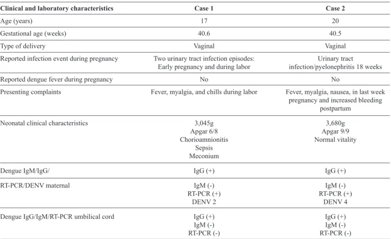
TABLE 1 - Clinical and laboratory characteristics of maternal and pregnancy outcomes.
Clinical and laboratory characteristics Case 1 Case 2
Age (years) 17 20
Gestational age (weeks) 40.6 40.5
Type of delivery Vaginal Vaginal Reported infection event during pregnancy Two urinary tract infection episodes: Urinary tract
Early pregnancy and during labor infection/pyelonephritis 18 weeks
Reported dengue fever during pregnancy No No
Presenting complaints Fever, myalgia, and chills during labor Fever, myalgia, nausea, in last week pregnancy and increased bleeding postpartum
Neonatal clinical characteristics 3,045g 3,680g
Apgar 6/8 Apgar 9/9
Chorioamnionitis Normal vitality
Sepsis Meconium
Dengue IgM/IgG/ IgG (+) IgG (+)
RT-PCR/DENV maternal IgM (-) IgM (-)
RT-PCR (+) RT-PCR (+)
DENV 2 DENV 4
Dengue IgG/IgM/RT-PCR umbilical cord IgG (+) IgG (+)
IgM (-) IgM (-)
RT-PCR (-) RT-PCR (-)
IgM: immunoglobulin M; IgG: immunoglobulin G; RT-PCR: reverse transcription-polymerase chain reaction; DENV: dengue virus. prescription of oral antibiotics to be taken at home. Laboratory
tests for dengue infection in the maternal blood sample showed
the following results: antidengue immunoglobulin G (IgG) indirect enzyme-linked immunosorbent assay (ELISA) (PanBio, Brisbane, Australia), positive; antidengue immunoglobulin M (IgM)-capture (PanBio and Focus Diagnostics, Cypress, CA) ELISA, negative; and DENV RT-PCR positive for DENV 2.
At birth, the neonate was hypotonic with generalized
cyanosis, intercostal retractions, nasal laring, and grunting.
The newborn was successfully resuscitated and intravenous antibiotic therapy was administered. He was discharged on the 7th day of life with a diagnosis of septicemia secondary to
chorioamnionitis. Blood and cerebrospinal luid cultures were
negative for dengue infection; however, the cord blood sample was IgG-positive, IgM-negative, and RT-PCR-negative.
Case 2
A 20-year-old, married woman in labor was admitted to our maternity ward in October 2011. She reported asthenia, nausea, myalgia, and fever in the seven days preceding labor.
This patient had been hospitalized at 18 weeks of pregnancy due to low back pain, fever, and headache. She was diagnosed with pyelonephritis and underwent antibiotic therapy. After discharge, she was referred to prenatal care. On admission for delivery, she had a satisfactory general condition and the obstetric examination showed 5-cm dilated cervix, intact
amniotic membranes, fetus in cephalic presentation, and
fetal auscultation of 140 beats per minute. The next day, she
delivered vaginally and gave birth to a healthy term, male infant weighing 3,680g with an Apgar score of 9 at the 1st and 5th min. At the macroscopic examination, the placenta was whole and normal. Twenty hours after delivery, the patient experienced dizziness, presyncope, asthenia, and an episode
of vaginal bleeding. Her hemoglobin level was 6.6g/dL. Complete patient recovery was achieved 46 h after delivery.
Both the mother and baby were discharged 2 days later.
The DENV-speciic laboratory tests carried out using the
maternal blood sample showed the following results:
IgG-ELISA, positive; IgM-IgG-ELISA, negative; and RT-PCR, positive for DENV 4. The cord blood sample was IgG-ELISA-positive, IgM-ELISA-negative, and RT-PCR-negative for dengue.
Table 1 presents the clinical and laboratory characteristics of both cases.
DISCUSSION
382
Rev Soc Bras Med Trop 49(3):380-382, May-June, 2016
REFERENCES
1. Guzman MG, Harris E. Dengue. The Lancet 2014; 385:453-465.
2. Teixeira MG, Siqueira JB, Ferreira GLC, Bricks L, Joint G.
Epidemiological Trends of dengue disease in Brazil (2000-2010): A Systematic Literature Search and Analysis. PLoS Negl Trop Dis
2013; 7:e2520.
3. Pouliot SH, Xiong X, Harville E, Paz-Soldan V, Tomashek KM, Breart G, et al. Maternal Dengue and Pregnancy Outcomes: a systematic review. Obstet Gynecol Surv 2010; 65:107-118.
4. Alvarenga CF, Silami VG, Brasil P, Boechat MEH, Coelho J, Nogueira RMR. Dengue during pregnancy: a study of thirteen
cases. Am J Infect Dis 2009; 5:288-293.
5. Leite RC, Souza AI, Castanha PMS, Cordeiro MT, Martelli CT, Ferreira, ALG, et al. Dengue infection in pregnancy and
transplacental transfer of anti-dengue antibodies in Northeast, Brazil. J Clin Virol 2014; 60:16-21.
6. Ribeiro CF, Lopes VGS, Brasil P, Coelho J, Muniz AG, Nogueira MR. Perinatal transmission of dengue: a report of 7 cases. J Pediatr
2013; 164:1514-1516.
7. Phongsamart W, Youksan S, Vanaprapa N, Chokephaibulkit K. Dengue virus infection in late pregnancy and transmission to the
infants. The Ped Infec Dis J 2008; 27:500-504.
8. Sinhabahu VP, Sathananthan R, Malavige GN. Perinatal transmission
of dengue: a case report. BMC Research Notes 2014; 7:795.
9. Cameron P, Simmons CP, Farrar JJ, Chau NV, Wills B. Dengue.
N Engl J Med 2012; 366:1423-1432.
10. Braga C, Luna C, Martelli CT, Souza WV, Cordeiro MT, Alexander
N, et al. Seroprevalence and risk factors for dengue infection in
socioeconomically distinct areas of Recife, Brazil. Acta Tropica
2010; 113:234-240.
11. Wing DA, Fassett MJ, Getahun D. Acute pyelonephritis in pregnancy: an 18-year retrospective analysis. Am J Obstet Gynecol
2014; 210:219.e1-6.
12. Aguiar M, Rocha F, Pessanha JEM, Mateus L, Stollenwerk N. Carnival or football, is there a real risk for acquiring dengue fever
in Brazil during holidays seasons? Sci Rep 2015; 5:8462.
In case 1 of the current study, abundant amniotic luid and
thick meconium with fetid odor was reported during labor and chorioamnionitis was diagnosed. These manifestations were also reported by Phongsamart et al.(7) and Sinhabahu et al.(8) in parturients in Thailand and Sri Lanka, respectively, and
these parturients were conirmed to have virological dengue. Therefore, their indings reinforce the evidence that such clinical
signs may constitute one of the manifestations of dengue fever during childbirth.
Postpartum hemorrhage and low hemoglobin level were seen in the parturient in our Case 2.Hemorrhagic manifestations of various intensities such as vaginal bleeding or surgical wounds bleeding in caesarean sections, with or without thrombocytopenia during labor or at the time of delivery have also been described by other authors in previous studies on pregnant women with dengue fever(3) (4).
The concordance of our clinical indings with those of other
case reports in Brazil(4) and other countries(3) provide further evidence that these symptoms are associated with dengue infection during late pregnancy. Consequently, these data warrant the need for analytical studies, preferentially in prospective longitudinal studies in pregnant women at risk of dengue infection in order to
better deine the prognostic factors and clinical manifestations of
dengue and other arboviruses in this population.
The detection of viral ribonucleic acid (RNA) in maternal
blood samples by using RT-PCR is indicative of the acute phase of dengue infection. The detection of antidengue IgG
(a serological marker detected approximately 10-15 days after infection) in the maternal and cord blood samples, in both
our cases, is indicative of prior exposure of the mothers to the dengue virus and transplacental transfer of the maternal antidengue IgG to the neonate. These laboratory results indicate
that both mothers had secondary infections of dengue (viremic phase) at delivery. The occurrence of dengue reinfection is
recognized as a risk of progression to severe dengue(9).
In our study, DENV RNA or antidengue IgM (a serological marker of recent dengue) was not detected by RT-PCR or ELISA, respectively, in the umbilical cord blood samples of both cases, conirming the absence of vertical transmission. Vertical transmission of DENV seems to be a rare event in endemic
areas, but several cases have been described in mothers-infants pairs in Brazil(7) and other countries(3). According to a previous
study, DENV-2 serotype is most often associated with vertical
transmission(7).This serotype was detected in the parturient in Case 1, and her newborn showed clinical manifestations
described in cases of DENV vertical transmission(7) (8).
The serological diagnosis of dengue is conirmed by the detection of a speciic IgM antibody or the increase in IgG
antibody titers that occurs in consecutive samples during the acute and convalescent phases(1). In neonates, the detection of antidengue IgM antibodies in umbilical cord blood samples
is indicative of transplacental transfer of DENV, whereas the
detection of antidengue IgG in these samples is indicative of the transfer of maternal IgG antibodies(3). On the other hand,
the detection of antidengue IgG antibodies and DENV RNA during the acute phase of the disease (viremia) in mothers,
as observed in the two cases reported here, is indicative of secondary infection(1).
Although both parturients exhibited signs and symptoms of dengue fever(1) and were living in a highly endemic area(10), the possibility of dengue fever was not considered during medical assistance. Since urinary tract infection during pregnancy is one of the most frequent causes of fever(11), the report of this symptom by the women possibly directed the clinical rationale for this diagnoses. Therefore, our report of two parturients with dengue highlights the need to include dengue fever as one of the possible causes of acute febrile syndrome or bleeding events during pregnancy or childbirth, especially in the months of the highest incidence of dengue in Brazil, i.e., January to July(12). It is also important to perform laboratory tests for the rapid diagnosis of dengue fever in pregnant women in the health care units, especially during periods of peak occurrence of the disease.
Conlict of interest