Meta-Analysis of the Detection of Plant
Pigment Concentrations Using Hyperspectral
Remotely Sensed Data
Jingfeng Huang1, Chen Wei1,2, Yao Zhang1, George Alan Blackburn3, Xiuzhen Wang4*, Chuanwen Wei1, Jing Wang1
1Institute of Agricultural Remote Sensing & Information Application, Zijingang Campus, Zhejiang University, Hangzhou, China,2Zhejiang Meteorological Service Center, Hangzhou, China,3Lancaster Environment Centre, Lancaster University, Lancaster, United Kingdom,4Institute of Remote Sensing and Earth Sciences, Hangzhou Normal University, Hangzhou, China
*wxz0516@sina.com
Abstract
Passive optical hyperspectral remote sensing of plant pigments offers potential for under-standing plant ecophysiological processes across a range of spatial scales. Following a num-ber of decades of research in this field, this paper undertakes a systematic meta-analysis of 85 articles to determine whether passive optical hyperspectral remote sensing techniques are sufficiently well developed to quantify individual plant pigments, which operational solutions are available for wider plant science and the areas which now require greater focus. The find-ings indicate that predictive relationships are strong for all pigments at the leaf scale but these decrease and become more variable across pigment types at the canopy and landscape scales. At leaf scale it is clear that specific sets of optimal wavelengths can be recommended for operational methodologies: total chlorophyll and chlorophyllaquantification is based on reflectance in the green (550–560nm) and red edge (680–750nm) regions; chlorophyllbon the red, (630–660nm), red edge (670–710nm) and the near-infrared (800–810nm); caroten-oids on the 500–580nm region; and anthocyanins on the green (550–560nm), red edge (700–710nm) and near-infrared (780–790nm). For total chlorophyll the optimal wavelengths are valid across canopy and landscape scales and there is some evidence that the same applies for chlorophylla.
Introduction
A pigment is a material that changes the spectral distribution of reflected or transmitted light as the result of wavelength-selective absorption which is determined by the physical properties of the pigment itself. Plant pigments play an important role in light capture, photosystem pro-tection, and in various growth and development functions. The photosynthetic pigments con-trol the amount of solar radiation absorbed by a leaf and thus determine photosynthetic potential and primary production [1,2]. Pigment concentrations are also related to plant stress (excess direct sunlight, UV–B irradiation, low temperature, water stress, nitrogen deficiencies and so on) and senescence (e.g., [3–9]). Therefore, accurate measurements of the temporal OPEN ACCESS
Citation:Huang J, Wei C, Zhang Y, Blackburn GA, Wang X, Wei C, et al. (2015) Meta-Analysis of the Detection of Plant Pigment Concentrations Using Hyperspectral Remotely Sensed Data. PLoS ONE 10 (9): e0137029. doi:10.1371/journal.pone.0137029
Editor:Benedicte Riber Albrectsen, Umeå Plant Science Centre, Umeå University, SWEDEN
Received:April 12, 2015
Accepted:August 11, 2015
Published:September 10, 2015
Copyright:© 2015 Huang et al. This is an open access article distributed under the terms of the Creative Commons Attribution License, which permits unrestricted use, distribution, and reproduction in any medium, provided the original author and source are credited.
Data Availability Statement:All relevant data are within the paper and its Supporting Information file.
Funding:Our work was supported by grants from the National Natural Science Foundation of China (41171276), National Key Technology R&D Program of China (SQ2012BAJY3429), and Ph.D. Programs Foundation of Ministry of Educational of China (200100101110035). The funders had a role in data collection and analysis.
dynamics and spatial variations of pigment concentration using remotely sensed data can pro-vide a basis for monitoring physiological and ecological processes [10,11].
The spectral absorbance properties of pigments offer the possibility of using measurements of reflected radiation as a non-destructive method for quantifying pigments. Different
approaches have arisen recently to remotely estimate pigment concentrations from a wide vari-ety of wavelengths and sensor types. These studies produced variable results, and none have been demonstrated to have satisfactory performance under all growth and environmental con-ditions. These inconsistencies may stem from the fact that the experimental results are influ-enced by a number of factors including different species, experimental conditions and analytical methods used [11].
Recent review articles have attempted to assimilate knowledge in this field of passive optical hyperspectral remote sensing with the sun as energy source. Blackburn [10] reviewed the devel-oping technologies and analytical methods for quantitative estimation of pigment across a range of spatial scales using passive optical hyperspectral remote sensing. Ustinet al. [11] appraised the most widely used methodologies for retrieving pigment information with hyper-spectral data at the leaf scale. However, it has been demonstrated that traditional qualitative reviewers may subjectively select their preferred studies when faced with conflicting results on a single question [12]. In contrast, it has been argued that meta-analysis can take the results from primary research articles and quantitatively analyze and synthesize these data in an attempt to arrive at more robust conclusions. As such, meta-analysis review papers make the shift from a narrative-driven to a data-driven approach [13,14].
Glass [15] published the first article to lay out the essential rationale of meta-analysis. As a fully general set of methods, meta-analysis has been widely applied to the integration of litera-tures in many areas of empirical science, including ecology [14]. This form of analysis has, for example, been used to determine the response of biodiversity to intensive biomass production, the effects of elevated CO2on plant–arthropod interactions, the influence of plant invasion on
carbon and nitrogen cycles and the causes and consequences of variations in leaf mass per area [16–19]. Today, many findings and advances are being made not only by those who do primary research studies, but also by those who use meta-analysis to discover the latent meaning of existing research literatures [13]. Recently, meta-analysis has been employed in remote sensing research. Garbulskyet al. [20] performed a meta-analysis to assess the use of the photochemical reflectance index (PRI) as an indicator of radiation use efficiencies at the leaf, canopy and eco-system scales for different time scales and vegetation types. Zolkoset al. [21] conducted a meta-analysis of publications on LiDAR remote sensing estimation of terrestrial aboveground biomass. These investigations show that meta-analysis can be used to systematically integrate the results from a collection of studies, and through statistical comparison, assess the relation-ships between remotely sensed measurements and variables of interest.
Here, a meta-analysis of data from a wide selection of studies reporting the passive optical hyperspectral remote sensing of pigments was used to quantify the development of this scien-tific field, identify optimal wavelengths for retrieval of individual pigments and evaluate the strength of the relationships between pigment concentration and remotely sensed data across pigment types and scales.
Materials and Methods
2.1 Study selection and data extraction
with the terms reflectance, estimation, quantification, retrieval, prediction and remote sensing. More than 4500 citations were collected as a result of this initial search.
Then the abstracts of these articles were reviewed and considered for inclusion in the meta-analysis. The following criteria were applied to ensure homogeneity in methodology. First, the studies had to include a chemical measurement of pigment concentration (total chlorophyll, chlorophylla, chlorophyllb, carotenoids, xanthophyll, carotene or anthocyanins). Second, the article had to report the quantification of pigments using remotely sensed data. Third, the authors must have provided the following statistical information: (1) coefficient of determina-tion for the reladetermina-tionships between pigment concentradetermina-tion and remotely sensed measurements; (2) the wavelength(s) used to estimate pigment concentration; and (3) training sample sizes.
2Based on the first two decision rules, 135 articles were selected. According to the final cri-terion, 50 studies were excluded because of insufficient statistical information. Finally, 85 articles were used in the meta-analysis, which reported results at different spatial and tempo-ral scales and from a wide range of vegetation types between 1977 and 2014. The number of studies selected at various stages is shown in the flow diagram inFig 1. Some studies reported multiple results for different pigment types or vegetation types. Different types of sensors were used in these studies, from spectrophotometers and hand-held spectroradiometers to satellite sensors. All the sensors were working in reflectance mode. Within the selected arti-cles 44 were working at the leaf scale, 21 at the canopy scale, 15 at the landscape scale, 2 at the leaf and canopy scales, 1 at the leaf and landscape scales, and 2 covered the leaf, canopy and landscape scales. The term“canopy”refers to either a single plant or a monospecific stand where the experimental results are influenced by a number of controlling factors, such as orientation of leaves (leaf angle distribution;LAD), variations in number of leaf layers (LAI), presence of non-leaf elements, multiple scattering and areas of shadow [10,22], the term“landscape”refers to a mixed-species stand where the reflectance spectrum from air-borne and spaceair-borne sensors is subject to even more controlling factors, such as atmo-spheric conditions, instrucment sensitivity (signal-to-noise ratio) and spatial resolution. In
Fig 1. Selection of studies for inclusion in the meta-analysis.
total, the sample size from all the selected studies is 16100. The Preferred Reporting Items for Meta-Analyses is shown inS1 PRISMA Checklist.
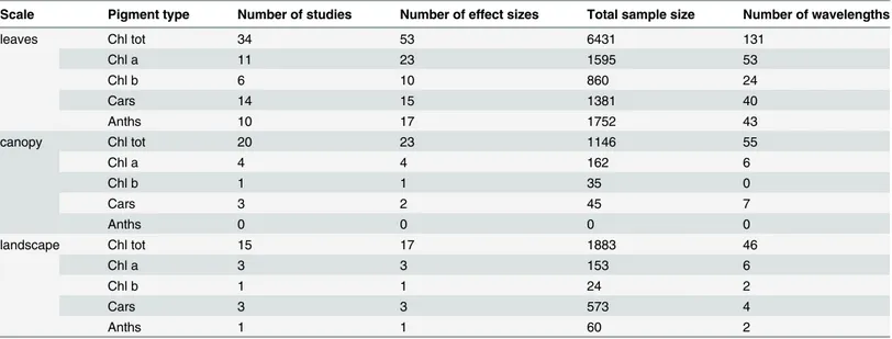
Relevant information was extracted from each study in the final set:①scales (leaf, canopy, landscape),②pigment types,③species,④wavelengths,⑤coefficient of determination,⑥ sample sizes,⑦sensors,⑧authors and⑨year of publication. In order to reduce human error in data extraction and coding, two sets of reviewers independently screened articles in accordance with those inclusion criteria discussed above, evaluated the quality and extracted the data from the eligible studies. The results from one group were cross-checked by the other group. Divergences of opinion about article selection and data extraction were settled by dis-cussion.Table 1is a summary of the studies contained in this research. This list is not exhaus-tive but it does cover most papers published related to quantification of pigments using remotely sensed data that met the selection criteria.Table 2provides a statistical summary of the data extracted from the studies included in the meta-analysis.
2.2 Statistical analysis of effect size
2.2.1 The calculation of effect size for each study. The coefficient of determination (R2) was used to evaluate the strength of relationships between spectral reflectance and pigment concentration in each article we selected. The value of R2, however, is affected by the number of selected wavelengths. The more wavelengths included in the model, be they relevant or not, the larger would be the R2[106]. The increase of R2is not without cost. The increasing number of selected wavelengths reduces the degrees of freedom, which reduces model robustness. The adjusted coefficient of determination was applied to correct for the degrees of freedom:
R2
A ¼1 ð1 R 2
Þn 1
n k ð1Þ
wherenis the sample size for each study,kis the number of independent variables in the linear or nonlinear model. Eq (1) shows thatR2
Ais always smaller thanR
2whenk>1, which means
the growth rate ofR2
Ais lower than that ofR
2as the number of parameters increase. This result
is straightforward and it has been shown that when the added parameter explains a significant amount of the behavior of the dependent variable,R2
Awill increase; otherwise,R
2
Awill decrease
[107]. SoR2
Awas chosen as the effect size statistic, the variance of effect size is calculated as
[108]:
Vi ¼ ð1 R2
AÞ
2
n 1 ; ð2Þ
The resulting data set was categorized by pigment type at the scales of leaf, canopy and land-scape to allow comparison.
2.2.2 Test of heterogeneity for effect sizes. It is important to assess the heterogeneity among the results from a collection of studies before computing the mean effect size [109]. Basically, there are two possible sources of heterogeneity in meta-analysis: methodological het-erogeneity and statistical hethet-erogeneity. To ensure homogeneity in methodology, we applied a series of criteria to identify the studies to be used in the meta-analysis (as described in section 2.1). Here theI2statistic was used to test for the statistical heterogeneity. TheI2statistic mea-sures the extent of true heterogeneity dividing the difference between the result of theQtest and its degrees of freedom by theQvalue itself [110]:
I2¼100%Qtot df
Qtot ð
3Þ
Table 1. A summary of the studies contained in this research that linked remotely sensed data with pigment.Specrad = spectroradiometer; Specpho = spectrophotometer; Chl tot = total chlorophyll; Chl a = chlorophylla; Chl b = chlorophyllb; Cars = carotenoids; Anths = anthocyanins.
Scale Pigment Type Year Species Sensor Reference
leaves Chl tot 1992 Amaranthus tricolor Specpho [23]
leaves Chl tot 1995 Slash pine Specrad [24]
leaves Chl tot 1995 Bigleaf maple Specrad [25]
leaves Chl tot 1996 Horse Chestnut, Norway maple,Cotoneaster, Tobacco Specpho [26]
leaves Chl tot 1996 Norway Maple, Horse Chestnut Specpho [27]
leaves Chl tot 1997 Norway Maple, Horse Chestnut, Fig, Cotoneaster, Tobacco,Oleander, Hibiscus, Vine, Rose
Specpho [28]
leaves Chl tot 1998 Tobacco, Horse Chestnut, Cotoneaster Specpho [29]
leaves Chl tot 1999 Beech tree, Elm tree,Wild vine shurb Specpho [30]
leaves Chl tot 1999 Bragg Soybean Specrad [31]
leaves Chl tot 2002 53 species Specrad [32]
leaves Chl tot 2002 Paper birch Specrad [33]
leaves Chl tot 2003 Bigleaf Maple, Horse Chestnut, Wild vine, Beech Specpho [34]
leaves Chl tot 2005 Cotton Specrad [35]
leaves Chl tot 2007 Winter wheat Specpho [36]
leaves Chl tot 2012 15 different species(Beech, Fraxinus lanuginosa, Acer Japonicum, Magnolia obovata and so on)
Specrad [37]
leaves Chl tot 2014 Douglasfir Specrad [38]
leaves Chl a 1994 Norway Maple, Horse Chestnut Specpho [39]
leaves Chl a 1994 Norway Maple, Horse Chestnut Specpho [40]
leaves Chl a 1996 Norway Maple, Horse Chestnut Specpho [41]
leaves Cars/Chl tot 1977 Cantaloupe, Corn, Spinach Cotton, Cucumber, tobacco, Head lettuce, Grain sorghum
Specpho [42]
leaves Cars/Chl tot 1992 Sunflower Specrad [43]
leaves Cars/Chl tot 1999 Norway Maple, Potato, Lemon, Apple, Coleus Specpho [7]
leaves Cars/Chl tot 2006 24 species of woody trees and shurbs Specpho [44]
leaves Anths/Cars/Chl
tot 1999 Quercus agrifolia, Pseudotsuga menziesii Specpho [45]
leaves Anths/Cars/Chl
tot 2003 Apple Specpho [46]
leaves Anths/Cars/Chl
tot 2004 Norway maple, Maize, Dogwood,Horse chestnut, Second-Wild vine shrub, Cotoneaster, Pelargonium zonale flush beech, Specpho [47]
leaves Chl tot/Anths 2014 Chilean strawberry Specrad [48]
leaves Cars/Chl a/ Chl b
1992 Soybean Specrad [49]
leaves Cars/Chl a/Chl b 1998 Beech, Oak, Maple, Sweet chestnut Specrad [50]
leaves Cars/Chl a/Chl b 2005 Rice Specrad [51]
leaves Chl tot/Chl a/Chl
b 1999 Norway Maple, Horse Chestnut, Beech, Oak Specrad [52]
leaves Chl tot/Chl a/Chl
b 2001 Croton, Elaeagnus, Japanese pittosporum,Benjaminfig Specrad [53] leaves Chl tot/Chl a/Chl
b 2010 Flowering cherry Specrad [54]
leaves Chl tot/Chl a 1996 Tobacco Specpho [55]
leaves Chl tot/Chl a 1999 Eucalyptus Specrad [56]
leaves Cars 2002 Norway maple, Horse chestnut,Second-flush beech Specpho [57]
leaves Cars 2009 Scot pine Specpho [58]
leaves Cars 2011 Bur oak, Sugar maple, LOPEX database Specrad [59]
Scale Pigment Type Year Species Sensor Reference
Table 1. (Continued)
leaves Anths 2001 Norway maple, Cotoneaster, Dogwood Specpho [60]
leaves Anths 2009 Grapevine Specrad [61]
leaves Anths 2009 European hazel, Siberian dogwood, Norway maple, Virginia creeper Specpho [62]
leaves Anths 2011 Grapevine Specrad [63]
leaves Anths 2011 Sweet cherries Specpho [64]
leaves Anths 2011 Norway maple, Horse chestnut, Beech,Virginia creeper, Dogwood Specpho&specrad [65]
Leaves/canopy Chl tot 2009 Maize Specpho [66]
Leaves/canopy Chl tot 2013 Irrigated maize Specrad [67]
Leaves/landscape Chl tot 2014 Black Spruce, Sugar maple Specrad&MERIS [68]
Leaves/canopy/
landscape Chl tot 2010 Winter Wheat, Winter Rapeseed Specrad [69]
Leaves/canopy/
landscape Cars/Chl tot 2000 Sugar maple Specrad [70]
canopy Chl tot 1990 Slash pine Airborne spectro [1]
canopy Chl tot 1994 pepper Specrad [71]
canopy Chl tot 2005 Maize, Soybean Specrad [72]
canopy Chl tot 2006 Rice Specrad [73]
canopy Chl tot 2007 Cotton Specrad [74]
canopy Chl tot 2008 Winter wheat, Corns Specrad [75]
canopy Chl tot 2008 Heterogeneous grassland Specrad [76]
canopy Chl tot 2008 Heterogeneous grassland Specrad [77]
canopy Chl tot 2008 Corn, Cotton Specrad [78]
canopy Chl tot 2010 Rice Specrad [79]
canopy Chl tot 2011 Rice Specrad [80]
canopy Chl tot 2012 Potato, Grassland Specrad [81]
canopy Chl tot 2013 Irrigated maize Specrad [82]
canopy Chl tot 2014 Winter wheat Specrad [83]
canopy Chl a 2003 Rice Specrad [84]
canopy Chl a 2007 Winter Wheat Specrad [85]
canopy Chl a/Chl b 2004 Winter wheat Specrad [86]
canopy Chl tot/Chl a 2006 Wheat Specrad [87]
canopy Cars/Chl tot 2010 Tall fescue Specrad [88]
canopy Cars 2008 Kermes oak Specrad [89]
canopy Cars 2008 Douglasfir Specrad [90]
landscape Chl tot 2002 Corn CASI [91]
landscape Chl tot 2003 Eucalypt CASI-2 [92]
landscape Chl tot 2004 Jack pine CASI [93]
landscape Chl tot 2004 Douglasfir MERIS [94]
landscape Chl tot 2007 Corn, Wheat CASI [95]
landscape Chl tot 2008 Rice, Cotton EO-1 [96]
landscape Chl tot 2008 Garlic, Alfalfa, Onion, Sunflower, Corn, Potato, Wheat, Vineyard, Sugar
beet PROBA/CHRIS [97]
landscape Chl tot 2010 Flax, Tea, Chestnut, Corn, Potato, Pine, Bamboo EO-1 [98]
landscape Chl tot 2010 Garlic, Onion, Corn, Alfalfa, Sugar beet, Sunflower, Potato, Vineyard,
Wheat PROBA/CHRIS [99]
landscape Chl tot 2014 London plane, Canary Island date palm, European nettle tree, White
mulberry CASI [100]
landscape Chl a 2004 Winter Wheat AVIS [101]
landscape Cars/Chl tot 2002 Quercus petrea, Pinus sylvestris CASI [102]
computed as [111]:
Qtot¼X
Ntot
i¼1
WiE2i
XNtot
i¼1
WiEi 0 @ 1 A 2
XNtot
i¼1
Wi
ð4Þ
whereWi¼1=vi,Eiis adjusted coefficient of determination (R 2
A).
TheI2statistic can be interpreted as the percentage of heterogeneous component in the total variability of effect size (Qtot), so the larger theI2statistic is, the stronger the heterogeneity is. If
I2exceeds 50%, the null hypothesis of homogeneity is rejected. TheI2statistic for different pig-ments at different scales were calculated, all the results were lower than 50%, the null hypothe-sis of homogeneity for this study was accepted.
2.2.3 The calculation of mean effect size for different pigments at different scales. In
contrast to studies based on original data, the unit of meta-analysis is the individual research study. Distinctive aspects of data analysis follow from this difference. The first complication is that the studies incorporated into the meta-analysis generally use different sample sizes and this controls the statistical properties of effect sizes [112]. From a statistical perspective, larger sample studies have less sampling error than smaller sample studies, thus more weight should be assigned to larger sample studies in the computation of the mean effect size. The other com-plication is inter-study variability, which is caused by the influence of an indeterminate number of characteristics that vary among the studies.
Table 1. (Continued)
landscape Chl a/Cars 2005 Rice PHI [103]
landscape Cars/Chl tot/Chl
a/Chl b 2008 Aspen, Birch, Spruce, Balsamfir CASI [104]
landscape Anths 2009 Austrocedrus chilensis forest Hyperion [105]
doi:10.1371/journal.pone.0137029.t001
Table 2. Summary statistics for the selected studies and extracted data for different pigment types at leaf, canopy and landscape scales.
Scale Pigment type Number of studies Number of effect sizes Total sample size Number of wavelengths
leaves Chl tot 34 53 6431 131
Chl a 11 23 1595 53
Chl b 6 10 860 24
Cars 14 15 1381 40
Anths 10 17 1752 43
canopy Chl tot 20 23 1146 55
Chl a 4 4 162 6
Chl b 1 1 35 0
Cars 3 2 45 7
Anths 0 0 0 0
landscape Chl tot 15 17 1883 46
Chl a 3 3 153 6
Chl b 1 1 24 2
Cars 3 3 573 4
Anths 1 1 60 2
Considering the two sources of variability discussed above, a random effects model was used to compute the weighted mean ofR2
Afor different pigment types. In contrast to afixed
effects model, the weight applied to each effect size in a random effects model must represent both subject-level sampling error and the additional random variance component [112]. As such, the mean effect size becomes a reasonable estimate of the true strength of the effect in the population. Because of the generality of the random effects model, it is the preferred strategy in meta-analysis [113]. The mean effect size is computed as:
Mrand ¼ X
Np
i¼1
WiðrandÞEi
X Np
i¼1
WiðrandÞ
ð5Þ
The variance is:
Vrand¼
1
X Np
i¼1
WiðrandÞ
ð6Þ
whereWiðrandÞ¼ 1
Viþs2,
s2¼ Qp
ðNp 1Þ
X Np
i¼1
Wi
X Np
i¼1
W2
i
X Np
i¼1
Wi
,Wi¼ 1
Vi
,Qp¼ X
Np
i¼1
WiE
2
i
X Np
i¼1 WiEi
!2
X Np
i¼1
Wi
Ei,
is adjusted coefficient of determination (R2
A) andNpis the total number of effect sizes for a specific type of pigment at each different scales (Table 2). Using this approach the mean effect size of Chl tot, Chl a, Chl b, Cars and Anths at the scales of leaf, canopy and landscape were calculated.
A confidence interval gives the range of values within which the mean effect size is likely to be, it is useful in indicating the degree of precision of the estimate of the mean effect size. A 95% confidence interval is subsequently calculated as follows:
Conf95¼Mrand1:96SErand ð7Þ
whereSErand¼ ffiffiffiffiffiffiffiffiffi Vrand p
. If the confidence intervals of multiple mean effect sizes donot overlap, then there are significant differences between these mean effect sizes.
2.3 Optimal wavelengths for pigment quantification
A large number of narrow-band indices were proposed to measure plant pigments in the selected articles. These narrow band indices include difference vegetation index (NBDVI), ratio vegetation index (NDRVI), normalized difference vegetation index (NBNDVI), anthocya-nin reflectance index (ARI), soil-adjusted vegetation index (SAVI), perpendicular vegetation index (PVI) and so on. The wavelengths used in these studies are different and there is lack of agreement on optimal wavelengths for pigment quantification.
histogram each subset is represented by a rectangle whose height is equal to the count of obser-vations that fall into the wavelength interval. A quantile plot is a simple and effective way to compare different wavelength distributions. Letλi(i= 1toG) be the wavelengths sorted in
increasing order so thatλ
1is the smallest wavelength andλGis the largest. Each wavelength,λi,
is paired with a percentage,fi, which indicates that approximately 100fi% of the data are below
or equal to the value,λ i.
fi¼
i 0:5
G ði¼1;. . .;GÞ ð8Þ
In a quantile plot,λ
iis graphed againstfi. This allows us to compare different wavelength
distributions based on their quantiles [114].
Results
3.1 Quantifying the development of remote sensing of plant pigment
concentrations
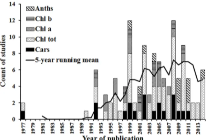
The number of studies used in the meta-analysis published over the period from 1977 to 2014 are shown inFig 2, along with the 5-year running mean which summarises the overall trajec-tory of development in this scientific field. After the first two studies were published in 1977 there were no other publications for 11 years, but then there was fast rate of growth from 1990 to 1999. The number of publications reached top in 1999 after which the publication rate stopped increasing, indicating that research in passive optical hyperspectral remote sensing of plant pigment concentrations is within a mature phase. The overall trajectory of publications shows three periods covering the origins, development and proliferation of research in this field. This trajectory corresponds to the developmental phases of hyperspectral instruments, which started with spectrophotometers and hand-held spectroradiometers enabling leaf and canopy-scale work. With the more recent advent of airborne and spaceborne imaging spec-trometers, more landscape scale analyses have become possible.
Despite this overall development in the field, there were substantial differences in research on different pigments. The first studies of total chlorophyll and carotenoids were published in 1977, followed by chlorophyllaand chlorophyllbin 1992 and anthocyanins in 1999. The growth rate of publications on chlorophylla, chlorophyllb, carotenoids and anthocyanins has
Fig 2. Histogram of numbers of selected studies published over time, showing the total in each year and the number focusing on each pigment type.The solid line is a 5-year running mean of the total number of studies.
been significantly lower than that for total chlorophyll. These differential rates of growth are perhaps indicative of the increased difficulty in quantifying the concentrations of individual photosynthetic and protective pigments remotely.
3.2 The relationships between pigment concentrations and remotely
sensed variables
The mean effect size for different pigments at the scales of leaf, canopy and landscape were cal-culated (Fig 3). At the leaf scale, the mean effect sizes were fairly consistent between different pigment types, varying from 0.87 to 0.93, while the difference in mean effect sizes between pig-ment types was statistically significant at the canopy and landscape scales. The mean effect size presented the highest value 0.93 (95% confidence interval, 0.92–0.95) for anthocyanins quanti-fication at the leaf scale, far higher than the result of 0.35 (95% confidence interval, 0.18–0.51) at the landscape scale. The mean effect size for total chlorophyll quantification was 0.88 (95% confidence interval, 0.87–0.89) at the leaf scale, 0.73 (95% confidence interval, 0.69–0.77) at the canopy scale and 0.79 (95% confidence interval, 0.76–0.82) at the landscape scale. The mean effect size for carotenoids was the lowest of the various pigments at 0.87 (95% confidence inter-val, 0.84–0.90) at the leaf scale, still higher than the result 0.80 (95% confidence interval, 0.71–
0.90) at the canopy scale and 0.85 (95% confidence interval, 0.76–0.94) at the landscape scale. The results show that these mean effect sizes varied across pigment types and scales. In general, the relationships are stronger at the leaf scale than those at the canopy and landscape scales.
Fig 3shows that the highest number of relationships published was for pigment quantifica-tions at the leaf scale. Pigment quantification at the canopy scale was less frequently reported in the literature and only a few studies were conducted at the landscape scale. This can be attributed to the limited availability and high costs of suitable airborne and spaceborne hyper-spectral instruments [20]. For each scale, the highest number of relationships published was for total chlorophyll quantification, followed by chlorophylla, carotenoids, chlorophyllband anthocyanins. These findings are consistent with previous studies [10,11].
3.3 Wavelength selection for pigment quantification using remotely
sensed data
3.3.1 Optimal wavelengths for chlorophyll quantification. There is a large quantity of
studies on the relationships between chlorophyll concentration and remotely sensed data. The distributions of wavelengths used at the three scales are shown inFig 4. It should be noted that
Fig 3. The mean effect size for pigment types at the scales of leaf, canopy and landscape.(The numbers of reported relationships found in the literature are shown in brackets, error bars represent 95% confidence intervals).
all of the wavelengths for pigment quantification were concentrated in the 350–950 nm region, except for total chlorophyll quantification at the canopy scale, which spread over 400–2400 nm. For comparison, wavelengths in the histograms and quantile plots were limited within the 350–950 nm region.
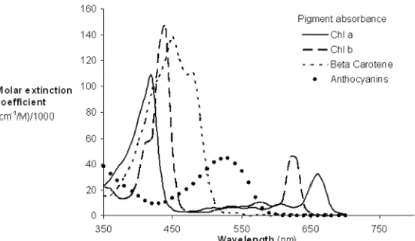
In general, the distribution of wavelengths displayed a double-peak feature, concentrated in the green (550–560 nm) and red edge (680–750 nm) regions rather than the main absorption wavelengths of chlorophyll (blue or red) (Fig 5). At the canopy scale, five wavelengths in the NIR to SWIR regions (1000–2400 nm) were also used for total chlorophyll quantification (not shown inFig 4B). This is due to the major influence of canopy structure in canopy reflectance and because leaf chlorophyll concentration was relatively stable in the particular studies [76,77].
The distribution of wavelengths proposed for chlorophyllaquantification at the leaf scale was similar to that of total chlorophyll, concentrated in the green and red edge ranges (Fig 6A). At the canopy and landscape scales, the number of wavelengths is limited and is difficult to identify the central tendency of wavelength distribution (Fig 6BandFig 6C).
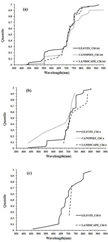
The distribution of wavelengths used for chlorophyllbquantification at the leaf scale were concentrated in the main absorption wavelength of chlorophyllb(red, 630–660 nm), the red edge (670–710 nm) and the NIR (800–810 nm) regions (Fig 7A). Only two wavelengths were selected at the landscape scale and could not be used for statistical inference (Fig 7B). The dis-tributions of wavelengths used for quantification of different pigments at different scales can be compared in the quantile plots (Fig 8). There were similar wavelength distributions for total chlorophyll quantification at the scales of leaf, canopy and landscape (Fig 8A). For chlorophyll athere were similar wavelength distributions at the leaf and canopy scales, but the landscape scale differed (Fig 8B), while a comparison across scales for chlorophyllbwas difficult due to a lack of data at scales other than the leaf (Fig 8C).
At the leaf scale, the wavelength distributions for total chlorophyll and chlorophylla quanti-fication were relatively similar while there were notable differences for chlorophyllb(Fig 9A). In the region 425–625 nm, the wavelengths used for chlorophyllaquantification were concen-trated in the region of the green peak in leaf reflectance (550nm), but the central tendency of wavelength distribution for chlorophyllbquantification was not obvious. In the red region, the wavelength distribution for chlorophyllaquantification was shifted to longer wavelengths than that of chlorophyllb(Fig 9A). The significant overlap in the absorption features of chloro-phyllaand chlorophyllb(Fig 5) and the low concentrations of chlorophyllbwith respect to chlorophyllain most leaves can present difficulties in defining optimal wavelengths for chloro-phyllbquantification. The absorption spectra of chlorophyllaand chlorophyllbboth display a double-peak feature; the absorption maxima of chlorophyllaare at 430 and 662 nm, and chlo-rophyllbhas peaks located at 453 and 642 nm (Fig 5). In the presence of carotenoids, it is diffi-cult to separately assess chlorophyllaand chlorophyllbfrom reflectance data in the blue region. However, in the red region, the wavelength position of maximum absorption by chloro-phyllais longer than that of chlorophyllb, which can be exploited for chlorophyllaand chlo-rophyllbdiscrimination (as seen inFig 9A). The capacity to use this approach to discriminate chlorophyllaand chlorophyllbis difficult to assess at the canopy and landscape scales due to the small number of studies on chlrophyll b (Fig 9B and 9C).
3.3.2 Optimal wavelengths for carotenoids quantification. At the leaf scale, the central
tendency of wavelength distribution was not obvious but was mainly concentrated in the 500–
3.3.3 Optimal wavelengths for anthocyanins quantification. Quantification of anthocya-nins from reflectance data has been given less attention by the passive optical hyperspectral remote sensing community than chlorophyll and carotenoids. Most studies have concentrated on the quantification of anthocyanins at the leaf scale, with some work at the landscape scale
Fig 4. Histogram of wavelengths for total chlorophyll quantification using remotely sensed data at leaf (a), canopy (b) and landscape (c) scales using an interval width of 10 nm.
but nothing at canopy level. At the leaf scale, the distribution of wavelengths used for quantify-ing anthocyanins was concentrated in the main absorption wavelength of anthocyanins (green, 550–560 nm), the red edge (700–710 nm) and the NIR (780–790 nm) ranges (Fig 12A). Simi-larly, the two wavelengths used to estimate anthocyanin concentration at the landscape scale were distributed in the green and red edge regions, respectively (Fig 12BandFig 13).
Discussion
This meta-analysis of 85 studies has demonstrated that remotely sensed variables are good esti-mators of plant pigment concentration. Most of the studies were conducted at the leaf scale, while pigment quantification at the canopy and landscape scales was less frequently reported. For each scale, most of the studies were conducted for total chlorophyll quantification, followed by chlorophylla, carotenoids, chlorophyllband anthocyanins. These findings are consistent with previous studies [10,11].
The strength of these relationships varied across pigments types and scales. In general, the relationships are stronger at the leaf scale than those at the canopy and landscape scales. At the leaf scale, the mean effect sizes were fairly consistent across different pigment types and were all greater than 0.87, while the difference in mean effect sizes between pigment types was statis-tically significant at the canopy and landscape scales. This result has been widely assumed, yet a quantitative evaluation has been lacking. At the leaf scale, the methodological basis for pig-ment quantification has been fully explored, which provides an important basis for developing estimation models at the canopy and landscape scales. The primary goal of most leaf scale pas-sive optical hyperspectral remote sensing studies has been to develop analytical approaches for pigment quantification that can be applied to data from airborne and spaceborne sensors [11]. At the canopy and landscape scales, the experimental results are influenced by a number of factors, which obscures the relationships between spectral reflectance and concentrations of individual pigments. The reflectance spectrum of a whole canopy is subject to canopy biophysi-cal attributes (e.g., orientation of leaves (leaf angle distribution;LAD), variations in number of leaf layers (LAI) and foliage clumping), presence of non-leaf elements (e.g., soil reflectance and the proportions of shadowed and sunlit background), anisotropic scattering of photons to interact with multiple surfaces such as leaves, woody material and soils, viewing geometry (e.g., sun and view zenith and azimuth angles) and illumination conditions (e.g., the ratio between direct and diffuse sunlight and atmospheric condition). It is the interaction of these factors,
Fig 5. Absorption spectra of the major plant pigments (reproduced from Blackburn, 2007).
including their potential covariance or unique behavior that drive variation in canopy and landscape reflectance characteristics in three-dimensional space [10,22].
It should be noted that part of the variability in effect sizes at the canopy scale may be entirely artifactual. These artifacts are common in experimental studies: studies vary in terms
Fig 6. Histogram of wavelengths for chlorophyllaquantification using remotely sensed data at leaf (a), canopy (b) and landscape (c) scales by an interval width of 10 nm.
of the quality of measurement; researchers make computational errors; people make typo-graphical errors in copying numbers from handwritten tables to computer; and sampling errors. With the advent of airborne and spaceborne imaging spectrometers, there have been opportunities to measure plant pigment concentrations at the landscape scale. The reflectance spectrum from airborne and spaceborne sensors is subject to even more controlling factors, notably, soil/litter surface reflectance, and vegetation structure. The range of controlling factors should be taken into account in subsequent analyses.
Table 2shows that the total sample size at the leaf scale is much more than that of canopy and landscape scales. The law of large numbers correctly states that large samples are reason-able representations of the population and parameter estimation is close to the real values when the sample size is large enough. Many researchers seem to believe that the same law applies to small samples and severely underestimate the amount of variability in findings that is caused by sampling errors. As a result, they erroneously expect statistics based on small sam-ples to be close to the real values [13]. At the canopy and landscape scales, the number of stud-ies and total sample size is limited, which influences the robustness and accuracy of effect sizes.
Despite the significant difference in effect sizes between different scales, it was found that the wavelength distribution for total chlorophyll quantification at the scales of leaf, canopy and landscape was similar, being concentrated in the green (550–560 nm) and red edge (680–750
Fig 7. Histogram of wavelengths for chlorophyllbquantification using remotely sensed data at leaf (a) and landscape (b) scales using an interval width of 10 nm.
nm) regions rather than the main absorption wavelength of chlorophyll (blue or red). The con-sistency in optimal wavelengths across scales can be attributed to several factors: (1) despite the many factors influencing reflectance at the canopy and landscape scales, it is the selective absorbance properties of pigments that determines the selection of wavelengths for pigment quantification, and (2) several estimation models derived at the leaf scale were directly applied
Fig 8. Quantile plots of the wavelengths used for the quantification of Chl tot (a), Chl a (b), and Chl b (c) at different scales.
to canopy and landscape scales. This suggests that the leaf-level study has provided an impor-tant basis for developing estimation models at the canopy and landscape scales.
At the leaf scale, the distribution of wavelengths used for chlorophyllaquantification was similar to that of total chlorophyll; the distribution of wavelengths for chlorophyllb
Fig 9. Quantile plot of the wavelengths used at leaf (a), canopy (b) and landscape (c) scales for the quantification of Chl tot, Chl a, and Chl b.
quantification was concentrated in the main absorption wavelength of chlorophyllb(red, 630–
660 nm), the red edge (670–710 nm) and the NIR (800–810 nm) regions; the central tendency of wavelength distribution for carotenoids quantification was not obvious, but was mainly con-centrated in the 500–580 nm region; for the estimation of anthocyanins, the distribution of
Fig 10. Histogram of wavelengths for carotenoids quantification using remotely sensed data at leaf (a), canopy (b) and landscape (c) scales using an interval width to 10 nm.
Fig 11. Quantile plot of the optimal wavelength for the quantification of Cars at different scales.
doi:10.1371/journal.pone.0137029.g011
Fig 12. Histogram of wavelengths for anthocyanins quantification using remotely sensed data at leaf (a) and landscape (b) scales using an interval width to 10 nm.
wavelengths was concentrated in the main absorption wavelength of anthocyanins (green, 550–560 nm), the red edge (700–710 nm) and the NIR (780–790 nm) ranges. In the present meta-analysis, the lack of studies reporting the quantification of carotenoids and anthocyanins at the canopy and landscape scales has hindered cross-scale comparisons (Fig 10;Fig 12). Con-sequently, it is not entirely clear if the optimal wavelengths for carotenoids and anthocyanins quantification at the leaf scale are necessarily the optimal wavelengths at the canopy and land-scape scales, where multiple scattering and other confounding effects may alter the spectral response of individual pigments, much in the way that pigment absorption peaks can vary depending upon their chemical and scattering medium. Therefore, more work may be needed to determine the optimal algorithms for airborne or spaceborne platforms.
It should be noted that the lack of statistical information in the studies (e.g., sample size and coefficient of determination) has hindered a more comprehensive cross-study comparison in the present research. When selecting the final set of studies, 50 studies were excluded due to the lack of statistical information. Insufficient statistical information can not only limit the research population covered by meta-analysis but also render the findings of the original study somewhat suspect. Thus, it is suggested that when conducting primary research, such informa-tion should include, but not be limited to, the sample size, the pertinent test statistic (e.g., r, t, or F), the unit of pigment concentration/content, the range of pigment concentrations/content, and estimation precision for pigment quantification (e.g. root mean squared error, RMSE).
This study has established the possibility of integrating the results of studies on the passive optical hyperspectral remote sensing of plant pigment concentrations across a range of vegeta-tion types and scales using a meta-analysis approach. Despite the robust models for pigment prediction at the leaf scale, the continuing challenge is to properly account for the multiple fac-tors introduced by scene components such as sunlit and shaded parts of tree crowns and gaps influencing the retrieved signal at the canopy and landscape scales. Recent work have illus-trated that, in addition to other influencing factors such as illumination geometry and atmo-spheric conditions, canopy architecture had an important control on the applicability of models for pigment prediction. Scanning LIDAR systems have only recently become widely available which enable the estimation of the range between the sensor and a target by recording the time during which the emitted laser pulse is reflected off an object and returns to the sensor
Fig 13. Quantile plot of the optimal wavelengths for the quantification of Anths at different scales.
[21]. LIDAR systems have the ability to directly measure spatial variations in canopy height and other aspects of the vertical structure of canopies. Given the high degree of structural com-plexity at the canopy and landscape scales, it would appear that the integration of vertical can-opy structural information provided by active LIDAR remote sensing with hyperspectral reflectance may has both a structural and physiological interpretation and improve the estima-tion of pigment concentraestima-tions over passive optical hyperspectral imagery alone [102].
Supporting Information
S1 PRISMA Checklist. PRISMA (Preferred Reporting Items for Systematic Reviews and Meta-Analyses) Checklist.
(DOC)
Acknowledgments
We acknowledge the contribution of Bao She, Weijiao Huang, Dilong Gan, Sujuan Wang and Zhewen Zhao for the literature database searches and associated support. The authors thank anonymous reviewers who provided very valuable comments also.
Author Contributions
Conceived and designed the experiments: JH. Performed the experiments: Chen Wei YZ XW Chuanwen Wei JW. Analyzed the data: Chen Wei. Contributed reagents/materials/analysis tools: Chen Wei. Wrote the paper: Chen Wei GAB JH.
References
1. Curran PJ, Dungan JL, Gholz HL. Exploring the relationship between reflectance red edge and chloro-phyll content in slash pine. Tree Physiol 1990; 7: 33–48. PMID:14972904
2. Filella I, Serrano L, Serra J, Penuelas J. Evaluating wheat nitrogen status with canopy reflectance indices and discriminant analysis. Crop Sci 1995; 35: 1400–1405.
3. Hendry GAF, Houghton JD, Brown SB. The degradation of chlorophyll: a biological enigma. New Phy-tol 1987; 107: 255–302.
4. Merzlyak MN, Gitelson A. Why and what for the leaves are yellow in autumn? On the interpretation of optical spectra of senescing leaves (Acerplatanoides L). J. Plant Physiol 1995; 145: 315–320.
5. Demmig—Adams B, Adams WW. The role of xanthophyll cycle carotenoids in the protection of photo-synthesis. Trends Plant Sci 1996; 1: 21–26.
6. Peñuelas J, Filella I. Visible and near-infrared reflectance techniques for diagnosing plant physiologi-cal status. Trends Plant Sci 1998; 3: 151–156.
7. Merzlyak MN, Gitelson AA, Chivkunova OB, Rakitin VY. Non- destructive optical detection of pigment changes during leaf senescence and fruit ripening. Physiol Plantarum 1999; 106: 135–141.
8. Chalker-Scott L. Environmental significance of anthocyanins in plant stress responses. Photochem Photobiol 1999; 70: 1–9.
9. Carter GA, Knapp AK. Leaf optical properties in higher plants: linking spectral characteristics to stress and chlorophyll concentration. Am J Bot 2001; 88: 677–684. PMID:11302854
10. Blackburn GA. Hyperspectral remote sensing of plant pigments. J. Exp Bot 2007; 58: 855–867. PMID:16990372
11. Ustin SL, Gitelson AA, Jacquemoud S, Schaepman M, Asner GP, Gamon JA, et al. Retrieval of foliar information about plant pigment systems from high resolution spectroscopy. Remote Sens Environ 2009; 113: S67–S77.
12. Hunter JE, Schmidt FL. Methods of meta-analysis: correcting error and bias in research findings. Los Angeles, USA: SAGE Publications; 2004. p. 33–34.
14. Curtis PS, Queenborough SA. Raising the standards for ecological meta–analyses. New Phytol 2012; 195: 279–281. doi:10.1111/j.1469-8137.2012.04207.xPMID:22702404
15. Glass GV. Primary secondary and meta-analysis of research. Educ Res 1976; 5: 3–8.
16. Verschuyl J, Riffell S, Miller D, Wigley TB. Biodiversity response to intensive biomass production from forest thinning in North American forests-A meta-analysis. Forest Ecol Manag 2011; 261: 221–232.
17. Robinson EA Ryan GD Newman JA A meta-analytical review of the effects of elevated CO2on
plant-arthropod interactions highlights the importance of interacting environmental and biological variables. New Phytol 2012; 194: 321–336. doi:10.1111/j.1469-8137.2012.04074.xPMID:22380757
18. Liao CZ Peng RH Luo YQ Zhou X H Wu X W Fang C M Chen J K Li B Altered ecosystem carbon and nitrogen cycles by plant invasion: a meta-analysis. New Phytol 2008; 177: 706–714. PMID: 18042198
19. Poorter H, Niinemets Ü, Poorter L, Wright IJ, Villar R. Causes and consequences of variation in leaf mass per area (LMA): a meta-analysis. New Phytol 2009; 182: 565–588. PMID:19434804
20. Garbulsky MF, Peñuelas J, Gamon J, Inoue Y, Filella I. The photochemical reflectance index (PRI) and the remote sensing of leaf canopy and ecosystem radiation use efficiencies: a review and meta-analysis. Remote Sens Environ 2011; 115: 281–297.
21. Zolkos SG, Goetz SJ, Dubayah R. A meta-analysis of terrestrial aboveground biomass estimation using lidar remote sensing. Remote Sens Environ 2013; 128: 289–298.
22. Asner GP. Biophysical and biochemical sources of variability in canopy reflectance. Remote Sens Environ 1998; 64: 234–253.
23. Curran PJ, Dungan JL, Macler BA, Plummer SE, Peterson DL. Reflectance spectroscopy of fresh whole leaves for the estimation of chemical concentration. Remote Sens Environ 1992; 39: 153–166.
24. Curran PJ, Windham WR, Gholz HL. Exploring the relationship between reflectance red edge and chlorophyll concentration in slash pine leaves. Tree Physiol 1995; 15: 203–206. PMID:14965977
25. Yoder BJ, Pettigrew-Crosby RE. Predicting nitrogen and chlorophyll content and concentrations from reflectance spectra (400–2500 nm) at leaf and canopy scales. Remote Sens Environ 1995; 53: 199– 211.
26. Gitelson AA, Merzlyak MN, Grits Y. Novel algorithms for remote sensing of chlorophyll content in higher plant leaves. In International Geoscience and Remote Sensing Symposium (IGARSS); Lincoln, NE, USA; May 1996. p. 2355–2357.
27. Gitelson AA, Kaufman YJ, Merzlyak MN. Use of a green channel in remote sensing of global vegeta-tion from EOS-MODIS. Remote Sens Environ 1996; 58: 289–298.
28. Gitelson AA, Merzlyak MN. Remote estimation of chlorophyll content in higher plant leaves. Int J Remote Sens 1997; 18: 2691–2697.
29. Gitelson AA, Merzlyak MN. Remote sensing of chlorophyll concentration in higher plant leaves. Adv Space Res 1998; 22: 689–692.
30. Gitelson AA, Buschmann C, Lichtenthaler HK. The chlorophyll fluorescence ratio F735/F700 as an accurate measure of the chlorophyll content in plants. Remote Sens Environ 1999; 69: 296–302.
31. Adams ML, Philpot WD, Norvell WA. Yellowness index: an application of spectral second derivatives to estimate chlorosis of leaves in stressed vegetation. Int J Remote Sens 1999; 20: 3663–3675.
32. Sims DA, Gamon JA. Relationships between leaf pigment content and spectral reflectance across a wide range of species leaf structures and developmental stages. Remote Sens Environ 2002; 81: 337–354.
33. Richardson AD, Duigan SP, Berlyn GP. An evaluation of noninvasive methods to estimate foliar chlo-rophyll content. New Phytol 2002; 153: 185–194.
34. Gitelson AA, Gritz Y, Merzlyak MN. Relationships between leaf chlorophyll content and spectral reflectance and algorithms for non–destructive chlorophyll assessment in higher plant leaves. J Plant Physiol 2003; 160: 271–282. PMID:12749084
35. Zhao DL, Reddy KR, Kakani VG, Read JJ, Koti S. Selection of optimum reflectance ratios for estimat-ing leaf nitrogen and chlorophyll concentrations of field-grown cotton. Agron J 2005; 97: 89–98.
36. Kochubey SM, Kazantsev TA. Changes in the first derivatives of leaf reflectance spectra of various plants induced by variations of chlorophyll content. J. Plant Physiol 2007; 164: 1648–1655. PMID: 17292510
38. Simic A, Chen JM, Leblanc SG, Dyk A, Croft H, Tian Han. Testing the top-down model inversion method of estimating leaf reflectance used to retrieve vegetation biochemical content within empirical approaches. IEEE J Sel Top Appl Earth Observ Remote Sens 2014; 7: 92–104.
39. Gitelson A, Merzlyak MN. Quantitative estimation of chlorophyll-a using reflectance spectra: experi-ments with autumn chestnut and maple leaves. J. Photoch Photobio B 1994; 22: 247–252.
40. Gitelson AA, Merzlyak MN. Spectral reflectance changes associated with autumn senescence of Aes-culus hippocastanum LandAcer platanoides LLeaves Spectral features and relation to chlorophyll estimation. J. Plant Physiol 1994; 143: 286–292.
41. Gitelson AA, Merzlyak MN. Signature analysis of leaf reflectance spectra: algorithm development for remote sensing of chlorophyll. J. Plant Physiol 1996; 148: 494–500.
42. Thomas JR, Gausman HW. Leaf reflectance vs Leaf chlorophyll and carotenoid concentrations for eight crops. Agron J 1977; 69: 799–802.
43. Gamon JA, Peñuelas J, Field CB. A narrow-waveband spectral index that tracks diurnal changes in photosynthetic efficiency. Remote Sens Environ 1992; 41: 35–44.
44. Levizou E, Manetas Y. Photosynthetic pigment contents in twigs of 24 woody species assessed by in vivo reflectance spectroscopy indicate low chlorophyll levels but high carotenoid/chlorophyll ratios. Environ Exp Bot 2007; 59: 293–298.
45. Gamon JA, Surfus JS. Assessing leaf pigment content and activity with a reflectometer. New Phytol 1999; 143: 105–117.
46. Merzlyak MN, Solovchenko AE, Gitelson AA. Reflectance spectral features and non–destructive esti-mation of chlorophyll carotenoid and anthocyanin content in apple fruit. Postharvest Biol Tec 2003; 27: 197–211.
47. Gitelson AA, Merzlyak MN. Non-destructive assessment of chlorophyll carotenoid and anthocyanin content in higher plant leaves: principles and algorithms. In Remote Sensing for Agriculture and the Environment. Stamatiadis S, LynchJ JM, Schepers JS, Eds. Ella, Greece: OECD; 2004. p. 78–94.
48. Garriga M, Retamales JB, Romero-Bravo S, Caligari PD, Lobos GA. Chlorophyll anthocyanin and gas exchange changes assessed by spectroradiometry in Fragaria chiloensis under salt stress. J. Integr Plant Biol 2014; 56: 505–15. doi:10.1111/jipb.12193PMID:24618024
49. Chappelle EW, Kim MS, McMurtrey JE III. Ratio analysis of reflectance spectra (RARS): an algorithm for the remote estimation of the concentrations of chlorophyll a chlorophyll b and carotenoids in soy-bean leaves. Remote Sens Environ 1992; 39: 239–247.
50. Blackburn GA. Spectral indices for estimating photosynthetic pigment concentrations: a test using senescent tree leaves. Int J Remote Sens 1998; 19 657–675.
51. Chen L, Huang JF, Wang FM. Retrieval of pigment contents in rice leaves and panicles using hyper-spectral data by artificial neuron network models. In International Geoscience and Remote Sensing Symposium (IGARSS); Seoul, Korea; July 2005. p. 1416–1419.
52. Blackburn GA. Relationships between spectral reflectance and pigment concentrations in stacks of deciduous broadleaves. Remote Sens Environ 1999; 70: 224–237.
53. Maccioni A, Agati G, Mazzinghi P. New vegetation indices for remote measurement of chlorophylls based on leaf directional reflectance spectra. J. Photoch Photobio B 2001; 61: 52–61.
54. Imanishi J, Nakayama A, Suzuki Y, Imanishi A, Ueda N, Morimoto Y, et al. Nondestructive determina-tion of leaf chlorophyll content in two flowering cherries using reflectance and absorptance spectra. Landsc Ecol Eng 2010; 6: 219–234.
55. Lichtenthaler HK, Gitelson A, Lang M. Non-destructive determination of chlorophyll content of leaves of a green and an aurea mutant of tobacco by reflectance measurements. J. Plant Physiol 1996; 148: 483–493.
56. Datt B. Visible/near infrared reflectance and chlorophyll content in eucalyptus leaves. Int J Remote Sens 1999; 20: 2741–2759.
57. Gitelson AA, Zur Y, Chivkunova OB, Merzlyak MN. Assessing carotenoid content in plant leaves with reflectance spectroscopy. Photochem Photobiol 2002; 75: 272–281. PMID:11950093
58. Filella I, Porcar-Castell A, Munne-Bosch S, Back J, Garbulsky MF, Penuelas J. PRI assessment of long-term changes in carotenoids/chlorophyll ratio and short–term changes in de-epoxidation state of the xanthophyll cycle. Int J Remote Sens 2009; 30: 4443–4455.
59. Garrity SR, Eitel JUH, Vierling LA. Disentangling the relationships between plant pigments and the photochemical reflectance index reveals a new approach for remote estimation of carotenoid content. Remote Sens Environ 2011; 115: 628–635.
61. Steele MR, Gitelson AA, Rundquist DC, Merzlyak MN. Nondestructive estimation of anthocyanin con-tent in grapevine leaves. Am J Enol Viticult 2009; 60: 87–92.
62. Gitelson AA, Chivkunova OB, Merzlyak MN. Nondestructive estimation of anthocyanins and chloro-phylls in anthocyanic leaves. Am J Bot 2009; 96: 1861–1868. doi:10.3732/ajb.0800395PMID: 21622307
63. Qin JL, Rundquist D, Gitelson A, Tan Z, Steele M. A non-linear model of nondestructive estimation of anthocyanin content in grapevine leaves with Visible/Red-infrared hyperspectral. In International Con-ference on Computer and Computing Technologies in Agriculture; Beijing, China; October 2011. p. 47–62.
64. Pappas CS, Takidelli C, Tsantili E, Tarantilis PA, Polissiou MG. Quantitative determination of anthocy-anins in three sweet cherry varieties using diffuse reflectance infrared fourier transform spectroscopy. J. Food Compos Anal 2011; 24: 17–21.
65. Vina A, Gitelson AA. Sensitivity to foliar anthocyanin content of vegetation indices using green reflec-tance. IEEE Geosci Remote Sens Lett 2011; 8: 464–468.
66. Ciganda V, Gitelson A, Schepers J. Non-destructive determination of maize leaf and canopy chloro-phyll content. J. Plant Physiol 2009; 166: 157–167. doi:10.1016/j.jplph.2008.03.004PMID: 18541334
67. Schlemmer M, Gitelson A, Schepers J, Ferguson R, Peng Y, Shanahan J, et al. Remote estimation of nitrogen and chlorophyll contents in maize at leaf and canopy levels. Int J Appl Earth Obs 2013; 25: 47–54.
68. Croft H, Chen JM, Zhang Y. The applicability of empirical vegetation indices for determining leaf chlo-rophyll content over different leaf and canopy structures. Ecol Complex 2014; 17: 119–130.
69. Ju CH, Tian YC, Yao X, Cao WX, Zhu Y, Hannaway D. Estimating leaf chlorophyll content using red edge parameters. Pedosphere 2010; 20: 633–644.
70. ZarcoTejada PJ. Hyperspectral remote sensing of closed forest canopies: estimation of chlorophyll fluorescence and pigment content. PhD thesis, York University, Toronto, Canada 2000.
71. Filella I, Penuelas J. The red edge position and shape as indicators of plant chlorophyll content bio-mass and hydric status. Int J Remote Sens 1994; 15: 1459–1470.
72. Gitelson AA, Vina A, Ciganda V, Rundquist DC, Arkebauer TJ. Remote estimation of canopy chloro-phyll content in crops. Geophys Res Lett 2005; 32: 1–4.
73. Yang XH, Huang JF, Wang FM, Wang XZ, Yi QX, Wang Y. Science letters: a modified chlorophyll absorption continuum index for chlorophyll estimation. J. Zhejiang Univ 2006; 7: 2002–2006.
74. Zhao DH, Huang LM, Li JL, Qi JG. A comparative analysis of broadband and narrowband derived veg-etation indices in predicting LAI and CCD of a cotton canopy. Isprs J Photogramm 2007; 62: 25–33.
75. Wu CY, Niu Z, Tang Q, Huang WJ. Estimating chlorophyll content from hyperspectral vegetation indi-ces: modeling and validation. Agr Forest Meteorol 2008; 148: 1230–1241.
76. Darvishzadeh R, Skidmore A, Schlerf M, Atzberger C, Corsi F, Cho M. LAI and chlorophyll estimation for a heterogeneous grassland using hyperspectral measurements. ISPRS J Photogramm 2008; 63: 409–426.
77. Darvishzadeh R, Skidmore A, Schlerf M, Atzberger C. Inversion of a radiative transfer model for esti-mating vegetation LAI and chlorophyll in a heterogeneous grassland. Remote Sens Environ 2008; 112: 2592–2604.
78. Haboudane D, Tremblay N, Miller JR, Vigneault P. Remote estimation of crop chlorophyll content using spectral indices derived from hyperspectral data. IEEE T Geosci Remote 2008; 46: 423–437.
79. Liu ML, Liu XN, Li M, Fang MH, Chi WX. Neural-network model for estimating leaf chlorophyll concen-tration in rice under stress from heavy metals using four spectral indices. Biosystems Eng 2010; 106: 223–233.
80. Xu X, Gu X, Song X, Li C, Huang W. Assessing rice chlorophyll content with vegetation indices from hyperspectral data. In International Conference on Computer and Computing Technologies in Agricul-ture; Beijing, China; October 2011. p. 296–303.
81. Clevers JGPW, Kooistra L. Using Hyperspectral Remote Sensing Data for Retrieving Canopy Chloro-phyll and Nitrogen Content. IEEE J Sel Top Appl Earth Observ Remote Sens 2012; 5: 574–583.
82. Clevers JGPW, Gitelson AA. Remote estimation of crop and grass chlorophyll and nitrogen content using red-edge bands on Sentinel-2 and -3. Int J Appl Earth Obs 2013; 23: 344–351.
83. Vincini M, Amaducci S, Frazzi E. Empirical Estimation of Leaf Chlorophyll Density in Winter Wheat Canopies Using Sentinel–2 Spectral Resolution. IEEE T Geosci Remote 2014; 52: 3220–3235.
85. Li J, Jiang JB, Chen YH, Wang YY, Su W, Huang WJ. Using hyperspectral indices to estimate foliar chlorophyll a concentrations of winter wheat under yellow rust stress. New Zeal J Agr Res 2007; 50: 1031–1036.
86. Zhao X, Liu SH, Wang JD, Tian ZK. A method for estimating chlorophyll content of wheat from reflec-tance spectra. In International Geoscience and Remote Sensing Symposium (IGARSS); Anchorage, AK, USA; September 2004. p. 4504–4507.
87. Bannari A, Khurshid KS, Staenz K, Schwarz J. Wheat crop chlorophyll content estimation from ground–based reflectance using chlorophyll indices. In International Geoscience and Remote Sens-ing Symposium (IGARSS); Denver, CO, USA; July 2006. p. 112–115.
88. Yang F, Li JL, Gan XY, Qian YR, Wu XL, Yang Q. Assessing nutritional status ofFestuca arundinacea by monitoring photosynthetic pigments from hyperspectral data. Comput Electron Agr 2010; 70: 52– 59.
89. Peguero-Pina JJ, Morales F, Flexas J, Gil-Pelegrin E, Moya I. Photochemistry remotely sensed physi-ological reflectance index and de-epoxidation state of the xanthophyll cycle inQuercus coccifera under intense drought. Oecologia 2008 156: 1–11. doi:10.1007/s00442-007-0957-yPMID: 18224338
90. Hall FG, Hilker T, Coops NC, Lyapustin A, Huemmrich KF, Middleton E, et al. Multi–angle remote sensing of forest light use efficiency by observing PRI variation with canopy shadow fraction. Remote Sens. Environ 2008; 112: 3201–3211.
91. Haboudane D, Miller JR, Tremblay N, Zarco-Tejada PJ, Dextraze L. Integrated narrow–band vegeta-tion indices for predicvegeta-tion of crop chlorophyll content for applicavegeta-tion to precision agriculture. Remote Sens Environ 2002; 81: 416–426.
92. Coops NC, Stone C, Culvenor DS, Chisholm LA, Merton RN. Chlorophyll content in eucalypt vegeta-tion at the leaf and canopy scales as derived from high resoluvegeta-tion spectral data. Tree Physiol 2003; 23: 23–31. PMID:12511301
93. Zarco-Tejada PJ, Miller JR, Harron J, Hu BX, Noland TL, Goel N, et al. Needle chlorophyll content estimation through model inversion using hyperspectral data from boreal conifer forest canopies. Remote Sens Environ 2004; 89: 189–199.
94. Dash J, Curran PJ. The MERIS terrestrial chlorophyll index. Int J Remote Sens 2004; 25: 5403–5413.
95. Haboudane D, Tremblay N, Vigneault P, Miller JR. Indices-based approach for crop chlorophyll con-tent retrieval from hyperspectral data. In International Geoscience and Remote Sensing Symposium (IGARSS); Barcelona, Spain; July 2007. p.3297–3300.
96. Rao NR, Garg PK, Ghosh SK, Dadhwal VK. Estimation of leaf total chlorophyll and nitrogen concen-trations using hyperspectral satellite imagery. J Agr Sci 2008; 146: 65–75.
97. Delegido J, Fernandez G, Gandia S, Moreno J. Retrieval of chlorophyll content and LAI of crops using hyperspectral techniques: application to PROBA/CHRIS data. Int J Remote Sens 2008; 29: 7107– 7127.
98. Wu CY, Han XZ, Niu Z, Dong JJ. An evaluation of EO-1 hyperspectral hyperion data for chlorophyll content and leaf area index estimation. Int J Remote Sens 2010; 31: 1079–1086.
99. Delegido J, Alonso L, González G, Moreno J. Estimating chlorophyll content of crops from hyperspec-tral data using a normalized area over reflectance curve (NAOC). Int J Appl Earth Obs 2010; 12: 165– 174.
100. Delegido J, Van Wittenberghe S, Verrelst J, Ortiz V, Veroustraete F, Valcke R, et al. Chlorophyll con-tent mapping of urban vegetation in the city of Valencia based on the hyperspectral NAOC index. Ecol Indic 2014; 40: 34–42.
101. Oppelt N, Mauser W. Hyperspectral monitoring of physiological parameters of wheat during a vegeta-tion period using AVIS data. Int J Remote Sens 2004; 25: 145–159.
102. Blackburn GA. Remote sensing of forest pigments using airborne imaging spectrometer and LIDAR imagery. Remote Sens Environ 2002; 82: 311–321.
103. Guan YN, Guo S, Liu JG, Zhang X. Algorithms for the estimation of the concentrations of chlorophyll a and carotenoids in rice leaves from airborne hyperspectral data. In Computational Science-ICCS 2005; Atlanta, GA, USA; May 2005. p. 908–915.
104. Thomas V, Treitz P, McCaughey JH, Noland T, Rich L. Canopy chlorophyll concentration estimation using hyperspectral and lidar data for a boreal mixedwood forest in northern Ontario Canada. Int J Remote Sens 2008; 29: 1029–1052.
106. Jacquemoud S, Verdebout J, Schmuck G, Andreoli G. Hosgood B. Investigation of leaf biochemistry by statistics. Remote Sens Environ 1995; 54: 180–188.
107. Cornell JA, Berger RD. Factors that influence the value of the coefficient of determination in simple lin-ear and nonlinlin-ear regression models. Phytopathology 1987; 77: 63–70.
108. Gurevitch J, Curtis PS, Jones MH. Meta-analysis in ecology. Adv Ecol Res 2001; 32: 199–247.
109. Hedges LV. Estimation of effect size from a series of independent experiments. Psychol Bull 1982; 92: 490–499.
110. Higgins J, Thompson SG. Quantifying heterogeneity in a meta-analysis. Stat Med 2002; 21: 1539– 1558. PMID:12111919
111. Hedges LV, Olkin I. Statistical methods for meta-analysis. Orlando, FL, USA: Academic Press; 1985. p. 31–34.
112. Lipsey MW, Wilson DB. Practical meta-analysis. London, UK: SAGE Publications; 2000. p. 112– 116.
113. Mosteller F, Colditz GA. Understanding research synthesis (meta-analysis). Annu Rev Publ Health 1996; 17 1–23.


