Induced by Calcitonin Gene-Related Peptide in a Mouse
Model of Neurofibromatosis
Stephanie White1", Blanca Marquez de Prado1,2", Andrew F. Russo2
, Donna L. Hammond1,3*
1Department of Anesthesia, University of Iowa, Iowa City, Iowa, United States of America,2Department of Molecular Physiology and Biophysics, University of Iowa, Iowa City, Iowa, United States of America,3Department of Pharmacology, University of Iowa, Iowa City, Iowa, United States of America
Abstract
This study examined whether mice with a deficiency of neurofibromin, a Ras GTPase activating protein, exhibit a nociceptive phenotype and probed a possible contribution by calcitonin gene-related peptide. In the absence of inflammation,Nf1+/2
mice (B6.129S6 Nf1,tm1Fcr./J) and wild type littermates responded comparably to heat or mechanical stimuli, except for a subtle enhanced mechanical sensitivity in female Nf1+/2
mice. Nociceptive phenotype was also examined after inflammation induced by capsaicin and formalin, which release endogenous calcitonin gene-related peptide. Intraplantar injection of capsaicin evoked comparable heat hyperalgesia and mechanical hypersensitivity inNf1+/2
and wild type mice of both genders. Formalin injection caused a similar duration of licking in maleNf1+/2
and wild type mice. FemaleNf1+/2
mice licked less than wild type mice, but displayed other nociceptive behaviors. In contrast, intraplantar injection of CGRP caused greater heat hyperalgesia inNf1+/2
mice of both genders compared to wild type mice. MaleNf1+/2
mice also exhibited greater mechanical hypersensitivity; however, femaleNf1+/2
mice exhibited less mechanical hypersensitivity than their wild type littermates. Transcripts for calcitonin gene-related peptide were similar in the dorsal root ganglia of both genotypes and genders. Transcripts for receptor activity-modifying protein-1, which is rate-limiting for the calcitonin gene-related peptide receptor, in the spinal cord were comparable for both genotypes and genders. The increased responsiveness to intraplantar calcitonin gene-related peptide suggests that the peripheral actions of calcitonin gene-related peptide are enhanced as a result of the neurofibromin deficit. The analgesic efficacy of calcitonin gene-related peptide receptor antagonists may therefore merit investigation in neurofibromatosis patients.
Citation:White S, Marquez de Prado B, Russo AF, Hammond DL (2014) Heat Hyperalgesia and Mechanical Hypersensitivity Induced by Calcitonin Gene-Related Peptide in a Mouse Model of Neurofibromatosis. PLoS ONE 9(9): e106767. doi:10.1371/journal.pone.0106767
Editor:Claudia Sommer, University of Wu¨rzburg, Germany
ReceivedFebruary 28, 2014;AcceptedAugust 8, 2014;PublishedSeptember 3, 2014
Copyright:ß2014 White et al. This is an open-access article distributed under the terms of the Creative Commons Attribution License, which permits unrestricted use, distribution, and reproduction in any medium, provided the original author and source are credited.
Funding:This work was supported by the Department of Defense through Grant NF073016 to DLH and Grant NF100114 to BMdP. The funders had no role in study design, data collection and analysis, decision to publish, or preparation of the manuscript.
Competing Interests:The authors have declared that no competing interests exist.
* Email: Donna-hammond@uiowa.edu
"SW and BMdP are co-first authors on this work.
Introduction
Neurofibromatosis 1 (NF1) is an autosomal dominant disorder that results in reduced levels of neurofibromin, a GTPase activating protein (GAP) involved in the regulation of Ras signaling (i.e. a Ras-GAP). This genetic disorder affects one in
3500 births worldwide – an incidence that equates to ,90,000
Americans and a million persons worldwide [1–4]. Nearly half of
these cases result from new mutations. As such,Nf1has one of the
highest rates of new mutations for any known single gene disorder [3,5]. One in four individuals with NF1 experience chronic bodily pain, as well as migraine and headache pain, over periods of months to years [1,6,7]. Severe pain also results from neurofibro-mas on spinal roots and malignant peripheral nerve sheath tumors [3]. The chronic nature of the pain, as well as its lancinating and paroxysmal character, contribute to the poor quality of life for patients with NF [8]. There is a great need for mechanistic based pharmacotherapies for the relief of pain in this patient population. Early studies by Hingtgen and colleagues focused attention on the possible role of calcitonin gene-related peptide (CGRP) in pain associated with NF1. CGRP is a key factor in peripheral
inflammation and in the production of nociception both in the spinal cord and in the periphery [9–12]. Interestingly, high densities of CGRP-immunoreactive fibers are present in
neurofi-bromas in patients [13]. Using a rodent model of NF1 (Nf1+/2
mice), Hingtgen and colleagues determined that haploid insuffi-ciency of neurofibromin is associated with an increased release of CGRP from cultured dorsal root ganglion (DRG) neurons and spinal cord slices [14], as well as increased excitability of primary afferent neurons that convey pain [15,16]. More recent studies
determined that the DRG of Nf1+/2 mice have increased
transcripts for Nav1.7 and Nav1.8, which are likely to underlie the enhanced excitability of primary afferent neurons [17], as well as increased N-type calcium currents that could contribute to enhanced release of neurotransmitter [18]. Collectively, these data suggest that an increased release of CGRP and decreased thresholds for activation of sensory neurons may underlie pain in NF1.
The aim of this study was to determine whether Nf1+/2 mice
Nociceptive responsiveness of male and female Nf1+/2 mice to heat and mechanical stimuli was determined in the absence of inflammation and after inflammation induced by exogenous administration of CGRP or by formalin or capsaicin, two agents that can release CGRP in the spinal cord and periphery. Ancillary studies quantitated levels of transcript for CGRP in the DRG and receptor activity-modifying protein-1 (RAMP1) in the spinal cord. RAMP1 associates with the calcitonin-like receptor to form the CGRP receptor [19], and is rate limiting for the activity of CGRP
[20]. The results indicate thatNf1+/2mice are not a robust animal
model for the pain experienced by NF patients, a conclusion that was also reached by O’Brien et al. [21]. Nonetheless, the finding that neurofibromin deficit is associated with an enhanced effect of CGRP in the periphery suggests that CGRP receptor antagonists may merit investigation for the treatment of pain in NF patients.
Materials and Methods
Experimental model
Mutation ofNf1 in one allele is sufficient for expression and
nearly complete penetrance of the disorder in humans. These
studies therefore used Nf1+/2 mice as a rodent model of NF1.
These mice exhibit an increased predisposition to develop tumors as they age [22,23]. Use of homozygous null mutant mice was not feasible as the mutation is embryonic lethal [22,24]. Moreover, no
definitive examples of patients homozygous for mutation of Nf1
have been identified [25].
Litter-matched wild type (WT) and Nf1+/2 (B6.129S6 Nf1,
tm1Fcr./J) mice of both genders (Jackson, Bar Harbor, Maine)
weighing 23–30 grams were used. Female mice were tested randomly throughout the estrous cycle. Mice were housed in groups of five on SoftZorb paper enrichment bedding in a temperature controlled room on a 12-hr light/dark cycle. All testing occurred during the light cycle from 9:00 to 15:00 hr. Mice
were euthanized at the conclusion of the experiments by CO2
inhalation. These experiments were approved by the University of Iowa Animal Care and Use Committee (ACURF 0711237) and were conducted in accordance with the guidelines of the National Institutes of Health Guide for the Care and Use of Laboratory Animals and the guidelines of the International Association for the Study of Pain. With the exception of the capsaicin test, mice were used once and received only one dose of an agent. Every effort was made to minimize the number of mice used and their suffering. In all experiments, the investigator was blinded to the genotype. Blinding to intraplantar (ipl) treatment was not possible because capsaicin and formalin both produce spontaneous pain behaviors, while CGRP produces erythema and inflammation upon injec-tion.
Heat threshold
Experiments were conducted to determine responsiveness to noxious heat, and whether heat hyperalgesia produced by ipl injection of CGRP or capsaicin differed by genotype or gender. Mice were acclimated to the testing environment for several hrs on two successive days and then allowed to move freely for one hr within a small Plexiglas enclosure that rested on an elevated glass
surface maintained at 25uC. On the third day, the mice were
acclimated to the environment for two more hrs and again allowed to acclimate to the testing chambers for another 30 min. A high intensity beam of light was then positioned under each hindpaw, and the latency to withdraw the hindpaw from the heat stimulus was recorded (UARD group, La Jolla, CA). If the mouse did not withdraw its paw within 20 sec, the test was terminated to prevent
tissue injury. Care was taken to ensure that the surface of the hindpaw was flush with glass to avoid a heat-sink effect [26].
The first set of experiments assessed basal sensitivity to a noxious heat stimulus. The radiant heat stimulus was delivered at
two different rates, 2.5uC/sec and 6.5uC/sec, as measured at the
surface. These rates corresponded to 0.6uC/sec and 1.0uC/sec, as
measured within the tissue. For both stimuli, the subdermal
temperature at which withdrawal occurred was 40–41uC, in
agreement with similar studies in the rat [27–29]. The lower rate of heating, which likely preferentially activates C-fibers, was used in the second set of experiments that assessed heat hyperalgesia. After determination of baseline paw withdrawal latency, an ipl
injection of 5mg CGRP, 0.01% capsaicin, or PBS was made into a
hind paw. Paw withdrawal latency was redetermined 15, 30, 45, and 60 min after CGRP and 15 min after capsaicin.
Data were expressed as the mean 6 S.E.M. Baseline paw
withdrawal latencies were compared using a two-way analysis of variance (ANOVA) in which genotype was one factor and intensity was the second factor. The effects of CGRP or capsaicin were compared to their vehicle using a two-way repeated measures ANOVA in which one factor was genotype and the repeated factor was time. The Holm-Sidak test was used to make comparisons among mean values for the different treatment
groups. A P,0.05 was considered significant in these and all
subsequent analyses.
Mechanical threshold
Additional experiments determined mechanical threshold and examined whether mechanical hypersensitivity after ipl injection of CGRP or capsaicin differed by genotype or gender. Mice were acclimated to the testing environment for several hrs on two successive days. On the third day, the mice were acclimated for two more hrs to the environment and then allowed to move freely for a further 30 min within a small Plexiglas enclosure with a mesh floor. Mechanical hypersensitivity was assessed with a series of von Frey filaments that ranged from 1.65–4.31 log mN (0.0008–2 g). The filaments were applied to the plantar surface of each hindpaw between the pads. Care was taken to vary the site of application in this area. Testing was initiated by a two-sec application of the 3.84 filament before and the 3.22 filament after ipl injection of CGRP or capsaicin with enough pressure to cause the filament to bend slightly. If the mouse withdrew or lifted the paw, filaments of successively lower force were applied until one that produced no response to five applications was identified. The next higher force filament was then applied for a total of five times and the percentage of responses was recorded for that filament. Filaments of successively higher force were applied in this manner until a filament was reached that produced withdrawal on all five presentations or the 4.31 filament was reached. Higher filaments were not used because they lifted the hindpaw before the filament
bent. After determination of basal threshold, either 5mg CGRP,
0.001 or 0.01% capsaicin or their respective vehicles were injected. Mechanical hypersensitivity was redetermined 5 and 15 min after CGRP or PBS, and 15 min after capsaicin or vehicle by which time capsaicin’s associated spontaneous pain behaviors had subsided.
Force-response curves were generated for both hindpaws and fit
by non-linear regression to determine an EF50 with 95%
confidence limits (CL) using Graphpad Prism. The minimum and maximum values were constrained to 0 and 100, respectively.
Comparisons of EF50values were made by F-test using Graphpad
Spontaneous pain behavior
Spontaneous pain behaviors evoked by ipl injection of 0.01% capsaicin or 2% formalin in one hindpaw were also quantified. In the case of capsaicin, the amount of time spent licking the injected paw was recorded in 5 min epochs for 15 min; spontaneous pain behaviors were essentially absent after 15 min. In the interest of making efficient use of each mouse and because the licking evoked by capsaicin was highly variable, each mouse received an injection of capsaicin in the left and right hindpaw, separated by a month. The duration of licking of each hindpaw was then averaged for that mouse. In the case of formalin, spontaneous pain behaviors were recorded for 90 min with a video camera and analyzed offline. The amount of time spent licking the paw, as well as the percentage of mice that exhibited guarding, flinching or unweighting of the injected paw, were quantified in 5 min epochs.
Duration of licking after capsaicin was expressed as the mean6
S.E.M. and compared between genotypes by two-way ANOVA for repeated measures. The Holm-Sidak test was used to compare group mean values. The duration of licking after formalin was analyzed in the same manner. In addition, the total time spent licking during phase one (0–5 min), phase two (15–50 min) and phase three (55–90 min) of the formalin test was calculated and compared by two-way ANOVA for repeated measures. Finally, guarding, flinching or unweighting of the injected paw after formalin were collectively scored as either present or absent. The percentage of mice that exhibited these behaviors was averaged over time for each of the three phases of the formalin test and compared by ANOVA.
Quantitation of CGRP and RAMP1 mRNA
Mice were euthanized by CO2inhalation followed by
decap-itation. The entire spinal cord and all DRG were rapidly removed from each mouse and stored in RNAlater (Ambion, Austin, TX). Total RNA was isolated from the tissue using the RNeasy kit according to the manufacturer’s directions (Qiagen, Valencia, CA) and DNA contamination removed by digestion with Amp Grade DNase I (Invitrogen, Carlsbad, CA). Briefly, less than 30 mg of tissue was disrupted and homogenized in RTL buffer containing
b-mercaptoethanol using a Tissue Tearor. The lysate was then
centrifuged and the supernatant saved in a clean tube. The RNA was precipitated using 1 volume of 70% ethanol and loaded on the
column. After one wash, 40ml of buffer containing 1 unit DNase I
was applied to the column and allowed to incubate for 15 min. The DNase was then removed by extensive washing. The RNA was eluted using RNase free water, and RNA concentration was determined by spectrometry. The RNA integrity number was determined for a subset of samples, and ranged between 8.1 and 8.6. Reverse transcription was performed using the Taqman
RT-PCR mix (Applied Biosystems, Carlsbad, CA) with 0.5mg RNA,
1X RT buffer, 5.5 mM MgCl2, 0.5 mM dNTP, 4 units RNase
inhibitor, 2.5mM random hexamers, 12.5 units Multiscribe
reverse transcriptase in 10ml at 25uC for 10 min, 48uC for
30 min and 95uC for 5 min. Q-PCR was performed using 50 ng
cDNA, 670 nM each primer (67 nM for CGRP primers) and 1X
IQtmSYB Green Supermix (BioRad, Hercules, CA) in 15ml. The
cycle conditions were: 95uC for 10 min, followed by 40 cycles of
95uC for 15 s and annealing/extension at 60uC for 30 s and 72uC
for 45 sec. Reactions were performed in triplicate and analyzed using a Bio-Rad MY-IQ thermocycler. At the end of
amplifica-Figure 1. Assessment of acute nociception inNf1+/2mice and
WT littermates.In the absence of injury,Nf1+/2mice did not exhibit
robust differences in sensitivity to noxious heat or tactile stimuli applied to the plantar surface of the hindpaw. (A) Paw withdrawal latency to low or high heating rates did not differ among genotypes or genders. Response latencies for both hindpaws were averaged to yield a single value for each mouse. Data are mean 6 SEM of 6–8 mice. (B) Mechanical sensitivity was slightly greater in femaleNf1+/2mice than wild type (WT) littermates (N = 18 in each group). (C) MaleNf1+/2
and WT mice did not differ in their mechanical sensitivity (n = 16 and 15,
respectively). Panels B and C were constructed using baseline data for the ipsilateral hindpaw of mice in the capsaicin and CGRP treatment groups. Data are expressed as the mean6SEM.
tion, a thermal melt curve was generated. The one sample that that did not yield a homogenous melt curve was excluded. Primers
for mRAMP1, CGRP, mGAPDH and b-actin have been
described [20]. During this work, it was discovered that the description of the reverse primer for mRAMP1 in Zhang et al. (2007) contained a typographical error and an extraneous
nucleotide. Although the correct primer (59
GCACTTGCT-GAAGTATCGATGG39) was used for that work, the textual errors were replicated when ordering primer for this study. Nonetheless, the PCR product yielded the correct cDNA as
verified by sequence analysis. Cycle thresholds (CT) for mRAMP1,
CGRP, GAPDH andb-actin were converted to absolute numbers
using standard curves generated with serial dilutions of
pGEM-QmRAMP1, pQmCGRP, pQmGAPDH and pQmb-actin
plas-mids, respectively. For RAMP1, the mean efficiency of the PCR
was 99.161.1% with a mean slope of23.3560.03. Forb-actin,
the mean efficiency was 94.861.0% with a mean slope of 2
3.4360.03. For CGRP, the efficiency of the PCR was 93.2% with
a mean slope of 23.299 and for GAPDH the efficiency of the
PCR was 101.0% with a mean slope of23.496.
Drugs and Vehicle Controls
All drugs were purchased from (Sigma, St Louis, MO) and prepared fresh each day. Calcitonin gene-related peptide was
dissolved in PBS, which served as its vehicle control. Capsaicin was dissolved in 5% Tween 80 and 5% ethanol and brought to volume with PBS. In the von Frey experiments, a few mice received PBS as the control and others received PBS with 5% Tween 80 and 5% ethanol as the control. As there was no difference in the effects of either vehicle, the data were pooled for statistical analysis. Formalin was diluted to 2% using PBS. All drugs were injected in the plantar surface of one hindpaw with a 33-gauge stainless
steel injector needle. Formalin was injected in a volume of 20ml,
while capsaicin or CGRP was injected in a volume of 10ml. Drug
delivery was monitored by following the movement of an air bubble in the tubing that connected the injector to the syringe pump.
Results
Nociceptive thresholds in the absence of injury
No differences in paw withdrawal latency were evident between the genotypes or by gender at either heating rate (Fig. 1A). With
respect to mechanical sensitivity, the EF50 (95% CL) values for
female WT andNf1+/2mice were 1.35 (1.3021.40) g and 1.16
(1.04–1.28) g, respectively (P,0.01). The small, but significant
increase in mechanical sensitivity of femaleNf1+/2mice could be
attributed to an enhanced responsiveness to lower force filaments
Figure 2. Intraplantar injection of 0.01% capsaicin or 5mg CGRP induced heat hyperalgesia.(A, B) The magnitude of heat hyperalgesia
determined 15 min after injection of capsaicin did not differ between genotypes in either females or males. * P,0.05 compared to baseline. (C, D) Paw withdrawal latency was decreased within 5 min of injection (arrow) of CGRP, but not PBS in females and males of both genotypes. Heat hyperalgesia in female and maleNf1+/2mice was greater than their WT littermates. The data for PBS are the pooled responses of two female and two male mice of each genotype. * P,0.05 compared to WT at the same time point. Data in all panels are expressed as the mean 6SEM of determinations in 6–8 mice of each gender and genotype.
(Fig. 1B). The EF50(95% CL) values for male WT and maleN1f+/
2mice were 1.32 (1.2321.42) and 1.38 (1.30–1.48) g, respectively
(P.0.5; Fig. 1C). Thus, with the exception of a small increase in
mechanical sensitivity in female Nf1+/2 mice, responsiveness to
heat and mechanical stimuli did not differ between the genotypes or between the genders. Subsequent experiments examined
whetherNf1+/2mice differed from their WT littermates in their
responses to inflammatory agents.
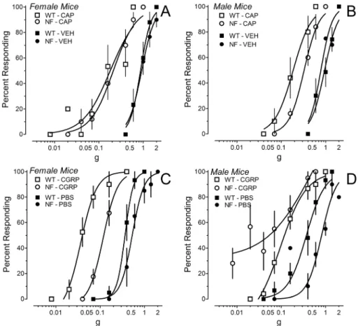
Figure 3. Mechanical hypersensitivity induced by intraplantar injection of 0.001% capsaicin (CAP) or 5mg CGRP.(A, B) Force-response
curves for the ipsilateral hindpaw after injection of CAP or vehicle (VEH) inNf1+/2(circles) or WT (squares) mice. Data are the mean
6SEM of responses by 5–7 mice of each gender and genotype. (C, D) Force-response curves for the ipsilateral hindpaw after injection of CGRP or PBS inNf1+/2 (circles) or WT (squares) mice. Data in panels C-D are the mean6SEM of responses by 4–8 mice of each gender and genotype, with the exception of CGRP inNf1+/2mice where 11 mice were tested.
doi:10.1371/journal.pone.0106767.g003
Table 1.Capsaicin evokes mechanical hypersensitivity in both wildtype andNf1+/2
mice.
Female WT Nf1+/2
Vehicle [6] Capsaicin [6] Vehicle [7] Capsaicin [5]
Baseline 1.43 (1.37–1.50) 1.37 (1.31–1.44) 1.57 (1.46–1.69) 1.52 (1.29–1.79)
After 0.87 (0.73–1.03)`
0.18 (0.11–0.29)`
** 0.92 (0.77–1.10)`
0.21 (0.15–0.29)` **
Male WT Nf1+/2
Vehicle [6] Capsaicin [5] Vehicle [4] Capsaicin [4]
Baseline 1.42 (1.36–1.48) 1.40 (1.29–1.52) 1.51 (1.40–1.63) 1.49 (1.36–1.63)
After 0.95 (0.78–1.15)`
0.18 (0.12–0.27)`
** 0.78 (0.60–1.01)`
0.32 (0.28–0.39)` **a
EF50and 95% confidence limits (g) of force–response curves for the ipsilateral hindpaw before and 15 min after intraplantar injection of 0.001% capsaicin or vehicle.
Curves were fit by non–linear regression with minimum constrained to 0 and maximum to 100. Numbers of mice in each group appear in brackets. *P,0.05,
** P,0.01 compared to vehicle at corresponding time point.
{
P,0.05, `
P,0.01 compared to baseline value.
aP
Heat hyperalgesia evoked by capsaicin or CGRP
Injection of 0.01% capsaicin in the hindpaw decreased paw
withdrawal latency to a similar extent in femaleNf1+/2and WT
mice (Fig. 2A). Capsaicin also significantly decreased paw
withdrawal latency in maleNf1+/2mice, but did not significantly
decrease paw withdrawal latency in male WT mice (P = 0.13;
Fig. 2B). Nonetheless, male WT andNf1+/2mice had
compara-ble paw withdrawal latencies after capsaicin (P.0.5). These results
indicate that the magnitude of capsaicin-induced heat hyperalgesia was comparable in both genotypes and genders.
Intraplantar injection of CGRP produced heat hyperalgesia in
Nf1+/2mice and WT littermates of both genders. FemaleNf1+/2
mice exhibited a significantly greater decrease in paw withdrawal latency than female WT mice 15 and 45 min after injection
(Fig. 2C). Male Nf1+/2 mice exhibited a significantly greater
decrease in paw withdrawal latency than male WT mice 45 min after injection (Fig. 2D). Intraplantar injection of PBS did not alter paw withdrawal latency of the ipsilateral hindpaw of either gender or genotype. Thus, unlike the heat hyperalgesia induced by capsaicin, the heat hyperalgesia induced by CGRP was greater in
both male and female Nf1+/2 mice compared to their
corre-sponding WT controls.
Mechanical sensitivity after capsaicin or CGRP
Injection of 0.01% capsaicin in the hindpaw dramatically shifted the force-response curves to the left such that the lowest filament (0.008 g, 1.65 log mN) elicited responses on more than 50% of its presentations in most mice. Although mechanical hypersensitivity did not differ between genotypes or gender, a ceiling effect could have disguised subtle differences. Fifteen min after injection of a ten-fold lower dose of capsaicin (0.001%)
female Nf1+/2 and WT mice exhibited equivalent mechanical
hypersensitivity, while maleNf1+/2mice exhibited less mechanical
hypersensitivity than male WT mice (Fig. 3B, Table 1). Injection of vehicle also produced a very small, but statistically significant leftward shift in the force-response curves compared to baseline (Table 1). Thus, in female mice, capsaicin induced an equivalent mechanical hypersensitivity in both genotypes, whereas male
Nf1+/2mice were less affected.
Intraplantar injection of CGRP or PBS also produced mechanical hypersensitivity of the ipsilateral hindpaw. Five min
after ipl injection of PBS or 5mg CGRP, force-response curves for
the ipsilateral paw were shifted left in both genotypes and genders (data not shown). The shift by CGRP was much greater than that by PBS (data not shown). Fifteen min later, the shift in force-response curves by PBS was greatly diminished but still significantly to the left of baseline values (Table 2 and Fig. 3C, D). In contrast, the dramatic leftward shift produced by CGRP
was sustained through 15 min (Table 2). In femaleN1f+/2mice,
CGRP induced less mechanical hypersensitivity than female WT
mice (Fig. 3C). However, male Nf1+/2 mice exhibited greater
mechanical hypersensitivity than male WT mice at very low filament forces (Fig. 3D). These results indicate that peripheral injection of CGRP produced mechanical hypersensitivity in both genotypes, and that the magnitude was a function of gender.
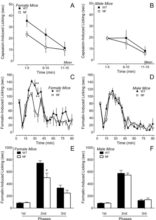
Spontaneous behavior after acute inflammatory stimuli
Injection of inflammatory agents such as capsaicin and formalin produced a variety of spontaneous pain behaviors. In the case of 0.01% capsaicin, the predominant behavior measured in the first
15 min was licking of the hindpaw. FemaleNf1+/2and WT mice
did not differ statistically in the duration of licking (simple main effect for genotype: P = 0.066; Fig. 4A). Male mice of each
genotype exhibited comparable durations of licking (P.0.6;
Fig. 4B). Similar conclusions were reached when the data were converted to area under the curve for the 15 min period. Thus, a deficit in neurofibromin neither facilitated nor inhibited capsaicin-induced spontaneous pain behaviors.
In the case of formalin, a constellation of behaviors was observed over 90 min that included guarding behaviors and unweighting of the affected hindpaw, as well as licking or flinching of the ipsilateral hindpaw. Interestingly, the duration of licking in
femaleNf1+/2 mice was significantly lower in the second phase
compared to female WT mice (Fig. 4C, E). There was no difference in the time spent licking in male mice of either genotype (Fig. 4D, F). Analysis of guarding, unweighting and flinching
behaviors revealed that female Nf1+/2 mice exhibited a much
higher incidence of these behaviors than female WT mice during the second and third phase (Fig. 5A). These behaviors are likely to
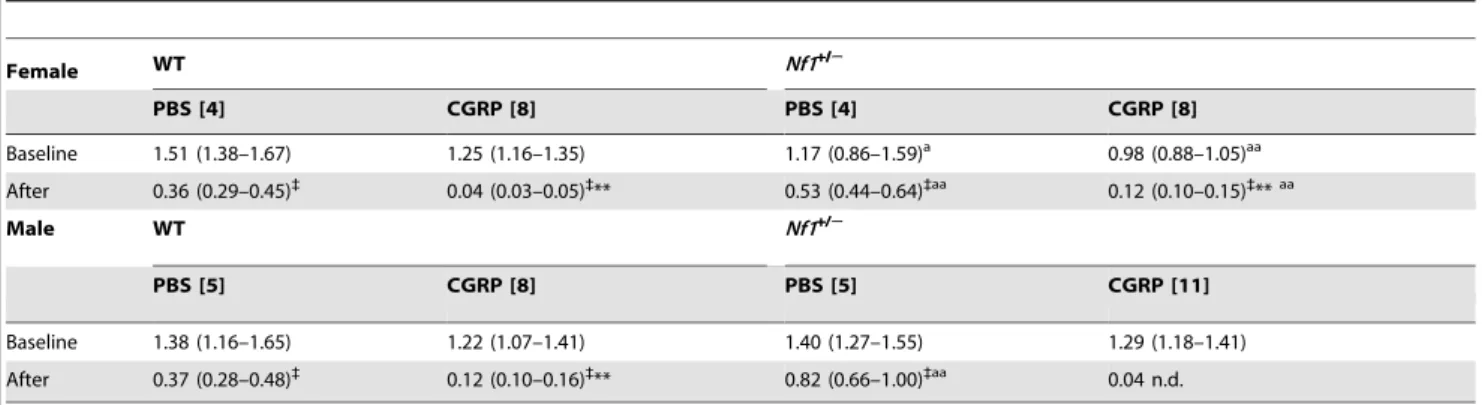
Table 2.Calcitonin gene-related peptide evokes mechanical hypersensitivity in both wildtype and Nf1+/2mice.
Female WT Nf1+/2
PBS [4] CGRP [8] PBS [4] CGRP [8]
Baseline 1.51 (1.38–1.67) 1.25 (1.16–1.35) 1.17 (0.86–1.59)a 0.98 (0.88–1.05)aa
After 0.36 (0.29–0.45)`
0.04 (0.03–0.05)`
** 0.53 (0.44–0.64)`aa 0.12 (0.10–0.15)` **aa
Male WT Nf1+/2
PBS [5] CGRP [8] PBS [5] CGRP [11]
Baseline 1.38 (1.16–1.65) 1.22 (1.07–1.41) 1.40 (1.27–1.55) 1.29 (1.18–1.41)
After 0.37 (0.28–0.48)`
0.12 (0.10–0.16)`
** 0.82 (0.66–1.00)`aa 0.04 n.d.
EF50and 95% confidence limits (g) determined for force-response curves for the ipsilateral hindpaw before and 15 min after intraplantar injection of 5mg CGRP or PBS
Curves were fit by non-linear regression with minimum constrained to 0 and maximum to 100. Numbers of mice in each group appear in brackets. *P,0.05,
** P,0.01 compared to vehicle at corresponding time point.
{
P,0.05, `
P,0.01 compared to baseline value.
aP,0.05, aaP
have interfered with licking. There was no significant difference in
the incidence of these behaviors between male Nf1+/2 and WT
mice (Fig. 5B, D). Formalin increased paw thickness by the same
extent in all genotypes and genders (by 0.860.1, 0.960.1, 0.960.1
and 0.760.1 mm, n = 8 each group; P.0.5). Contralateral paw
thickness ranged between 1.3 and 1.4 mm.
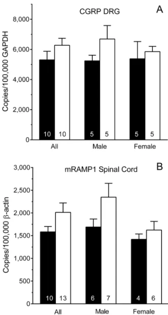
CGRP and RAMP1 Transcripts
Figure 6A illustrates levels of CGRP mRNA in the DRG of
Nf1+/2mice and their WT littermates. There were no significant
differences when the genders were combined for analysis (All; P = 0.2 Student’s t-test). Two-way analysis indicated that CGRP transcript levels did not differ between genotypes within a gender, or between genders within a genotype (P.0.2 each factor). Figure 6B illustrates levels of RAMP1 mRNA in the spinal cord of
Nf1+/2and WT littermates (Fig. 6B). Levels of RAMP1 transcript
did not differ between genotypes when genders were combined for analysis (All; P.0.1; Student’s t-test). Two-way analysis indicated that RAMP1 transcript levels did not differ between genotypes within a gender, or between genders within a genotype (P.0.05
Figure 4. Duration of licking of the ipsilateral hindpaw after intraplantar injection of inflammatory irritants.Panels A and B illustrate
the time course of licking after intraplantar injection of 0.01% capsaicin in the hindpaw of female or male mice, respectively. Panels C and D illustrate the time course of licking after intraplantar injection of 2% formalin in female or male mice, respectively. Panels E and F present the total duration of licking in phases 1 (0–5), 2 (15–55 min) and 3 (55–90 min) of the formalin test in female and male mice, respectively. Data are the mean6S.E.M. of determinations in 6–8 mice of each gender and genotype. * P,0.05, ** P,0.01 compared to corresponding WT littermates.
each factor). The difference between maleNf1+/2and WT mice was not statistically significant (P = 0.1; Student’s t-test).
Discussion
This extensive characterization of the nociceptive responses of
male and femaleNf1+/2mice was prompted by (1) the enhanced
excitability of primary afferent neurons inNf1+/2mice [16], (2)
the increased release of CGRP from sensory neurons of Nf1+/2
mice [14], and (3) the well-established role of CGRP as a nociceptive neurotransmitter in the periphery and spinal cord [9–
12]. The results indicate thatNf1+/2mice did not differ from WT
mice in responsiveness to acute heat stimuli delivered at a rate that
preferentially activated either Ad- or C-fibers [27,28]. They also
did not differ from WT mice in responsiveness to innocuous mechanical stimuli with the exception of a subtle enhanced
mechanical sensitivity in femaleNf1+/2mice. Given that neither
content nor the basal release of CGRP from cultured DRG
neurons and spinal cord slices differed between WT andNf1+/2
mice [14], it is perhaps not unexpected that WT andNf1+/2mice
did not differ in their responsiveness to brief heat or mechanical stimuli in the absence of inflammation. O’Brien et al. reached a similar conclusion in their recent survey of the responsiveness of
Nf1+/2mice to noxious heat or itch-producing stimuli [21].
Nociceptive phenotype in inflammatory models
Intraplantar injection of capsaicin releases CGRP from the central and peripheral terminals of primary afferent neurons
[14,30–33]. Neither female nor maleNf1+/2 mice differed from
their WT littermates with respect to heat hyperalgesia induced by
ipl capsaicin. FemaleNf1+/2mice also did not differ from their
WT littermates in the magnitude of mechanical hypersensitivity
that developed, and maleNf1+/2mice exhibited only slightly less
mechanical hypersensitivity than WT littermates. These findings were unexpected given that capsaicin evokes greater release of
CGRP from the terminals of nociceptive afferents inNf1+/2mice
than WT mice [14].
The formalin test was used to assess nociceptive phenotype in a model of more prolonged inflammation and as a non-reflexive measure of nociceptive behaviors. Formalin-evoked pain behaviors are also dependent on CGRP [34]. Formalin directly activates transient receptor potential (TRP), subfamily A, member 1 channels (TRPA1) [35,36], and TRP channel, subfamily V, member 1 (TRPV1) channels [37] in DRG neurons. In mice, many TRPV1-immunoreactive primary afferent neurons coex-press TRPA1 [38,39]. Formalin is therefore likely to cause a central and peripheral release of CGRP similar to that caused by
capsaicin. Indeed, male Nf1+/2 mice did not differ from WT
littermates in either the duration of licking or other nociceptive
Figure 5. Intraplantar injection of formalin induced spontaneous pain behaviors in both genders and genotypes.Time course of
guarding, flinching or unweighting of the hindpaw after intraplantar injection of formalin in (A) female or (B) male mice. The percentage of female
Nf1+/2
mice that exhibited these behaviors was higher than WT littermates, but did not differ in male mice of either genotype. Percentage of (C) female or (D) male mice exhibiting guarding, flinching or unweighting of the hindpaw averaged for the first phase (0–5 min), second phase (15– 55 min) and third phase (55–90 min) of the formalin test. Six to eight mice of each gender and genotype were tested. * P,0.05, ** P,0.01 compared to corresponding WT littermate.
behaviors in agreement with the report by O’Brien et al. [21].
FemaleNf1+/2mice showed less licking than WT mice, but this
observation is not necessarily indicative of a reduced nociception because these mice also exhibited additional competing behaviors that suggested that formalin was more noxious.
Subsequent experiments injected CGRP into the hindpaw to limit the site of action to the periphery. A more consistent phenotype of exacerbated nociception emerged in this model.
Both male and female Nf1+/2 mice exhibited greater heat
hyperalgesia than their respective WT littermates after ipl injection
of CGRP. Male Nf1+/2 mice also exhibited greater mechanical
hypersensitivity their WT littermates, although female Nf1+/2
mice exhibited less mechanical hypersensitivity than their WT littermates. It is well established that CGRP increases its own
synthesis, and most likely its own release from sensory neurons [20,40]. Although levels of transcript (this study) and protein [14]
for CGRP were equivalent in the DRG ofNf1+/2and WT mice,
an enhanced release of endogenous CGRP from the peripheral
terminals of primary afferents inNf1+/2mice cannot be excluded.
Another mechanism that may be responsible for the enhanced
nociceptive effects of peripherally administered CGRP inNf1+/2
mice involves invading macrophages and the subsequent release of
inflammatory cytokines [41]. Macrophages in Nf1+/2 mice may
express increased numbers of the CGRP receptor or RAMP1, or receptors of higher affinity or efficacy upon activation leading to increased release of cytokines. Additional studies will be required to test this hypothesis.
Nociceptive Phenotypes of Other Ras-GAP Deficient Mice
Recently, the nociceptive phenotype of another Ras-GAP deficient mouse was investigated. Mice with a heterozygous mutation for Synaptic GAP (SynGAP), a neuronal Ras-GAP, also did not differ from WT mice in their responsiveness to heat or mechanical stimuli in the absence of inflammation [42]. As
observed with Nf1+/2 mice, ipl injection of capsaicin induced
equivalent mechanical hypersensitivity in SynGAPdeficient and
WT mice [42]. Although capsaicin induced greater heat
hyper-algesia inSynGAPdeficient mice than WT mice, this result can be
attributed to the finding thatSynGAPmice have three-fold higher
levels of TRPV1 in the DRG [42]. It is not known whether the
DRG ofNf1+/2mice have higher levels of TRPV1 than WT mice.
However, given that the magnitude of capsaicin-induced heat hyperalgesia was similar in both genotypes, this is considered unlikely.
Conclusions
The results of this study do not support the hypothesis that a reduction in neurofibromin is associated with enhanced acute or inflammatory nociception, and confirm the conclusions of another
recent comprehensive analysis of male Nf1+/2 mice. [21]. The
present study extends this conclusion to additional models of
inflammatory injury and also includes female Nf1+/2 mice.
Gender is an important consideration given that many chronic pain conditions such as migraine and fibromyalgia are more prevalent in women than men [43,44].
This study also provides new information relevant to the ‘CGRP hypothesis’ posited by Hingtgen and colleagues. It determined that levels of transcript for CGRP were unchanged
in the DRG of Nf1+/2 mice, as were levels of transcript for
RAMP1 in the spinal cord. The finding of enhanced heat hyperalgesia in both genders and of mechanical hypersensitivity in
maleNf1+/2 mice after ipl injection of CGRP suggests that the
peripheral actions of CGRP may be enhanced as a result of neurofibromin deficit. This exacerbation may be the result of increased peripheral release of neurotransmitter from primary afferent neurons or the expression of CGRP receptors by invading macrophages that release proinflammatory cytokines in response to CGRP. As noted earlier, high densities of CGRP-immunore-active fibers are present in neurofibromas in patients [13]. Thus, consideration should be given to testing the efficacy of CGRP receptor antagonists developed for relief of migraine [45] in diminishing bodily pain and pain associated with nerve sheath tumors in NF patients.
Acknowledgments
We thank Dr. Christina Winford and Dr. Roxanne Walder for their assistance with troubleshooting the RT-qPCR experiments.
Figure 6. Levels of CGRP mRNA and RAMP1 mRNA.(A) Levels of
CGRP mRNA in the dorsal root ganglion (DRG) ofNf1+/2and WT mice did not differ by genotype or gender. (B) Levels of RAMP1 mRNA in the spinal cord ofNf1+/2and WT mice did not differ by gender or genotype. Data are expressed as mean 6 SEM copies normalized to 100,000 GAPDH for the DRG and to 100,000b-actin for spinal cord. Numbers in the columns are the number of mice. * P,0.05 compared to corresponding WT littermates.Nf1+/2mice (open bars); WT mice (solid bars)
Author Contributions
Conceived and designed the experiments: DLH AFR. Performed the experiments: SW BMdP. Analyzed the data: DLH SW BMdP.
Contrib-uted reagents/materials/analysis tools: AFR. Wrote the paper: DLH BMdP.
References
1. Riccardi VM (1992) Type 1 neurofibromatosis and the pediatric patient. Curr Probl Pediatr 22: 66–106; discussion 107.
2. Lakkis MM, Tennekoon GI (2000) Neurofibromatosis type 1. I. General overview. J Neurosci Res 62: 755–763.
3. Lee MJ, Stephenson DA (2007) Recent developments in neurofibromatosis type 1. Current Opin Neurol 20: 135–141.
4. McClatchey AI (2007) Neurofibromatosis. Ann Rev Pathol 2: 191–216. 5. Theos A, Korf BR (2006) Pathophysiology of neurofibromatosis type 1. Ann
Intern Med 144: 842–849.
6. Creange A, Zeller J, Rostaing-Rigattieri S, Brugieres P, Degos JD, et al. (1999) Neurological complications of neurofibromatosis type 1 in adulthood. Brain 122: 473–481.
7. Drouet A, Wolkenstein P, Lefaucheur JP, Pinson S, Combemale P, et al. (2004) Neurofibromatosis 1-associated neuropathies: a reappraisal. Brain 127: 1993– 2009.
8. Wolkenstein P, Zeller J, Revuz J, Ecosse E, Leplege A (2001) Quality-of-life impairment in neurofibromatosis type 1: a cross-sectional study of 128 cases. Arch Dermatol 137: 1421–1425.
9. Seybold VS (2009) The role of peptides in central sensitization. Hdbk Exper Pharmacol 451–491.
10. Yu L-C, Hou J-F, Fu F-H, Zhang Y-X (2009) Roles of calcitonin gene-related peptide and its receptors in pain-related behavioral responses in the central nervous system. Neurosci & Biobehav Rev 33: 1185–1191.
11. Van Rossum D, Hanisch U-K, Quirion R (1997) Neuroanatomical localization, pharmacological characterization and functions of CGRP, related peptides and their Receptors. Neurosci & Biobehav Rev 21: 649–678.
12. Benemei S, Nicoletti P, Capone JG, Geppetti P (2009) CGRP receptors in the control of pain and inflammation. Cur Opin Pharmacol 9: 9–14.
13. Vaalasti A, Suomalainen H, Kuokkanen K, Rechardt L (1990) Neuropeptides in cutaneous neurofibromas of von Recklinghausen’s disease. J Cutaneous Pathol 17: 371–373.
14. Hingtgen CM, Roy SL, Clapp DW (2006) Stimulus-evoked release of neuropeptides is enhanced in sensory neurons from mice with a heterozygous mutation of the Nf1 gene. Neuroscience 137: 637–645.
15. Wang Y, Duan JH, Hingtgen CM, Nicol GD (2010) Augmented sodium currents contribute to the enhanced excitability of small diameter capsaicin-sensitive sensory neurons isolated from Nf1+/mice. J Neurophysiol 103: 2085– 20894.
16. Wang Y, Nicol GD, Clapp DW, Hingtgen CM (2005) Sensory neurons from Nf1 haploinsufficient mice exhibit increased excitability. J Neurophysiol 94: 3670– 3676.
17. Hodgdon KE, Hingtgen CM, Nicol GD (2012) Dorsal root ganglia isolated from Nf1+/2mice exhibit increased levels of mRNA expression of voltage-dependent sodium channels. Neuroscience 206: 237–244.
18. Duan JH, Hodgdon KE, Hingtgen CM, Nicol GD (2014) N-type calcium current, Cav2.2, is enhanced in small-diameter sensory neurons isolated from Nf1+/2
mice. Neuroscience 270: 192–202.
19. Hay DL, Poyner DR, Sexton PM (2006) GPCR modulation by RAMPs. Pharmacol & Ther 109: 173–197.
20. Zhang Z, Winborn CS, Marquez de Prado B, Russo AF (2007) Sensitization of calcitonin gene-related peptide receptors by receptor activity-modifying protein-1 in the trigeminal ganglion. J Neurosci 27: 2693–2703.
21. O’Brien DE, Brenner DS, Gutmann DH, Gereau RWt (2013) Assessment of pain and itch behavior in a mouse model of neurofibromatosis type 1. J Pain 14: 628–637.
22. Jacks T, Shih TS, Schmitt EM, Bronson RT, Bernards A, et al. (1994) Tumour predisposition in mice heterozygous for a targeted mutation in Nf1. Nat Genetics 7: 353–361.
23. Riccardi VM, Womack JE, Jacks T (1994) Neurofibromatosis and related tumors. Natural occurrence and animal models. Am J Pathol 145: 994–1000. 24. Brannan CI, Perkins AS, Vogel KS, Ratner N, Nordlund ML, et al. (1994)
Targeted disruption of the neurofibromatosis type-1 gene leads to developmental abnormalities in heart and various neural crest-derived tissues. Genes & Develop 8: 1019–1029.
25. Vance JM, Pericak-Vance MA, Yamaoka LH, Speer MC, Rosenwasser GO, et al. (1989) Genetic linkage mapping of chromosome 17 markers and neurofibromatosis type I. Am J Human Gen 44: 25–29.
26. Hirata H, Pataky A, Kajander K, LaMotte RH, Collins JG (1990) A model of peripheral mononeuropathy in the rat. Pain 42: 253–255.
27. Yeomans DC, Proudfit HK (1994) Characterization of the foot withdrawal response to noxious radiant heat in the rat. Pain 59: 85–94.
28. Yeomans DC, Proudfit HK (1996) Nociceptive responses to high and low rates of noxious cutaneous heating are mediated by different nociceptors in the rat: electrophysiological evidence. Pain 68: 141–150.
29. McMullan S, Simpson DA, Lumb BM (2004) A reliable method for the preferential activation of C- or A-fibre heat nociceptors. J Neurosci Methods 138: 133–139.
30. Lundberg JM, Franco-Cereceda A, Alving K, Delay-Goyet P, Lou Y-P (1992) Release of Calcitonin Gene—Related Peptide from Sensory Neurons. Ann NY Acad Sci 657: 187–193.
31. Li D, Ren Y, Xu X, Zou X, Fang L, et al. (2008) Sensitization of primary afferent nociceptors induced by intradermal capsaicin involves the peripheral release of calcitonin gene-related Peptide driven by dorsal root reflexes. J Pain 9: 1155–1168.
32. Sun R-Q, Lawand NB, Willis WD (2003) The role of calcitonin gene-related peptide (CGRP) in the generation and maintenance of mechanical allodynia and hyperalgesia in rats after intradermal injection of capsaicin. Pain 104: 201–208. 33. Kilo S, Harding-Rose C, Hargreaves KM, Flores CM (1997) Peripheral CGRP release as a marker for neurogenic inflammation: a model system for the study of neuropeptide secretion in rat paw skin. Pain 73: 201–207.
34. Salmon AM, Damaj MI, Marubio LM, Epping-Jordan MP, Merlo-Pich E, et al. (2001) Altered neuroadaptation in opiate dependence and neurogenic inflammatory nociception in alpha CGRP-deficient mice. Nat Neurosci 4: 357–358.
35. McNamara CR, Mandel-Brehm J, Bautista DM, Siemens J, Deranian KL, et al. (2007) TRPA1 mediates formalin-induced pain. Proc Natl Acad Sci U S A 104: 13525–13530.
36. Macpherson LJ, Xiao B, Kwan KY, Petrus MJ, Dubin AE, et al. (2007) An ion channel essential for sensing chemical damage. J Neurosci 27: 11412–11415. 37. Tian LJ, Du YR, Xiao Y, Lv ZM, Yu YQ, et al. (2009) Mediating roles of the
vanilloid receptor TRPV1 in activation of rat primary afferent nociceptive neurons by formaldehyde. Acta physiol Sin 61: 404–416.
38. Story GM, Peier AM, Reeve AJ, Eid SR, Mosbacher J, et al. (2003) ANKTM1, a TRP-like channel expressed in nociceptive neurons, is activated by cold temperatures. Cell 112: 819–829.
39. Malin S, Molliver D, Christianson JA, Schwartz ES, Cornuet P, et al. (2011) TRPV1 and TRPA1 function and modulation are target tissue dependent. J Neurosci 31: 10516–10528.
40. Segond von Banchet G, Pastor A, Biskup C, Schlegel C, Benndorf K, et al. (2002) Localization of functional calcitonin gene-related peptide binding sites in a subpopulation of cultured dorsal root ganglion neurons. Neuroscience 110: 131–145.
41. Ma W, Quirion R (2006) Increased calcitonin gene-related peptide in neuroma and invading macrophages is involved in the up-regulation of interleukin-6 and thermal hyperalgesia in a rat model of mononeuropathy. J Neurochem 98: 180– 192.
42. Duarte DB, Duan JH, Nicol GD, Vasko MR, Hingtgen CM (2011) Reduced expression of SynGAP, a neuronal GTPase-activating protein, enhances capsaicin-induced peripheral sensitization. J Neurophysiol 106: 309–318. 43. Fillingim RB, King CD, Ribeiro-Dasilva MC, Rahim-Williams B, Riley JL 3rd
(2009) Sex, gender, and pain: a review of recent clinical and experimental findings. J Pain 10: 447–485.
44. Bartley EJ, Fillingim RB (2013) Sex differences in pain: a brief review of clinical and experimental findings. Br J Anaesth 111: 52–58.