Streptozotocin-induced diabetes mellitus
affects lysosomal enzymes in rat liver
G.B. Peres
1, M.A. Juliano
2, J.A.K. Aguiar
1and Y.M. Michelacci
1 1Departamento de Bioquı´mica, Escola Paulista de Medicina, Universidade Federal de Sa˜o Paulo, Sa˜o Paulo, SP, Brasil 2
Departamento de Biofı´sica, Escola Paulista de Medicina, Universidade Federal de Sa˜o Paulo, Sa˜o Paulo, SP, Brasil
Abstract
It has been previously shown that dextran sulfate administered to diabetic rats accumulates in the liver and kidney, and this could be due to a malfunction of the lysosomal digestive pathway. The aim of the present study was to evaluate the expression and activities of lysosomal enzymes that act upon proteins and sulfated polysaccharides in the livers of diabetic rats. Diabetes mellitus was induced by streptozotocin in 26 male Wistar rats (12 weeks old), while 26 age-matched controls received only vehicle. The livers were removed on either the 10thor the 30thday of the disease, weighed, and used to evaluate the activity,
expression, and localization of lysosomal enzymes. A 50-60% decrease in the specific activities of cysteine proteases, especially cathepsin B, was observed in streptozotocin-induced diabetes mellitus. Expression (mRNA) of cathepsins B and L was also decreased on the 10th, but not on the 30thday. Sulfatase decreased 30% on the 30thday, while glycosidases did
not vary (or presented a transitory and slight decrease). There were no apparent changes in liver morphology, and immunohistochemistry revealed the presence of cathepsin B in hepatocyte granules. The decrease in sulfatase could be responsible for the dextran sulfate build-up in the diabetic liver, since the action of sulfatase precedes glycosidases in the digestive pathway of sulfated polysaccharides. Our findings suggest that the decreased activities of cathepsins resulted from decreased expression of their genes, and not from general lysosomal failure, because the levels of glycosidases were normal in the diabetic liver.
Key words: Liver; Diabetes mellitus; Lysosomal enzymes; Cathepsin; Glycosidase; Sulfatase
Introduction
Streptozotocin-induced diabetes mellitus (STZ-DM) in rats leads to a marked decrease in the urinary excretion of glycosaminoglycans (1). Decreased urinary excretion of exogenous dextran sulfate also occurs in STZ-DM, with accumulation of dextran sulfate in liver and kidney (2). Because dextran sulfate is very soluble in water, its presence in liver and kidney 48 h after administration suggests intracellular localization of these molecules.
The concentration of glycosaminoglycan also increased in diabetic kidney (2,3), and there is evidence that both anabolic and catabolic pathways are involved (4,5). De-creased expression of lysosomal enzymes in diabetic kidney was recently reported (5), and autophagy was recently pro-posed as a therapeutic target for diabetic nephropathy (6).
Lysosomal enzymes are the main agents of the digestive process that takes place in autophagic vacuoles (reviewed in Ref. 7). Even though much of the pioneering
research on autophagy comes from 40 years of studies on liver and isolated hepatocytes (8), and although lyso-somes were discovered back in the 1950s by Christian de Duve (9), who also maintained a lifetime interest in the actions of insulin and glucagon on the liver (10), there are few studies on the expression and activities of lysosomal enzymes in the diabetic liver.
Most of the recent work on liver autophagy has focused on lipid digestion (lipophagy) (11,12) and glycogen diges-tion (13). There are few studies of liver lysosomal enzymes that act upon proteins and sulfated polysaccharides. The aim of the present study was to investigate the expression and activities of lysosomal hydrolases in the liver of rats with STZ-DM, in order to elucidate the mechanisms responsible for the dextran sulfate build-up. Two different stages of the disease were studied: 10 days (initial diabetic state) and 30 days (diabetic nephropathy).
Correspondence: Y.M. Michelacci, Disciplina de Biologia Molecular, Departamento de Bioquı´mica, Escola Paulista de Medicina, UNIFESP, Rua Treˆs de Maio, 100, 04044-020 Sa˜o Paulo, SP, Brasil. Fax: ++55-11-5573-6407. E-mail: yara.bioq@epm.br
Present address of J.A.K. Aguiar: Departamento de Bioquı´mica, Universidade Federal de Juiz de Fora, Juiz de Fora, MG, Brasil.
Material and Methods
Animals, urine, and tissue
The Ethics Committee of Escola Paulista de Medicina, UNIFESP approved the present research (CEP No. 0170/ 09), which was carried out in accordance with UNIFESP guidelines, and also in accordance with EC Directive 2010/63/EU for animal experiments.
Male Wistar rats (n=52), 12 weeks of age (275-360 g body weight), were randomly assigned to one of four groups: control day 10, diabetes (DM) day 10, control day 30, and diabetes day 30. Before the induction of DM, the animals were weighed, blood glucose was measured (Advantage Kit Roche, Switzerland), and the animals were placed in metabolic cages for 24-h urine collection. Diabetes was induced in the 26 rats of DM groups by a single intraperitoneal injection of 60 mg/kg body weight STZ. The drug was dissolved in 300mL 10 mM sodium citrate buffer, pH 4.5. These animals were fed standard laboratory chow and a 5% glucose solutionad libitumfor 72 h. Afterward, the glucose solution was replaced by water. Glycemia was measured 72 h after STZ adminis-tration, and also at the end of each experiment (either 10th or 30thday). Only animals that, at 72 h, presented blood glucose higher than 250 mg/dL were considered ‘‘dia-betic’’ (14). The 26 age-matched animals that served as controls received only 300mL buffer and were fed standard laboratory chow and waterad libitum.
At the end of each experiment, body weight was again measured, and the rats were placed in metabolic cages for 24-h urine collection. The 24-h urine volume was measured and the urine was centrifuged at 1000gfor 10 min at room temperature to remove debris and used for determination of creatinine, total protein, and albumin. Creatinine was quantified by the picric acid reaction under alkaline conditions (CELM creatinine kit, Brazil), total protein was measured by the pyrogallol red-molybdate complex method (Sensiprot, Labtest, Brazil) (15), and albumin was determined by two methods: radial immunodiffusion based on precipitation with rabbit antibodies against rat albumin (1), and ELISA using a Bethyl E110-125 Rat Albumin Quantification Set (USA). The results obtained for total protein and creatinine were published in Peres et al. (5). After the urine was collected, the rats were killed, and the livers were removed, weighed, and carefully cut into small fragments (,100 mg each). These fragments were used
for RNA extraction, measurement of enzyme activities, Western blotting, and quantification of total protein. The liver fragments were put into sterile tubes, frozen in liquid nitrogen, and stored at ––706C until use.
Liver enzyme activities
To measure the enzyme activities, liver samples (,100 mg) were disrupted in liquid nitrogen and
resus-pended in 1 mL 50 mM Tris-HCl buffer, pH 7.4, contain-ing 200 mM NaCl and 250 mM sucrose (16) plus 1 mL
0.2% Triton X-100. After standing for 10 min in an ice bath, debris was removed by centrifugation (12,000g, 10 min, 46C), and aliquots of the supernatant (100mL) were stored in sterile tubes at ––706C until use (tissue extracts).
Protease activities were quantified by fluorometric assays using either carbobenzoxy-Phe-Arg-7-amide-4-methylcoumarin (Z-FR-MCA; Sigma-Aldrich, USA) or e -NH2-caproyl-Cys(Bzl)-Cys(Bzl)-MCA (synthesized by Prof.
Dr. Maria A. Juliano) (17) as substrates. These substrates were used to quantify total cysteine proteases and ca-thepsin B, respectively. Incubation was carried out in dark microplates (Corning, USA), in 50 mM phosphate buffer, pH 6.3, containing 10 mM EDTA. The enzymes (50mg protein) were preactivated by incubation of tissue extract aliquots with 2 mM dithiothreitol (10 min, room tempera-ture), and the substrate was then added (20mM, 200mL final volume). The fluorescence produced upon hydrolysis of the substrates was measured every 20 s in a FlexStation 3 microplate reader (Molecular Devices, USA), using
lexcitation=380 nm and lemission=460 nm. The assays
were also performed in the presence of the following inhibitors: 1 mM phenylmethylsulfonyl fluoride (PMSF; inhibitor of serine proteases), 5mM E64 (irreversible inhibitor of cysteine proteases), and 1mM CA074 (irrever-sible inhibitor of cathepsin B).
The activities of b-D-glucuronidase, N-acetyl-b-D -glu-cosaminidase, N-acetyl-b-D-galactosaminidase, and
sul-fatases were measured by spectrophotometric assays using the following substrates: 4-nitrophenyl b-D
-glucur-onide, 4-nitrophenyl N-acetyl-b-D-glucosaminide, 4-nitro-phenyl N-acetyl-b-D-galactosaminide, and 4-nitrophenyl
sulfate, respectively (Sigma-Aldrich). All assays were performed on microplates, always as triplicates, and the incubation mixtures (150mL final volume) contained 50 mM sodium acetate buffer, pH 5.0, 2 mM substrate, and increasing amounts of tissue extracts (2-30mL containing 20-120mg protein). After 1 h of incubation at room temperature, the 4-nitrophenol released was solu-bilized by addition of 1 M NaOH (150mL), and the absorbance was measured immediately at l405 in a
microplate reader (Molecular Devices).
Quantification of liver protein and Western blotting
Protein was quantified in tissue extracts by a modified Lowry procedure with bicinchoninic acid (18) (BCA Protein Assay Kit, Thermo Scientific Pierce, USA), using bovine serum albumin as standard.
antibody. Specific bands were detected by secondary antibody (anti-rabbit IgG) conjugated with horseradish peroxidase (HRP) and enhanced chemiluminescence (ECL) substrate. Images were obtained with the MF-ChemiBIS gel documentation system, where each lane represents a pool of four animals for each group.
Color images (tetramethylbenzidine; KPL, USA) and chemiluminescence images (ECL Advance Western Blotting Detection Kit; GE Healthcare, USA) were obtained with the MF-ChemiBIS gel documentation system (DNR Bio Imaging Systems Ltd., Israel) with the GelCapture 7.0.6 software for WindowsTM.
RNA extraction and real-time reverse transcriptase-polymerase chain reaction (PCR)
Each liver fragment (,100 mg) was disrupted in liquid
nitrogen, homogenized in 1 mL QIAzol Lysis Reagent (QIAGEN, USA), and processed according to the manu-facturer’s instructions. Absorbances (A260/280and A260/230)
were measured (ND-1000, NanoDrop, USA), and samples with ratios lower than 1.8 and 1.7, respectively, were discarded. RNA integrity was also evaluated from 28S and 18S rRNA bands after agarose gel electrophoresis using Tris-borate-EDTA buffer, as previously described (20,21). To avoid contamination with genomic DNA, RNA samples were treated with DNase I (1 U/mg RNA, 30 min, 376C; Fermentas International, Canada) followed by 25 mM EDTA (1mL/enzyme unit, 10 min, 656C) to inactivate the DNase I.
One microgram of RNA was reverse-transcribed to complementary DNA (cDNA) with RevertAid M-MuLV (Fermentas International), and the resulting single-strand cDNA was amplified by quantitative PCR (qPCR) in a reaction mixture containing 0.4mM of each primer and 7.5mL SYBR Green (15mL final volume; Rotor-Gene SYBR Green PCR Kit, QIAGEN). The thermal cycling conditions were as follows: initial 5 min at 956C, followed by 40-45 cycles of denaturing at 956C for 5 s, annealing at 606C for 10 s, and extension at 956C for 10 min. Cycle number and cDNA concentration were adjusted so that amplified products remained within the linear range of the PCR. PCR amplification was conducted on a Corbett Rotor-Gene 6000 (QIAGEN). Each PCR was done in duplicate.
Relative gene expression was calculated by the 2–DDCT
method developed by Livak and Schmittgen (22). In this
method, it is assumed that the expression of a reference gene (housekeeping gene) is independent of external factors and that its expression is quite constant. In the present paper, two genes were used as references: ribosomal protein 29S (RPS29) andb-actin (ACTB). Other genes were tested, but they varied in the diabetic liver, compared with controls, and therefore were not used. The PCR primers (Bioneer Corp., USA) are shown in Table 1.
Histology and immunohistochemistry
For histology and immunohistochemistry, one rat in each group was anesthetized with 10% chloral hydrate (4 mL/kg body weight) and perfused with filtered saline (150 mL, 12 mL/min) followed by 4% formalin in 0.1 M phosphate buffer, pH 7.4 (500 mL, 12 mL/min). The liver was removed, cut, dehydrated, embedded in paraffin, and cut in 4-mm sections. These sections were transferred to silane-coated microscope slides and dewaxed as pre-viously described (23).
Liver sections were stained with either hematoxylin and eosin or toluidine blue, and images were obtained with a Zeiss Axiolab microscope (Carl Zeiss MicroImaging GmbH, Germany), equipped with an AxioCam MRc digital camera and AxioVision software.
For immunohistochemistry, the dewaxed slides were transferred to 200 mL prewarmed 10 mM sodium citrate buffer, pH 6.0 (956C). Antigen retrieval was 30 min at 956C, 20 min on the bench, and 5 min under running water. Endogenous peroxidase was blocked by 3% hydrogen peroxide (10 min, 10 times), followed by running water (10 min) and phosphate-buffered saline (PBS; 3 min, 3 times). Nonspecific protein binding was blocked by 200mL 1% bovine serum albumin (Cat. #A3059, Sigma-Aldrich) and 2% fetal calf serum in a moist chamber at room temperature. The primary antibody was rabbit anti-cathepsin B (Cat. #06-480; EMD Millipore, USA) diluted 1:100 in blocking solution. After an overnight incubation at 46C and three 5-min washes in PBS, HRP-conjugated goat anti-rabbit IgG, secondary antibody (Cat.#12-348, EMD Millipore) diluted 1:200 in blocking solution was added. After a 90-min incubation and three 5-min washes in PBS, nickel-enhanced diaminobenzidine (Cat. #54-74-00, HistoMark Orange Peroxidase System; KPL) was used as HRP substrate. Then, the tissue sections were rinsed under running water and counterstained by Harris modified hematoxylin solution (Cat. #HHS16, Sigma-Aldrich) for
Table 1. PCR primers.
Enzyme Forward Reverse
Cathepsin B 59-GCTATCCCTCTGGAGCATGGAAC-39 59-GACGGGAGCCATTGACATGGT-39 Cathepsin L 59-AGGCAATCAGGGCTGTAATGGAG-39 59-CGTTAGCCACAGCATACTCAGCTC-39
b-D-glucuronidase 59-CAGTGCTTCCACAGCGATGGA-39 59-GTGATGTCAGCCTCAAAGGGGAG-39
b-actin 59-GGATGACGATATCGCTGCGCT-39 59-CTGACCCATACCCACCATCACAC-39
30 s. Excess hematoxylin was removed by 0.1% HCl in 70% ethanol, followed by a 5-min rinse under running water. Slides were dehydrated and mounted in Entellan (Cat.#14800, Merck, Germany) and sealed with clear nail polish. Negative controls were incubated with blocking solution at each step.
Statistical analysis
Statistical analysis was performed with the PASW Statistics software (SPSS Statistics) for WindowsTM
(version 18.0.0). The Shapiro-Wilk test was used to evaluate data for normality, and data were standardized (z-score) when the parametric distribution was not observed. Bootstrap (a resampling method) was also used to check for the stability of the experimental data. Results are reported as means±SD, except for enzyme kinetics, when means±95% confidence interval are reported. Differences between groups were analyzed by ANOVA. Values of P,0.05 were considered to be significant, except where otherwise stated.
Results
Glycemia, albuminuria, and liver weight
The body weight of the animals at the beginning (day zero) and at the end (either 10thor 30th day) of the ex-periment, glycemia, urine volume, albuminuria, and liver weight are reported in Table 2. Two animals of the DM day 10 group did not become diabetic (glycemia,250 mg/dL) and were excluded, and three animals of the DM day 30 group died before the end of the experimental period. In contrast to normal controls, which gained weight during the experimental period, all the diabetic rats showed a progressive and significant loss of body weight. In the
controls, glycemia was maintained in the normal range during the entire experimental period, but in the diabetic rats glycemia, as well as urine volume, were very high on days 10 and 30. Significant albuminuria (evaluated by two different methods) appeared only on day 30. Liver weight did not vary in the diabetic rats, compared with the controls, but liver/body weight increased on day 30.
Activity of liver proteases
The kinetics of formation of fluorescent product (MCA) from two different substrates [Z-FR-MCA and e-NH2
-caproyl-Cys(Bzl)-Cys(Bzl)-MCA] is shown in Figure 1. The kinetics was measured in the presence and absence of the inhibitors PMSF, E64, and CA074, which inhibit serine proteases, cysteine proteases, and cathepsin B, respec-tively. PMSF had no effect on enzymatic activities of the substrates used here. In contrast, enzyme activities were very low in the presence of E64, indicating that cysteine proteases were the main enzymes acting on the substrates. Z-FR-MCA was the substrate for cathepsin B and also for other cysteine proteases, while e-NH2
-caproyl-Cys(Bzl)-Cys(Bzl)-MCA was the substrate mainly for cathepsin B (inhibited by CA074).
Cathepsin B corresponds to about one-half of total cysteine proteases in rat liver, both normal and diabetic (Figure 2), and all cysteine protease activities were decreased in the diabetic liver compared with normal activity, on both the 10thand 30thdays.
Activities of glycosidases and sulfatase
In contrast to the proteases, only the specific activity of
b-D-glucuronidase was decreased, and only on the 10thday of STZ-DM (Figure 3). The specific activities ofN-acetyl-b
-D-glucosaminidase and N-acetyl-b-D-galactosaminidase
Table 2. Body and liver weights, and glycemia of rats 10 and 30 days after streptozotocin-induced diabetes mellitus (DM) and their matched controls.
10 Days 30 days
Control (n=13) DM (n=11) Control (n=13) DM (n=10)
Body weight (g)
Initial 307 ± 15 313 ± 33 300 ± 22 326 ± 28
Final 326 ± 19 282 ± 32* 325 ± 19 247 ± 6*
Glycemia (mg/dL)
Initial 105 ± 9 111 ± 13 108 ± 13 111 ± 10
Final 116 ± 28 461 ± 108* 108 ± 21 456 ± 81*
24-h urine volume (mL) 9.1 ± 1.2 97.0 ± 8.4* 8.0 ± 2.1 84.6 ± 11.9*
Urinary albumin (mg/24 h)
RID 0.66 ± 0.36 0.89 ± 0.19 0.48 ± 0.23 2.70 ± 1.85*
ELISA 1.67 ± 0.94 2.03 ± 0.60 1.31 ± 0.70 7.78 ± 2.95**
Liver weight (g) 8.9 ± 0.7 10.3 ± 1.5 9.9 ± 0.7 9.3 ± 1.3
Liver/body weight (610-2) 2.81 ± 0.33 3.35 ± 0.24 3.33 ± 0.20 3.79 ± 0.10*
did not vary in the diabetic liver, compared with normal activity. Decreased specific activity of sulfatases was observed only on the 30thday of DM.
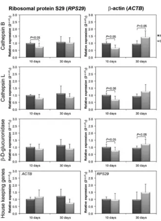
Liver expression (mRNA) of cathepsin B, cathepsin L, andb-D-glucuronidase (qPCR)
The expression of three lysosomal enzymes (cathep-sin B, cathep(cathep-sin L, andb-D-glucuronidase) was analyzed by qPCR in normal and diabetic rat liver. The results are reported as 2–DDCT relative to the two housekeeping
genes,RPS29andACTB(Figure 4).
Figure 1.Kinetics of cysteine protease activities in diabetic (DM) and normal (NL) rat livers. Tissue extracts (2-6mL containing 50mg protein) were preactivated by incubation of tissue extract aliquots with 2 mM dithiothreitol (10 min, room temperature). The following substrates were then added (20mM, 200mL final volume): Z-FR-MCA (substrate for cysteine proteases), or e -NH2-caproyl-Cys(Bzl)-Cys(Bzl)-MCA (substrate for cathepsin B). The fluorescence produced upon hydrolysis of the substrates was measured every 20 s in FlexStation 3 (Molecular Devices, USA), usinglexc=380 nm andlemi=460 nm. The assays were also performed in the presence of inhibitors: 1 mM phenylmethyl-sulfonyl fluoride (PMSF, inhibitor of serine-proteases), 5mM E64 (irreversible inhibitor of cysteine-proteases), and 1mM CA074 (irreversible inhibitor of cathepsin B). A maximum of 10% substrate consumption was considered, and each point repre-sents the mean±95% confidence interval of four replicates for all animals in each group.
Figure 2.Specific activities of cysteine proteases in diabetic (DM) and normal (NL) rat livers. The assays were performed as described in Figure 1, except that specific activities are shown (enzyme units/mg protein, U/mg). The following substrates were used: Z-FR-MCA (substrate for cysteine proteases), or e-NH2 -caproyl-Cys(Bzl)-Cys(Bzl)-MCA (substrate for cathepsin B). One enzyme unit was defined as the amount of enzyme that produces 1 nmol product/min. Data are reported as means±SD. Statistically significant differences between NL and DM are reported as P,0.05 (ANOVA).
The relative expression of the two housekeeping genes used here did not vary on the 10th day of DM compared with controls, indicating that both can be used as reference. Nevertheless, on the 30thday, the expres-sion ofACTB decreased (relative toRPS29) or RPS29
increased (relative to ACTB) in DM (Figure 4), indicat-ing that at least one of them is not a good reference gene.
On the 10thday of DM, relative toACTB, expression of the three enzymes decreased, whereas, relative to
RPS29, only the expression of cathepsin B decreased. In contrast, on the 30th day, relative to ACTB, the expression of cathepsin B and b-D-glucuronidase increased, whereas, relative toRPS29, they did not vary. It is possible that the observed increase in expression of cathepsin B and b-D-glucuronidase relative to ACTB is only apparent, sinceACTBdecreased relative toRPS29. Furthermore, this apparent increase does not correlate to the specific activities of the enzymes (see Figures 2 and 3), suggesting that, in the diabetic liver,RPS29is a better housekeeping gene thanACTB.
Histology, immunohistochemistry, and Western blotting
No changes were observed in the general histological organization of the tissue (Figure 5). Figure 6 shows that, upon toluidine blue staining, metachromatic cells appeared both in normal and diabetic liver (perivascular), and cytoplasmatic granules appeared in all hepatocytes, both normal and diabetic. Tiny granules were also stained by immunohistochemistry for cathepsin B (Figure 7). The specificity of the antibodies was analyzed by Western blotting, which revealed the expected bands of pro-cathepsin B (,40 kDa) and native cathepsin B (26 and
30 kDa).
Discussion
Although much of the pioneering research on autoph-agy comes from studies in liver, and lysosomes were first described in liver (9), there are few studies on the expression and activities of lysosomal enzymes that act upon protein and sulfated polysaccharides in the diabetic liver. In 1974, Amherdt et al. (24) reported an increase in hepatic lysosome volume in severe diabetes induced by 100 mg/kg STZ, primarily due to autophagosomes. In 1978, Dice et al. (25) showed that the degradation of proteins is accelerated in the liver of insulin-deficient rats, and, in 1988, Jorda´ et al. (26) reported a decrease in the half-life of liver mitochondrial ATPase in the severe diabetic state, possibly associated with hepatic autoph-agy. These data indicate that the liver autophagy pathway seems to be implicated in diabetic complications. Never-theless, the activities and expression of lysosomal enzymes were not measured.
Figure 4.Expression (mRNA) of cathepsin B, cathepsin L, and
b-D-glucuronidase in diabetic (DM) and normal (NL) rat livers. The expression of mRNA was normalized either by ribosomal protein S29 (RPS29) orb-actin (ACTB). Data are reported as means±SD. Statistically significant differences between NL and DM are reported as P,0.05 (ANOVA).
In this article, we report a significant decrease in the specific activities of cysteine proteases, especially cathep-sin B, in the liver of rats with STZ-DM, on the 10thand 30th days of the disease. The decrease in liver cysteine protease activities was much greater than that of kidney (liver: 50-60% decrease; kidney: 11-15% decrease, see Ref. 5 for comparison), although in liver the specific activities of these enzymes were much lower (liver: 2.3-2.7 U/mg, Figure 2; kidney: 12.5-14.1 U/mg) (5). Decreases in the expressions (mRNA) of cathepsin B and cathepsin L were also observed on the 10th day after DM induction, suggesting that the
lower gene expression may be one of the mechanisms responsible for the lower enzyme activities.
Conversely, the picture concerning sulfated polysac-charides was very different. The specific activities of most glycosidases were higher in liver than in kidney (5) as follows:
b-D-glucuronidase, liver: 10.2-11.5, kidney: 2.6-3.1 U/mg;N -acetyl-b-D-glucosaminidase, liver: 7.7, kidney: 3.5-3.9 U/mg; N-acetyl-b-D-galactosaminidase, liver: 1.1-1.5, kidney:
2.2-2.5 U/mg. Nevertheless, in contrast to kidney, most activities did not vary in STZ-DM relative to control. A transitory and slight decrease in the expression and activity ofb-D-glucuronidase (8%) was observed only on the 10thday of diabetes, while the activities ofN-acetyl-b-D
-glucosamini-dase andN-acetyl-b-D-galactosaminidase did not vary. It is noteworthy that the specific activity of liver sulfatase was found to have decreased 30% on the 30th
day after DM induction. This decrease could be respon-sible for the previously reported accumulation of dextran sulfate in the diabetic liver (2), since the action of sulfatases precedes the action of glycosidases in the digestive pathway of sulfated polysaccharides (27).
Figure 6.Optical microscopy of diabetic (DM) and normal (NL) rat livers. The experiment was performed as described in Figure 5, except that the liver sections were stained by toluidine blue. Note the metachromatic cells around vessels (arrowheads), and the cytoplasmatic granules (arrows) in hepatocytes.
There were no apparent changes in liver morphology, suggesting that the observed effects on lysosomal proteases and sulfatases were not a consequence of STZ toxicity. Immunohistochemistry for cathepsin B showed cytoplasmatic granules in hepatocytes, corrobor-ating the lysosomal location for these enzymes. The specificity of the antibodies was confirmed by Western blotting, which revealed the presence of the expected bands. Toluidine blue staining also showed cytoplasmatic granules in hepatocytes, both in normal and diabetic liver. Taken together, our results suggest that STZ-DM leads to decreased cathepsin and sulfatase activities in liver. It seems that this decrease is not a consequence of general lysosome failure, since the activities of glycosi-dases did not concurrently decrease in the diabetic liver. Also, it is not a consequence of general STZ liver toxicity, since no histological changes were visible.
Degradation of sulfated polysaccharides is usually initiated by endoglycosidases (matrix or lysosomal), which produce sulfated poly- and oligosaccharides as products, and these are the substrates for sulfatases. Only desulfated products are susceptible to the action of
exoglycosidases that remove specific sugars, one by one, from the nonreducing end of the molecule. If one of these lysosomal enzymes is reduced (or absent), partially degraded molecules accumulate in lysosomes, and may impair cell function. Stuffed lysosomes may appear as cytoplasmatic granules in histological analysis, especially with toluidine blue staining.
The decrease in sulfatase activity observed in the present study explains the previously reported (2) dextran sulfate build-up in the diabetic liver.
Acknowledgments
The authors want to express their gratitude to Prof. Dr. Ivarne L.S. Tersariol for helpful discussion on kinetics and specificity of cathepsin B, to Prof. Dr. Miriam Galvonas Jasiulionis for her supportive assistance in the qPCR experiments, and to Prof. Dr. Manuel de Jesus Simo˜es for help with the histology preparation and analysis. Research supported by FAPESP (#2009/11817-2, #2010/16022-5, #2013/07109-8), CNPq (# 08642/2010-4), and CAPES.
References
1. Cadaval RA, Kohlman O, Michelacci YM. Urinary excretion of glycosaminoglycans and albumin in experimental diabetes mellitus.Glycobiology2000; 10: 185-192, doi: 10.1093/glycob/ 10.2.185.
2. de Lima CR, Aguiar JA, Michelacci YM. Reduced urinary excretion of sulfated polysaccharides in diabetic rats.Biochim Biophys Acta2005; 1741: 30-41, doi: 10.1016/j.bbadis.2004. 10.001.
3. Michelacci YM, Cadaval RA, Rovigatti RM, Kohlman O. Renal and urinary glycosaminoglycans in an experimental model of chronic renal failure in rats.Exp Nephrol2001; 9: 40-48, doi: 10.1159/000020706.
4. Hadad SJ, Michelacci YM, Schor N. Proteoglycans and glycosaminoglycans synthesizedin vitroby mesangial cells from normal and diabetic rats.Biochim Biophys Acta1996; 1290: 18-28, doi: 10.1016/0304-4165(95)00183-2. 5. Peres GB, Juliano MA, Simoes MJ, Michelacci YM.
Lysosomal enzymes are decreased in the kidney of diabetic rats.Biochim Biophys Acta2013; 1832: 85-95, doi: 10.1016/ j.bbadis.2012.09.011.
6. Tanaka Y, Kume S, Kitada M, Kanasaki K, Uzu T, Maegawa H, et al. Autophagy as a therapeutic target in diabetic nephropathy.Exp Diabetes Res2012; 2012: 628978, doi: 10.1155/2012/628978.
7. de Duve C, Wattiaux R. Functions of lysosomes.Annu Rev Physiol 1966; 28: 435-492, doi: 10.1146/annurev.ph.28. 030166.002251.
8. Yin XM, Ding WX, Gao W. Autophagy in the liver. Hepatology2008; 47: 1773-1785, doi: 10.1002/hep.22146. 9. de Duve C, Pressman BC, Gianetto R, Wattiaux R, Appelmans F. Tissue fractionation studies. 6. Intracellular distribution patterns of enzymes in rat-liver tissue.Biochem J1955; 60: 604-617.
10. de Duve C. My love affair with insulin.J Biol Chem2004; 279: 21679-21688, doi: 10.1074/jbc.X400002200. 11. Singh R, Kaushik S, Wang Y, Xiang Y, Novak I, Komatsu M,
et al. Autophagy regulates lipid metabolism.Nature2009; 458: 1131-1135, doi: 10.1038/nature07976.
12. Papackova Z, Palenickova E, Dankova H, Zdychova J, Skop V, Kazdova L, et al. Kupffer cells ameliorate hepatic insulin resistance induced by high-fat diet rich in mono-unsaturated fatty acids: the evidence for the involvement of alternatively activated macrophages.Nutr Metab2012; 9: 22, doi: 10.1186/1743-7075-9-22.
13. Kalamidas SA, Kondomerkos DJ. Autophagosomal glyco-gen-degrading activity and its relationship to the general autophagic activity in newborn rat hepatocytes: The effects of parenteral glucose administration. Microsc Res Tech 2010; 73: 495-502, doi: 10.1002/jemt.20788.
14. Reckelhoff JF, Tygart VL, Mitias MM, Walcott JL. STZ-induced diabetes results in decreased activity of glomerular cathepsin and metalloprotease in rats.Diabetes1993; 42: 1425-1432, doi: 10.2337/diab.42.10.1425.
15. Watanabe N, Kamei S, Ohkubo A, Yamanaka M, Ohsawa S, Makino K, et al. Urinary protein as measured with a pyrogallol red-molybdate complex, manually and in a Hitachi 726 automated analyzer.Clin Chem1986; 32: 1551-1554. 16. Laplante A, Liu D, Demeule M, Annabi B, Murphy GF,
Daloze P, et al. Modulation of matrix gelatinases and metalloproteinase-activating process in acute kidney rejec-tion. Transpl Int 2003; 16: 262-269, doi: 10.1111/j.1432-2277.2003.tb00297.x.
Pept Res1996; 9: 92-96.
18. Smith PK, Krohn RI, Hermanson GT, Mallia AK, Gartner FH, Provenzano MD, et al. Measurement of protein using bicinchoninic acid. Anal Biochem 1985; 150: 76-85, doi: 10.1016/0003-2697(85)90442-7.
19. de Lima CR, de Arimatea dos Santos Junior J, Nazario AC, Michelacci YM. Changes in glycosaminoglycans and pro-teoglycans of normal breast and fibroadenoma during the menstrual cycle.Biochim Biophys Acta2012; 1820: 1009-1019, doi: 10.1016/j.bbagen.2012.04.010.
20. Sambrook J, Russel DW. Molecular cloning: a laboratory manual. Cold Spring Harbor: CSH Laboratory Press; 2001. 21. Fleige S, Pfaffl MW. RNA integrity and the effect on the real-time qRT-PCR performance. Mol Aspects Med2006; 27: 126-139, doi: 10.1016/j.mam.2005.12.003.
22. Livak KJ, Schmittgen TD. Analysis of relative gene expression data using real-time quantitative PCR and the 2(-Delta Delta C(T)) Method.Methods2001; 25: 402-408, doi: 10.1006/meth.2001.1262.
23. Robertson D, Savage K, Reis-Filho JS, Isacke CM. Multiple
immunofluorescence labelling of formalin-fixed paraffin-embedded (FFPE) tissue.BMC Cell Biol2008; 9: 13, doi: 10.1186/1471-2121-9-13.
24. Amherdt M, Harris V, Renold AE, Orci L, Unger RH. Hepatic autography in uncontrolled experimental diabetes and its relationships to insulin and glucagon.J Clin Invest1974; 54: 188-193, doi: 10.1172/JCI107742.
25. Dice JF, Walker CD, Byrne B, Cardiel A. General character-istics of protein degradation in diabetes and starvation.Proc Natl Acad Sci U S A1978; 75: 2093-2097, doi: 10.1073/ pnas.75.5.2093.
26. Jorda´ A, Perez-Pastor E, Portoles M. Effect of streptozo-tocin-diabetes on rat liver mitochondrial adenosine tripho-sphatase turnover.Biochem J1988; 251: 621-624. 27. Freeze HH. Genetic disorders of glycan degradation. Chapter