Assessment of the Relationship between the
Shape of the Lateral Meniscus and the Risk of
Extrusion Based on MRI Examination of the
Knee Joint
Arkadiusz Szarmach1, Piotr Luczkiewicz2, Monika Skotarczak1, Mariusz Kaszubowski3, Pawel J. Winklewski4*, Jaroslaw Dzierzanowski5, Maciej Piskunowicz6,
Edyta Szurowska1, Bogusław Baczkowski2
12-nd Department of Radiology, Medical University of Gdansk, Gdansk, Poland,22-nd Clinic of Orthopaedics and Kinetic Organ Traumatology, Medical University of Gdansk, Gdansk, Poland, 3Department of Economic Sciences, Faculty of Management and Economics, Gdansk University of Technology, Gdansk, Poland,4Institute of Human Physiology, Medical University of Gdansk, Gdansk, Poland,5Department of Neurosurgery, Medical University of Gdansk, Gdansk, Poland,61-st Department of Radiology, Medical University of Gdansk, Gdansk, Poland
Abstract
Background
Meniscus extrusion is a serious and relatively frequent clinical problem. For this reason the role of different risk factors for this pathology is still the subject of debate. The goal of this study was to verify the results of previous theoretical work, based on the mathematical mod-els, regarding a relationship between the cross-section shape of the meniscus and the risk of its extrusion.
Materials and Methods
Knee MRI examination was performed in 77 subjects (43 men and 34 women), mean age 34.99 years (range: 18–49 years), complaining of knee pain. Patients with osteoarthritic
changes (grade 3 and 4 to Kellgren classification), varus or valgus deformity and past inju-ries of the knee were excluded from the study. A 3-Tesla MR device was used to study the relationship between the shape of the lateral meniscus (using slope angle, meniscus-carti-lage height and meniscus-bone angle) and the risk of extrusion.
Results
Analysis revealed that with values of slope angle and meniscus-bone angle increasing by one degree, the risk of meniscus extrusion raises by 1.157 and 1.078 respectively. Also, an increase in meniscus-cartilage height by 1 mm significantly elevates the risk of extrusion. At the same time it was demonstrated that for meniscus-bone angle values over 42 degrees and slope angle over 37 degrees the risk of extrusion increases significantly.
a11111
OPEN ACCESS
Citation:Szarmach A, Luczkiewicz P, Skotarczak M, Kaszubowski M, Winklewski PJ, Dzierzanowski J, et al. (2016) Assessment of the Relationship between the Shape of the Lateral Meniscus and the Risk of Extrusion Based on MRI Examination of the Knee Joint. PLoS ONE 11(7): e0159156. doi:10.1371/ journal.pone.0159156
Editor:Ara Nazarian, Harvard Medical School/ BIDMC, UNITED STATES
Received:February 16, 2016
Accepted:June 28, 2016
Published:July 14, 2016
Copyright:© 2016 Szarmach et al. This is an open access article distributed under the terms of the Creative Commons Attribution License, which permits unrestricted use, distribution, and reproduction in any medium, provided the original author and source are credited.
Data Availability Statement:All relevant data are within the paper and its Supporting Information files.
Funding:AS received funding from Medical University of Gdansk. The other authors received no specific funding for this work.
Competing Interests:The authors have declared
Conclusions
This was the first study to demonstrate a tight correlation between slope angle, meniscus-bone angle and meniscus-cartilage height values in the assessment of the risk of lateral meniscus extrusion. Insertion of the above parameters to the radiological assessment of the knee joint allows identification of patients characterized by an elevated risk of develop-ment of this pathology.
Introduction
The menisci are fibrocartilaginous disks that play an important role in force distribution across the knee. In knees with osteoarthritis, the menisci are often ruptured or even completely mac-erated which might progress to their external shift (extrusion)[1–4]. The disruption of the meniscus causes the inability to generate hoop stress during load transmission and reduces mechanical protection of the cartilage. For this reason, meniscal extrusion is a risk factor for osteoarthritis progression and knee symptoms [3–6]. Although meniscal tears are often not associated with symptoms, meniscal extrusion is strongly associated with frequency of knee pain because it may mechanically stress the joint capsule with pain receptors [4,7]. Although there is a strong association between degenerative radial meniscal tears and extrusion it has been recently demonstrated that meniscal tear is the only predisposing factor to extrusion [8,9]. Other factors reported to contribute to the external shift of the meniscus are a varus alignment of the lower limb [10], joint space narrowing [5,11,12] and tibial and meniscal mor-phology [13]. Clinical studies are often not sufficient to establish predictors of meniscal extru-sion because their cross-sectional and retrospective nature leaving the causality analysis in the sphere of speculation [3,14,15,16]. In our previous work we presented a three dimensional finite element analysis of meniscal deformation and stress distribution in the human knee joint [17]. We have shown that changes of meniscal geometry could restrict extrusion of the torn lat-eral meniscus. Our analysis suggested that increase of slope angle defining the meniscus cross-section geometry could increase the force acting in radial direction which contributes to extru-sion motion. Because the phenomenon described in this paper was not validated in clinical test we decided to confirm these findings in radiological trial.
Materials and Methods
Study participants
The experimental protocol and the study were approved by the ethics committee of the Medical University of Gdansk. All volunteers gave written informed consent to participate in the study.
Between October 2014 and September 2015 we performed MRI examination in 77 patients (34 women–44.16%, and 43 men–55.84%) aged 18–49 years (mean: 34.99 ± 7.98), who were complaining of knee joint disorder.
The analysis only included young (aged 20–50 years), healthy patients, with proper joint alignment, and without previous injury or diagnosed knee joint disease, in whom MRI exami-nation failed to reveal any pathology.
MRI protocol
All knees were examined on a 3 T MRI scanner (Achieva TX, Philips Medical System, Eindho-ven, The Netherlands) using a 16- channel extremity coil in the axial, sagittal and coronal planes with following parameters:
The spin-echo acquisition images were acquired using the following parameters: T1 coronal plane (slice thickness: 3.5 mm, FOV: 180x152x88 mm, inter-slice gap: 0.5 mm, TE: 20 ms, TR: 662 ms, matrix: 432x296) and STIR coronal plane (slice thickness: 3 mm, inter- slice gap: 1 mm, FOV: 180x151x87 mm, TE30 ms, TR/TI: 3636/190 ms, matrix size: 512x512 pixels).
The spin-echo proton density weighted acquisition images with following parameters: axial (slice thickness: 3.5 mm, FOV: 190x157x96 mm, inter-slice gap: 0.5 mm, TE: 30 ms, TR: 2888 ms, matrix: 456x259) and sagittal (slice thickness: 3.5 mm, FOV: 170x170x92 mm, inter-slice gap: 0.5 mm, TE: 30 ms, TR: 1411 ms, matrix size: 512x512 pixels).
T2-weighted images were also acquired using the following parameters for axial images: slice thickness: 3.5 mm, FOV: 190x157x96 mm, inter-slice gap: 0.5 mm, TE: 12 ms, TR: 435 ms, matrix size: 512x512 pixels).
For this study were analyzed only coronal (T1 and STIR) scans in the level of the intercondy-lar eminence because they provide optimal delineation of the meniscus body. Other sequences were used for an overall assessment of the knee.
MRI interpretations
The process of reviewing the 77 MR images was performed.
The extent of lateral meniscus extrusion was evaluated using a professional workstation (MR Workspace) with professional software (Extended MR workspace 2.6.3.5, 2013).
On the coronal image at the mid-point of the lateral femoral condyle, extrusion of the lateral meniscus from the margin of the lateral tibial plateau was evaluated.
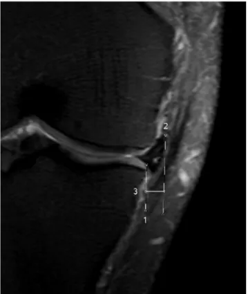
The measurement was performed by first drawing a vertical line intersecting the peripheral margin of the lateral tibial plateau at the point of transition from horizontal to vertical; the length of another line extending from the first line to the outer margin of the meniscus was defined as meniscal extrusion (Fig 1).
Patients were divided into two groups: in the absence of extrusion of meniscus (N: 48), and with the extrusion (N: 29).
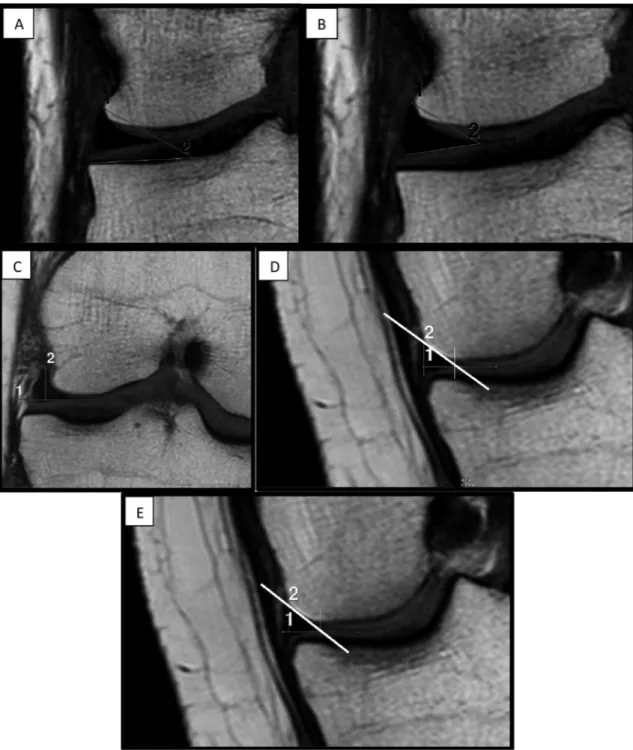
The following parameters were used in the analysis of the shape of the lateral meniscus (Fig 2A–2E):
1. MBA (meniscus-bone angle) (expressed in degrees)—angle between tibial plateau and supe-rior margin of the meniscus (Fig 2A),
2. MCA (meniscus-cartilage angle) (expressed in degrees)—angle between superior and infe-rior margin (maximum meniscal height) of the meniscus (Fig 2B),
3. MCH (meniscus-cartilage height) (expressed in millimeters)–distance between the top of the shaft and a line drawn along the superior margin of tibial cartilage (Fig 2C),
4. slope A (expressed in degrees)–equal to the arctangent of the quotient of maximum meniscal height (2) to its width (1), determined according to the following equation: slope A = arctan (2/1) (Fig 2D),
All MRI studies were assessed retrospectively by two radiologists (AS, MS) with, respec-tively, ten years and four years of experience in assessment of musculoskeletal system examina-tions. The raters did not know the purpose of the study; they were blind to study participant characteristics and study objectives. Two radiologists examined each MRI study and performed measurements by reaching a consensus. All data and related metadata underlying the findings reported in this article is available as Supporting Information (S1 Table).
Statistical Analysis
All data were presented as a mean ± standard deviation. Slope A and slope angle values were calculated as arctangent function of ratio respectively height A or height MCH to the length of the meniscus. This provided slopes in degrees. Differences between mean values in groups with (E) and without (NE) extrusion were examined by Welch’s t test for independent samples or Mann-Whitney U test if necessary. Normality assumption was verified using the W Shapiro-Wilk test. Linear interdependence between quantitative variables was assessed by Pearson's correlation coefficient values.
To find out which of the presented variables were associated with the risk of extrusion, the logistic regression was used. After identifying the risk factors receiver operating characteristic (ROC) analysis was used to determine the power of the predictor and cut-off point designated by Youden’s index.
Additionally, Kaplan-Meier's survival function was performed where survival time was set as given risk factor and censoring indicator as the presence of extrusion (dichotomous variable). The median of this survival function gave information when (for which value) the probability of extrusion presence is equal to its non-presence.
Fig 1. Meniscal extrusion is defined as the greatest distance (line No 3) from the most peripheral aspect of the meniscus (line No 2) to the border of the tibial plateau (line No 1).
The level of significance was set atα= 0.05. All calculated p-values were for two-tailed tests. All raw data were analyzed using statistical software Statistica 12.5 (StatSoft, Inc. 2014).
Results
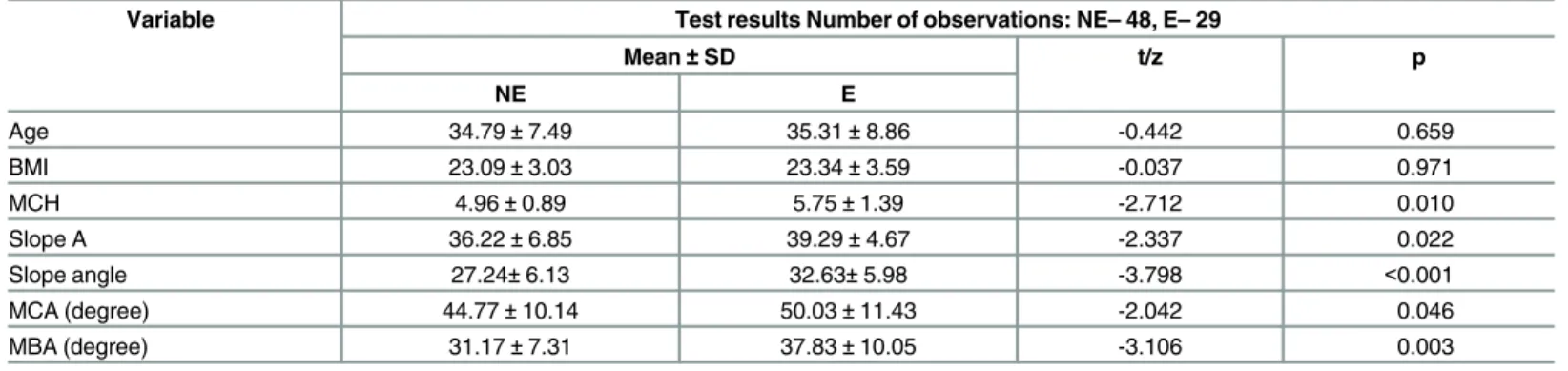
The analysis revealed statistically significant differences between mean values of MCH (p = 0.010), MCA (p = 0,046), MBA (p = 0.003), slope A (0.022) and slope angle (p<0.001) in
Fig 2. A-E. The central slice of a coronal MRI imaging of the knee focused on lateral compartment: A) meniscus-bone angle, B) meniscus-cartilage angle, C) meniscus-cartilage height, D) slope A, E) slope angle.
groups with (E) and without (NE) extrusion (Table 1). None of BMI and Age differed significantly.
Strong positive correlation (Table 2) existed between the pairs slope A and MCA (r = 0.815, p<0.001), MCA and MBA (r = 0.789, p<0.001), MBA and slope angle (r = 0.767, p<0.001), slope angle and MCA (r = 0.698, p<0.001).
Statistically significant (p<0.001) logit model (Table 3) suggests that mainly MBA (p = 0.024) and little MCH (p = 0.065) determine the risk of extrusion. The growth of MBA about 1 degree a risk of extrusion increases 1.078 times and with the growth of 1 mm of MCH the risk grows 1.593 times. The total predictability of this model is 67.53%.
Table 1. Test results for difference between the groups NE and E.
Variable Test results Number of observations: NE–48, E–29
Mean±SD t/z p
NE E
Age 34.79±7.49 35.31±8.86 -0.442 0.659
BMI 23.09±3.03 23.34±3.59 -0.037 0.971
MCH 4.96±0.89 5.75±1.39 -2.712 0.010
Slope A 36.22±6.85 39.29±4.67 -2.337 0.022
Slope angle 27.24±6.13 32.63±5.98 -3.798 <0.001
MCA (degree) 44.77±10.14 50.03±11.43 -2.042 0.046
MBA (degree) 31.17±7.31 37.83±10.05 -3.106 0.003
Groups with (E) and without (NE) extrusion; SD- standard deviation, BMI—body mass index, MCH—meniscus-cartilage height; MCA—meniscus-cartilage
angle.
doi:10.1371/journal.pone.0159156.t001
Table 2. Corelation Coeficients matrix for MCH, MCA, MBA, Slope A and Slope angle.
Variable Pearson’s Corelation Coeficients.
MCH MCA MBA Slope A Slope angle
MCH 1.000 0.175 0.397 0.229 0.647
MCA 0.175 1.000 0.789 0.815 0.698
MBA 0.397 0.789 1.000 0.650 0.767
Slope A 0.229 0.815 0.650 1.000 0.781
Slope angle 0.647 0.698 0.767 0.781 1.000
MCH—meniscus-cartilage height; MCA—meniscus-cartilage angle; MBA—meniscus-bone angle.
doi:10.1371/journal.pone.0159156.t002
Table 3. Logistic regression results—model 1.
N = 77 Logistic regression (logit) N of 0's: 29 1's: 48 Chi^2(2) = 14,156 p = 0,00084
Const. B0 MCH MBA
Estimate -5.546 0.466 0.075
Standard Error 1.599 0.252 0.033
Wald's Chi-square 12.031 3.414 5.085
p-value 0.001 0.065 0.024
Odds ratio 1.593 1.078
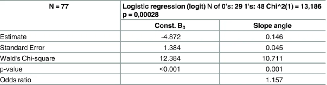
Next statistically significant (p<0.001) logit model (Table 4) takes into account only slope angle (p = 0.001). Rising the slope angle with 1 degree increases the risk of extrusion about 1.157 times. The total predictability of this model is 68.83%.
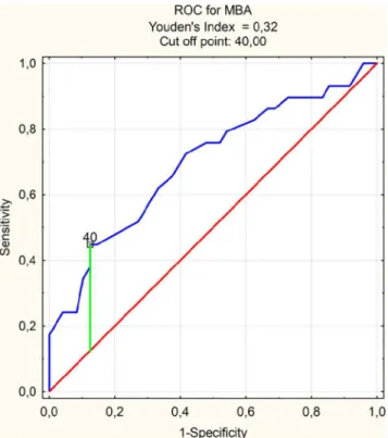
The ROC analysis indicated that previously specified by logistic regression extrusion risk factors like MBA (AUC = 0.697) and slope angle (AUC = 0.729) had prediction ability at an acceptable level (Table 5). The cut-off point for MBA was 40.00 (Fig 3) while sensitivity and specificity at this level respectively 0.448 and 0.875. Similarly for slope angle cut off point 27.70 (Fig 4) marked sensitivity at 0.828 and specificity as 0.542.
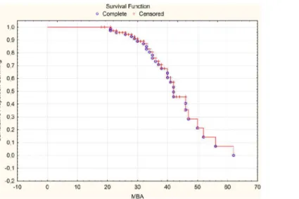
The median values of survival functions estimated for MBA (Fig 5) and slope angle (Fig 6) were approximately respectively 420 and 370. Because the growth of MBA and slope angle increases the risk of extrusion presence then for angles greater than median values it is more likely to occur.
Discussion
The goal of this work was to verify the results of Luczkiewicz et al. [17] regarding a relationship between the shape of the meniscus and the risk of its extrusion. In that study, based on a math-ematical model of a knee joint using finite element method, a tight correlation was demon-strated between an increase of the angle of inclination of the superior meniscal surface (slope angle) and increase in forces acting in radial direction, which is responsible for extrusion in the medial part of the meniscus.
Although findings were consistent with intuitive expectations, the mathematical model did not possess physical validation. For that reason, we decided to conduct an additional clinical study. Previous studies have shown that extrusion can be associated with joint space narrowing, radial meniscal tear and varus deformity [4]. Therefore, we excluded patients with the above-mentioned pathologies from our study group.
We demonstrated a correlation between the value of slope angle, MBA and MCH and the probability of lateral meniscus extrusion, which confirmed the results of earlier studies based on mathematical analyses (finite element method) [17].
Table 4. Logistic regression results—model 2.
N = 77 Logistic regression (logit) N of 0's: 29 1's: 48 Chi^2(1) = 13,186 p = 0,00028
Const. B0 Slope angle
Estimate -4.872 0.146
Standard Error 1.384 0.045
Wald's Chi-square 12.384 10.711
p-value <0.001 0.001
Odds ratio 1.157
doi:10.1371/journal.pone.0159156.t004
Table 5. ROC analysis results for MBA and Slope angle.
ROC analysis results
AUC SE z p Cut off point
MBA 0.697 0.063 3.101 0.002 40.0°
Slope angle 0.729 0.059 3.907 <0.001 27.7°
ROC—receiver operating characteristic; MBA—meniscus-bone angle.
Fig 3. ROC and cut off point for MBA.ROC—receiver operating characteristic.
doi:10.1371/journal.pone.0159156.g003
Fig 4. ROC and cut off point for Slope angle.ROC—receiver operating characteristic.
The risk factors for meniscal extrusion are quite well described in the literature
[1,4,13,14,18,19,20,21]. Nevertheless, the vast majority of authors focused on the assessment of the influence of meniscal tears or change in biomechanical conditions of the knee joint on the risk of development of this pathology [4,13,14,18].
Nakamura et al. evaluated an association between the shape of tibial spurs and lateral menis-cal displacement. They observed a relationship between tibial morphology and menismenis-cal luxa-tion only in the medial compartment [13].
Sturnick et al. [18] demonstrated a relationship between the geometry of the posterior meniscal horn and anterior cruciate ligament (ACL) injury. They revealed that increased slope of the articular cartilage and reduced height of the posterior horn of the meniscus were associ-ated with elevassoci-ated risk of non-contact ACL injury. The study findings suggest a direct link between the geometry of the meniscus with articular cartilage and knee laxity. While previous
Fig 5. Survival Function for MBA.MBA—meniscus-bone angle.
doi:10.1371/journal.pone.0159156.g005
Fig 6. Survival Function for Slope angle.
reports focused on the analyses of posterior meniscal horn geometry, those analyses concern geometry of the central part of the meniscus.
In our study, we identified a new parameter in the radiological assessment of the knee joint, such as the slope angle. It is most useful in predicting the risk of lateral meniscus extrusion. According to the ROC analysis the cut-off point for this parameter is 27 degrees (Fig 4), while the risk of extrusion increases significantly over the value of 37 degrees (Fig 6). Taking into consideration a dynamic character of this phenomenon of meniscal luxation, measurements of the above parameter can find practical applications in the assessment of the risk of instability, which correlates clinically with patients’pain perception in degenerative meniscal lesions [4,14,9,22]. Degenerative meniscal tears are not directly associated with symptoms [7] in spite of meniscal tears with extrusion which are strongly associated with knee pain [4]. The parame-ter evaluated by our team could be used as an additional risk factor for estimation of extrusion in patients with degenerative damage to the meniscus. Moreover, this parameter appears useful in selecting the best-suited allograft for meniscal transplantation. Meniscal graft extrusion is a common problem in the meniscus-transplanted knee [23]. One hypothesized cause of this phe-nomenon is over-sizing of the allograft [24]. Nowadays, method used for the estimation of meniscal graft size relies on measurements of width and length of meniscal allograft [25]. We believe that the results of our work allow for the introduction of the additional parameter that allows a better fit of the allograft to the shape of the uninjured knee meniscus.
MBA was another analyzed parameter. ROC curve determines the cut-off point at 40 degrees (Fig 3). Above 42 degrees the risk of lateral meniscus extrusion grows significantly (Fig 5).
Meniscal- cartilage height (MCH) was an important factor (although statistically less signifi-cant than the others) influencing the probability of extrusion. We showed that an increase in this parameter by 1 mm raises the risk of extrusion by 1.593 (Table 3).
Sturnick et al. demonstrated a tight correlation between the risk of ACL injury and meniscal height (MCH) as well as middle-cartilage slope (MCS) value in the lateral knee joint compart-ment [18]. These results are concordant with our study. Even though our analyses did not con-cern assessment of meniscal horns (we studied the core) or the risk factors for ACL injury, we confirmed (after Sturnick) the importance of a parameter describing the probability of knee joint pathology in the lateral compartment, such as meniscal- cartilage height (MCH).
The above analyses seem to confirm that lateral meniscus protrusion is a multifactorial pro-cess, involving not only its shape, but also structure and position of the tibia. Also, one should not forget about an important risk factor for meniscal dislocation, such as joint injury.
We believe that study group selection may be one of the limitations of our study. In our analysis we only included young patients with normal BMI, and no medical history of knee injury or other disorders. Meniscal extrusion, although indisputably harmful from the bio-mechanical point of view, is often asymptomatic in the healthy population. In our study menis-cal extrusion occurred in approximately 37%. According to available literature, this result does not differ from the average for the population of young, healthy people [2].
survival function is based on a similar reasoning as in material engineering. Increasing a factor (in our case, "slope") eventually leads to the occurrence of the event ("extrusion").
Nevertheless, the results of our studies gave similar conclusions we regard that problem of determining the risk factors of meniscal extrusion is still open.
Conclusions
The results of this study are further confirmed by the outcome of the mathematical model, which indicates a correlation between the shape of the meniscus and the risk of its displace-ment. According to our analysis the rise of the slope angle, MBA and MCH defining the menis-cus cross-section geometry, could increase the risk of meniscal extrusion.
We believe that the above-mentioned parameters may be useful, not only in the assessment of the risk of meniscal extrusion, particularly with coexisting degenerative changes but also in the better selection of the allograft for meniscal transplantation.
Supporting Information
S1 Table.(XLS)
Author Contributions
Conceived and designed the experiments: AS PL MS MK PJW JD MP ES BB. Performed the experiments: AS PL MS. Analyzed the data: AS PL MS MK. Contributed reagents/materials/ analysis tools: AS PL MS MP. Wrote the paper: AS PL MS MK PJW JD MP ES BB.
References
1. Hwang SH, Jung KA, Lee WJ, Yang KH, Lee DW, Carter A, et al. Morphological changes of the lateral meniscus in end stage lateral compartment osteoarthritis of the knee. Osteoarthritis and cartilage. 2012; 20: 110–116. doi:10.1016/j.joca.2011.11.005PMID:22133800
2. Rennie WJ, Finlay DB. Meniscal extrusion in young athletes: associated knee joint abnormalities. AJR Am J Roentgenol. 2006; 186: 791–794. PMID:16498108
3. Lee DH, Lee BS, Kim JM, Yang JS, Cha EJ, Park JH et al: Predictors of degenerative medial meniscus extrusion: radial component and knee osteoarthritis. Knee Surg Sports Traumatol Arthrosc. 2011; 19: 222–229. doi:10.1007/s00167-010-1274-2PMID:20890696
4. Wenger A, Englund M, Wirth W, Hudelmaier M, Kwoh K, Eckstein F et al. Relationship of 3D meniscal morphology and position with knee pain in subjects with knee osteoarthritis: a pilot study. Eur Radiol. 2012; 22: 211–20. doi:10.1007/s00330-011-2234-zPMID:21842432
5. Bloecker K, Wirth W, Guermazi A Hunter DJ, Resch H, Hochreiter J et al. Relationship between medial meniscal extrusion and cartilage loss in specific femorotibial subregions: Data from the osteoarthritis initiative. Arthritis Care & Research. 2015; 67: 1545–1552.
6. Englund M, Niu J, Guermazi A, Roemer FW, Hunter DJ, Lynch JA, et al. Effect of meniscal damage on the development of frequent knee pain, aching, or stiffness. Arthritis Rheum. 2007; 56: 4048–4054.
PMID:18050201
7. Englund M, Guermazi A, Gale D,Hunter DJ, Aliabadi P, Clancy M, et al. Incidental meniscal findings on knee MRI in middle-aged and elderly persons. N Engl J Med. 2008; 359: 1108–1115. doi:10.1056/
NEJMoa0800777PMID:18784100
8. Costa CR, Morrison WB, Carrino JA. Medial meniscus extrusion on knee MRI: is extent associated with severity of degeneration or type of tear? Am J Roentgenol. 2004; 183: 17–23.
9. Sharma L, Eckestein F, Song J, Guermazi A, Prasad P, Almagor O, et al. Relationship of meniscal dam-age, meniscal extrusion and joint laxity to subsequent cartilage loss in osteoarthritic knees. Arthritis Rheum. 2008; 58: 1716–1726. doi:10.1002/art.23462PMID:18512777
11. Gale DR, Chaisson CE, Totterman SM, Schwartz RK, Gale ME, Felson D. Meniscal subluxation: asso-ciation with osteoarthritis and joint space narrowing. Osteoarthritis Cartilage. 1999; 7: 526–532. PMID:
10558850
12. Hunter DJ, Zhang YQ, Tu X, Lavalley M, Niu JB, Amin S, et al. Change in joint space width: hyaline articular cartilage loss or alteration in meniscus? Arthritis Rheum. 2006; 54: 2488–2495. PMID:
16868968
13. Nakamura N, Sumen Y, Sakaridani K, Exham H, Ochi M. Relationship between the shape of tibial spurs on X-ray and meniscal changes on MRI in early osteoarthritis of knee. Magnetic Resonance Imaging 2006; 24: 1143–1148. PMID:17071336
14. Wenger A, Wirth W, Hudelmaier M, Noebauer-Huhmann I, Trattnig S, Bloecker K, et al. Meniscus body position, size, and shape in persons with and persons without radiographic knee osteoarthritis: quanti-tative analyses of knee magnetic resonance images from the osteoarthritis initiative. Arthritis Rheum. 2013; 65: 1804–11. doi:10.1002/art.37947PMID:23529645
15. Chan WP, Huang GS, Hsu SM, Chang YC, Ho WP. Radiographic joint space narrowing in osteoarthritis of the knee: relationship to meniscal tears and duration of pain. Skeletal Radiol. 2008; 37: 917–922.
doi:10.1007/s00256-008-0530-8PMID:18594811
16. Raynauld JP, Martel-Pelletier J, Berthiaume MJ, Beaudoin G, Choquette D, Haraoui B. Long term eval-uation of disease progression through the quantitative magnetic resonance imaging of symptomatic knee osteoarthritis patients: correlation with clinical symptoms and radiographic changes. Arthritis Res Ther. 2006; 8: 21–33.
17. Luczkiewicz P, Daszkiewicz K, Witkowski W, Chroscielewski J, Zarzycji W. Influence of meniscus shape in the cross sectional plane on the knee contact mechanics. J Biomech. 2015; 48: 1356–1363.
doi:10.1016/j.jbiomech.2015.03.002PMID:25892539
18. Sturnick DR, Van Gorder R, Vacek PM, DeSarno MJ, Gardner-Morse MG, Tourville TW. Tibial Articular Cartilage and Meniscus Geometries Combine to Influence Female Risk of Anterior Cruciate Ligament Injury. J Orthop Res 2014; 32: 1487–1494. doi:10.1002/jor.22702PMID:25099246
19. Arno S, Walker PS, Bell CP, Krasnokutsky S, Samuels J, Abramson SB, et al: Relation between carti-lage volume and meniscal contact in medial osteoarthritis of the knee. The Knee. 2012; 19: 896–901.
doi:10.1016/j.knee.2012.04.005PMID:22560645
20. Khan N, McMahon P, Obaid H. Bony morphology of the knee and non-traumatic meniscal tears: Is there a role for meniscal impingement? Skeletal Radiol. 2014; 43: 955–962. doi:
10.1007/s00256-014-1867-9PMID:24722655
21. Hudek R, Fuchs B, Regenfelder, Koch PP. Is Noncontact ACL Injury Associated with the Posterior Tib-ial and Meniscal Slope? Clin Orthop Relat Res. 2011; 469: 2377–2384. doi:
10.1007/s11999-011-1802-5PMID:21318628
22. Sharma L, Song J, Felson DT, Cahue S, Shamiyeh E, Dunlop DD. The role of knee alignment in dis-ease progression and functional decline in knee osteoarthritis. JAMA 2001; 286: 188–95. PMID:
11448282
23. Lee BS, Kim JM, Bin SI, Kim KA, Bim SU. Patient-related risk factors for the extrusion of lateral menis-cal allograft transplants. Arthroscopy. 2015; 31: 699–706. doi:10.1016/j.arthro.2014.10.016PMID:
25530512
24. Jang SH, Kim JG, Ha JG, Shim JC. Reducing the size of the meniscal allograft decreases the percent-age of extrusion after meniscal allograft transplantation. Arthroscopy. 2011; 27: 914–22. doi:10.1016/j.
arthro.2011.02.017PMID:21693346
25. Pollard ME, Kang Q, Berg EE. Radiographic sizing for meniscal transplantation. Arthroscopy. 1995; 11: 684–7. PMID:8679029
26. Basu B, Tiwari D, Kundu D, Prasad R. Is Weibull distribution the most appropriate statistical strength distribution for brittle materials, Ceramics International. 2009; 35: 237–246.