Mutation on Intestinal Colonization, Translocation, and
Induction of Immunopathology in Gnotobiotic IL-10
Deficient Mice
Markus M. Heimesaat1*, Raimond Lugert2, Andre´ Fischer1, Marie Alutis1, Anja A. Ku¨hl3, Andreas E. Zautner2,4, A. Malik Tareen2, Ulf B. Go¨bel1, Stefan Bereswill1
1Department of Microbiology and Hygiene, Charite´ - University Medicine Berlin, Berlin, Germany,2Department of Medical Microbiology, University Medical Center Go¨ttingen, Go¨ttingen, Germany,3Department of Pathology/Research Center ImmunoSciences (RCIS), Charite´ - University Medicine Berlin, Berlin, Germany,4Department of Clinical Chemistry/UMG-Laboratory, University Medical Center Go¨ttingen, Go¨ttingen, Germany
Abstract
Background: Although Campylobacter jejuni infections have a high prevalence worldwide and represent a significant socioeconomic burden, the underlying molecular mechanisms of induced intestinal immunopathology are still not well understood. We have recently generated a C. jejuni mutant strain NCTC11168::cj0268c, which has been shown to be involved in cellular adhesion and invasion. The immunopathological impact of this gene, however, has not been investigatedin vivoso far.
Methodology/Principal Findings:Gnotobiotic IL-10 deficient mice were generated by quintuple antibiotic treatment and perorally infected withC. jejunimutant strain NCTC11168::cj0268c, its complemented version (NCTC11168::cj0268c
-comp-cj0268c), or the parental strain NCTC11168. Kinetic analyses of fecal pathogen loads until day 6 post infection (p.i.) revealed that knockout ofcj0268cdid not compromise intestinalC. jejunicolonization capacities. Whereas animals irrespective of the analysedC. jejunistrain developed similar clinical symptoms of campylobacteriosis (i.e. enteritis), mice infected with the NCTC11168::cj0268cmutant strain displayed significant longer small as well as large intestinal lengths indicative for less distinctC. jejuniinduced pathology when compared to infected control groups at day 6 p.i. This was further supported by significantly lower apoptotic and T cell numbers in the colonic mucosa and lamina propria, which were paralleled by lower intestinal IFN-cand IL-6 concentrations at day 6 following knockout mutant NCTC11168::cj0268cas compared to parental strain infection. Remarkably, less intestinal immunopathology was accompanied by lower IFN-csecretion inex vivobiopsies taken from mesenteric lymphnodes of NCTC11168::cj0268cinfected mice versus controls.
Conclusion/Significance:We here for the first time show that thecj0268cgene is involved in mediatingC. jejuniinduced immunopathogenesis in vivo. Future studies will provide further deep insights into the immunological and molecular interplays betweenC. jejuniand innate immunity in human campylobacteriosis.
Citation:Heimesaat MM, Lugert R, Fischer A, Alutis M, Ku¨hl AA, et al. (2014) Impact ofCampylobacter jejuni cj0268cKnockout Mutation on Intestinal Colonization, Translocation, and Induction of Immunopathology in Gnotobiotic IL-10 Deficient Mice. PLoS ONE 9(2): e90148. doi:10.1371/journal.pone.0090148
Editor:Dipshikha Chakravortty, Indian Institute of Science, India
ReceivedDecember 25, 2013;AcceptedJanuary 31, 2014;PublishedFebruary 25, 2014
Copyright:ß2014 Heimesaat et al. This is an open-access article distributed under the terms of the Creative Commons Attribution License, which permits unrestricted use, distribution, and reproduction in any medium, provided the original author and source are credited.
Funding:This work was supported by grants from the German Research Foundation (DFG) to RL (GR906/13-1, CampyGerm), UBG (GO363/12-1, CampyGerm; SFB633, TP A7), SB and AF (SFB633, TP A7), AAK (SFB633, TP Z1), MMH (SFB633, TP B6), MA (SFB633, Immuco), the Deutsche Akademische Austauschdienst (DAAD) to AMT, and from the German Federal Ministry of Education and Research (BMBF) to SB (TP1.1). The funders had no role in study design, data collection and analysis, decision to publish, or preparation of the manuscript.
Competing Interests:Markus M. Heimesaat and Stefan Bereswill currently serve as academic editors for this journal. This does not alter the authors’ adherence to all the PLOS ONE policies on sharing data and materials.
* E-mail: markus.heimesaat@charite.de
Introduction
Campylobacter jejuni is the most important cause of bacterial diarrhea in developing as well as in industrialized countries. The characteristic features of the disease vary from watery to bloody diarrhea accompanied by abdominal cramps and fever. In rare cases complications such as the Guillain-Barre´ syndrome might arise post infection (p.i.) [1,2]. Although many virulence factors of
C. jejunihave been described yet, the overall image of this bacterial infection is still incomplete [3,4].
A successful infection with C. jejuni requires adherence of the pathogen to host cells and several proteins of C. jejuni that contribute to this initial interaction have been characterized in the past. MOMP, CadF and FlpA, for instance, were shown to possess fibronectin-binding properties whereby specifically CadF and FlpA initiate the remodelling of the actin cytoskeleton via the activation of integrin receptors to allow internalization ofC. jejuni
to be important for the invasion of Caco2 cells by the pathogen [15,16,17]. Thereby, we could show that the invasion-relevant phenotype of Cj0268c is due to its adherence mediating function, not only in C. jejuni but also when this protein is expressed heterologously inE. coli[17]. However, the functional relevance of Cj0268c for the interaction of C. jejuni with the host immune system has not been demonstrated so far.
To address this we here applied the gnotobiotic murine IL-102/
2 infection model. In order to eradicate the colitogenic stimuli
derived from the conventional intestinal microbiota, IL-102/2
mice were subjected to a broad-spectrum antibiotic treatment for at least 3 months starting immediately after weaning [18]. UponC. jejuniinfection gnotobiotic IL-102/2mice get readily colonized by
the pathogen and display acute enterocolitis within one week p.i.mimicking severe campylobacteriosis in humans, whereas gnotobiotic or with human microbiota reassociated wildtype mice display intestinal pro-inflammatory immune responses but no overt clinical symptoms such as bloody diarrhea upon C. jejuni
infection [18]. We here for the first time investigated i) the colonization capacities and ii) clinical as well as iii) intestinal pro-inflammatory immune cell and cytokine responses upon infection of gnotobiotic IL-102/2 mice with the C. jejuni mutant strain
NCTC11168::cj0268c, its complemented version NCTC11168::cj0268c-comp-cj0268c and the parental strain NCTC11168.
Results
Impact of Cj0268c onC. jejuniColonization Capacity in Infected Gnotobiotic IL-102/2Mice
Given that the murine commensal gut microbiota is essential for the physiological host resistance againstC. jejuniinfection [19], we generated gnotobiotic IL-102/2 mice by quintuple antibiotic
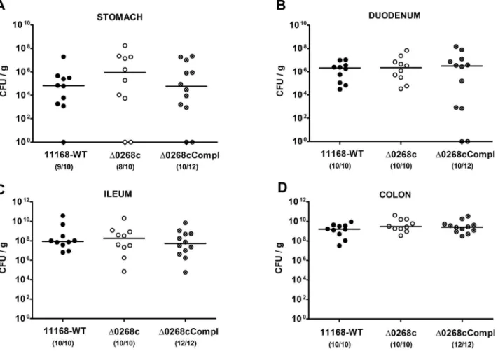
treatment for at least 3 months (refer to [20,21]) to investigate the colonization capacity of C. jejuni mutant strain NCTC11168::cj0268c. Following peroral infection on two consec-utive days with a comparable challenge of 109viable mutantC. jejuni NCTC11168::cj0268c, its complemented version NCTC11168::cj0268c-comp-cj0268c, or the parental strain NCTC11168, each in the stationary phase (not shown), gnotobi-otic mice were readily colonized with comparably high loads of 109to 1010colony forming units (CFU) of either strain per g feces over time until day 6 p.i. (Fig. 1). In addition, when luminal samples were taken from the entire gastrointestinal (GI) tract on the day of necropsy (day 6 p.i.), either C. jejunistrain could be cultured from the stomach, duodenum, ileum and colon, with the highest loads in the large intestine of approximately 109to 1010 CFU per g luminal content (Fig. 2). Thus, deficiency of the
cj0268cgene did not impact gastrointestinal colonization capacities ofC. jejuniin gnotobiotic IL-102/2mice upon peroral infection.
strains at defined time points did not differ.
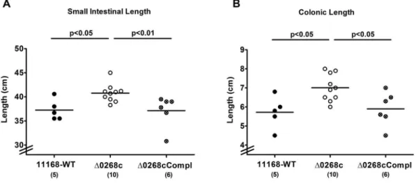
Given that acute intestinal inflammation is accompanied by a significant shortening of the intestinal tract [18,20,22], we determined the absolute lengths of the small as well as large intestines at day 6 p.i. Interestingly, gnotobiotic IL-102/2 mice
infected with the C. jejuni mutant strain NCTC11168::cj0268c
displayed longer small intestines (approximately 10% mean difference; Fig. 4A) and colons (approximately 20% mean difference; Fig. 4B) as compared to mice infected with the parental strain NCTC11168 (p,0.05) or complemented strain NCTC11168::cj0268c-comp-cj0268c(p,0.05 and p,0.01, respec-tively;Fig. 4AB) indicative for significantly less distinct intestinal pathology. Furthermore, viable bacteria of theC. jejuni parental strain NCTC11168 and the complemented strain NCTC11168::cj0268c-comp-cj0268ccould be cultured from mes-enteric lymphnodes (MLNs) in 20.00% (2 out of 10) and 8.33% (1 out of 12) of infected animals at day 6 p.i., respectively, whereas the mutant strain NCTC11168::cj0268c did not translocate into MLNs at all (not shown). Furthermore, virtually no pathogenic translocation to extra-intestinal compartments could be detected given that spleen, liver, kidney and cardiac blood were exclusively
C. jejuniculture-negative (not shown). Taken together, uncompro-mised colonization capacities of C. jejuni lacking cj0268c were accompanied by comparable induction of gross disease (clinical symptoms of ulcerative enterocolitis). Longer small and large intestines as well as lower translocation frequencies in C. jejuni
mutant strain NCTC11168::cj0268cinfected gnotobiotic IL-102/2
mice, however, hint towards less pronounced intestinal immuno-pathology caused by absence of thecj0268cgene.
Impact of Cj0268c on Induction of Intestinal Pro-inflammatory Immune Responses inC. jejuniInfected Gnotobiotic IL-102/2Mice
We further assessed the immunopathological responses of mice upon infection with the C. jejuni knockout mutant NCTC11168::cj0268c. Irrespective of the strain, gnotobiotic mice displayed comparable histopathological changes in hematoxylin and eosin (H&E) stained colonic paraffin sections at day 6 p.i. (not shown). Given that apoptosis is a commonly used diagnostic marker in the histopathological evaluation and grading of intestinal disease [21] and a key feature of C. jejuni induced ulcerative enterocolitis in gnotobiotic IL-102/2 mice [18], we quantitatively assessed caspase-3+
Figure 1. Kinetic survey ofC. jejuniknockout mutant NCTC11168::cj0268ccolonization in gnotobiotic IL-102/2mice.Gnotobiotic IL-102/2mice were generated by antibiotic gut decontamination and perorally infected withC. jejuniNCTC11168 (11168-WT, closed circles;A), mutant strain NCTC11168::cj0268c(D0268c, open circles;B), or the complemented strain NCTC11168::cj0268c-comp-cj0268c(D0268cCompl, crossed circles;C) as described (see methods). The intestinal colonization capacities over time were determined by quantification of liveC. jejuniin fecal samples applying cultural analysis (CFU, colony forming units) starting two days until six days post infection as indicated on the x-axis. Medians (black bars) are indicated and numbers of animals harbouring the respectiveC. jejunistrain out of the total number of analyzed animals given in parentheses. Data shown were pooled from three independent experiments.
doi:10.1371/journal.pone.0090148.g001
Figure 2.C. jejuniknockout mutant NCTC11168::cj0268ccolonization along the gastrointestinal tract of gnotobiotic IL-102/2mice.
Gnotobiotic IL-102/2mice were generated by antibiotic gut decontamination and perorally infected withC. jejuniNCTC11168 (11168-WT, closed circles), mutant strain NCTC11168::cj0268c(D0268c, open circles), or the complemented strain NCTC11168::cj0268c-comp-cj0268c(D0268cCompl, crossed circles) as described (see methods). The pathogen densities in distinct compartments of the gastrointestinal tract were determined by quantification of liveC. jejuniin luminal samples taken from stomach, duodenum, ileum, and colon at day 6 p.i. by cultural analysis (CFU, colony forming units). Medians (black bars) are indicated and numbers of animals harbouring the respectiveC. jejunistrain out of the total number of analyzed animals given in parentheses. Data shown were pooled from three independent experiments.
positive cell numbers in the colonic mucosa of the former (p,0.05;
Fig. 5A).
Given that recruitment of pro-inflammatory immune cell populations to sites of inflammation is a hallmark of human campylobacteriosis [21], we next quantitatively assessed the influx of innate and adaptive immune as well as effector cell populations into the large intestine by applying in situimmunohistochemical staining of colonic paraffin sections. FollowingC. jejuniinfection, a marked influx of CD3+cells (i.e. T lymphocytes) into the colonic
mucosa and lamina propria could be detected until day 6 p.i.
(Fig. 5B). This increase, however, was significantly less pro-nounced in mice infected with the knockout mutant NCTC11168::cj0268c as compared to parental strain NCTC11168 and complemented strain NCTC11168::cj0268c -comp-cj0268c infected control animals (p,0.01 and p,0.05, respectively;Fig. 5B). Irrespective of theC. jejunistrain, infected mice displayed comparable increases of Foxp3+
regulatory T cells, B220+
B lymphocytes, MPO7+
neutrophils, and F4/80+
macro-phages and monocytes in the colonic mucosa at day 6 p.i. as compared to naı¨ve animals (p,0.0005–0.0001;Fig. 5C–F).
Figure 3. Kinetic survey of clinical symptoms followingC. jejuniknockout mutant NCTC11168::cj0268cinfection of gnotobiotic
IL-102/2mice.Gnotobiotic IL-102/2mice were generated by antibiotic gut decontamination and perorally infected withC. jejuniNCTC11168
(11168-WT, closed circles, n = 10; A), mutant strain NCTC11168::cj0268c(D0268c, open circles, n = 11; B), or the complemented strain NCTC11168::cj0268c -comp-cj0268c(D0268cCompl, crossed circles; n = 12; C) as described (see methods). Disease activity before and followingC. jejuniinfection was assessed daily by applying a standardized clinical scoring system. Means (black bars) and levels of significance (P-values) determined by the Mann-Whitney-U test are indicated. Data shown were pooled from three independent experiments.
doi:10.1371/journal.pone.0090148.g003
Figure 4. Intestinal lengths following C. jejuni knockout mutant NCTC11168::cj0268c infection of gnotobiotic IL-102/2 mice.
We next determined intestinal pro-inflammatory cytokine expression levels uponC. jejuniinfection. Lower colonic apoptotic cell and T lymphocyte counts were accompanied by lower IL-6 and IFN-c protein concentrations in ex vivo colonic biopsies obtained from gnotobiotic IL-102/2 mice six days following
infection with the knockout mutant NCTC11168::cj0268c as compared to the parental strain NCTC11168 (Fig. 6A, B). The impact ofcj0268cin mediatingC. jejuniinduced immunopathology was further underlined by lower IFN-clevels inex vivobiopsies of draining mesenteric lymphnodes in mutant strain NCTC11168::cj0268c as compared to parental strain NCTC11168 infected mice (p,0.05;Fig. 6C). Whereas intestinal pro-inflammatory cytokine levels in complemented and wildtype strain infected mice did not differ, a trend towards higher intestinal IL-6 and IFN-cconcentrations six days following complemented as compared to knock-out mutant strain infection could be observed. Given high standard deviations in the respective groups, however, the differences did not reach statistical significance (Fig. 6).
Taken together,cj0268cgene deficiency does not alterC. jejuni
NCTC11168 colonization capacities in vivo. In addition, the Cj0268c protein is involved in mediating C. jejuniinduced acute
enteritis as indicated by i.) less shrinkage of the small as well as large intestines, ii.) less abundance of colonic epithelial apoptotic cells, iii.) less distinct T lymphocyte infiltrations in the colonic mucosa and iv.) less pro-inflammatory cytokine secretion at intestinal tissue sites including mesenteric lymphnodes of gnoto-biotic IL-102/2 mice infected with the knockout mutant strain
NCTC11168::cj0268cwhen compared to control animals.
Discussion
We have recently shown that theC. jejuniprotein Cj0268c is an important prerequisite for pathogen adhesion and invasion of host cellsin vitro[17]. In the present study we investigated the impact of
cj0268cinC. jejuniinduced immunopathology in vivo. To prevent conventionally colonized IL-102/2 mice from spontaneous chronic colitis due to antigenic stimuli derived from the intestinal microbiota, mice were subjected to at least 3 months broad-spectrum antibiotic treatment starting immediately after weaning [18]. Upon peroralC. jejuniinfection gnotobiotic IL-102/2 mice develop non-selflimiting ulcerative enterocolitis within one week p.i. mimicking severe campylobacteriosis in immuno-compro-mized patients [18]. Here, kinetic analyses revealed that until day
Figure 5. Impact of thecj0268cgene on colonic inflammatory and immune cell responses following infection of gnotobiotic IL-102/
2mice.Gnotobiotic IL-102/2mice were generated by antibiotic gut decontamination and perorally infected withC. jejuniNCTC11168 (11168-WT,
closed circles), mutant strain NCTC11168::cj0268c (D0268c, open circles), or the complemented strain NCTC11168::cj0268c-comp-cj0268c (D0268cCompl, crossed circles) as described (see methods). The average numbers of apoptotic cells (positive for caspase-3, panel A), T lymphocytes (positive for CD3, panel B), regulatory T cells (Treg, positive for Foxp3, panel C), B lymphocytes (positive for B220, panel D), neutrophils (positive for MPO7, panel E), and macrophages/monocytes (positive for F4/80, panel F) from at least six high power fields (HPF, 400x magnification) per animal were determined microscopically in immunohistochemically stained colon sections at day 6 p.i. uninfected animals (Naı¨ve; open diamonds) served as negative controls. Numbers of analyzed animals are given in parentheses. Means (black bars) and levels of significance (P-values) determined by the Mann-Whitney-U test are indicated. Data shown were pooled from three independent experiments.
6 following peroral infection, mice harboured high intestinal loads of the knockout mutant strain NCTC11168::cj0268c, which were comparable to those detected in mice upon infection with the parental strain NCTC11168 or the complemented version NCTC11168::cj0268c-comp-cj0268c. Hence, knockout of the
cj0268c gene did neither compromise infection capacities in vitro
[17] nor in vivo. Of note, genetic complementation clearly demonstrates that the generated NCTC11168::cj0268cknock-out mutant strain is not polar. Remarkably, mice infected with the mutant strain NCTC11168::cj0268c displayed significantly less severe immunopathology in the intestinal tract as compared to mice infected with the parental strain NCTC11168 or the complementedC. jejunistrain NCTC11168::cj0268c-comp-cj0268c
as indicated by a plethora of results. First, knockout mutant strain NCTC11168::cj0268c infected gnotobiotic IL-102/2 mice dis-played less shrinkage of the small as well as large intestines which is a rather rough, but reliable indicator for less pronounced intestinal pathology [20,22,23,24]. Second, this was further supported by less abundance of caspase-3+
cells in the colonic mucosa given that apoptosis is a commonly used diagnostic marker in the histopath-ological evaluation and grading of intestinal disease [21] and a key feature of C. jejuniinduced ulcerative enterocolitis in gnotobiotic IL-102/2mice [18]. Third, T lymphocytes well known to play a pivotal role in induction and perpetuation of C. jejuni induced immunopathology in mice [18,21,25,26,27] were infiltrating the intestinal mucosa and lamina propria of gnotobiotic IL-102/2
mice following infection with knockout mutant NCTC11168::cj0268c to a far lesser extent as compared to the applied control strains. Fourth, virtually no translocation of viable
C. jejuni from the intestinal tract to MLNs was observed upon infection with mutant strain NCTC11168::cj0268c. Fifth, expres-sion of pro-inflammatory cytokines such as IL-6 and IFN-cwas more than 50% lower inex vivobiopsies derived from colon and MLNs upon infection with the mutant versus the parental strain. In our previous work where we independently studied theC. jejuni -induced immunopathological sequelae in two other murine C. jejuni infection models we could unequivocally demonstrate that severity of campylobacteriosis was paralleled by up-regulated
expression levels of IFN-cand IL-6 in both, the colon and MLNs [18,21,25], further supporting significance of the results presented here. It is tempting to speculate that the decreased intestinal IFN-c
and IL-6 levels following mutant as compared to parental strain infection might be indicative for shifted intestinal T cell populations in the absence of cj0268c which needs to further unraveled.
Irrespective of theC. jejunistrain, however, infected gnotobiotic IL-102/2 mice developed comparable clinical symptoms of enteritis over time in the presented study, which was contrasting the less pronounced immunopathological outcome in the intestinal tract. Despite the observation of comparable clinical symptoms upon infection with the knockout mutant strain NCTC11168::cj0268c, one needs to take into account that the clinical picture of a disease is rather the sum of different effects resulting from several levels of immunopathological mechanisms. Furthermore, thecj0268cgene is by far not the only factor involved in adhesion and invasion and subsequent induction of immuno-pathology [28]. Nevertheless, severity ofC. jejuniinduced enteritis can vary considerably between infected human individuals and range from very mild, sublatent and self-limiting complaints to severe symptoms such as abdominal cramps, fever, and bloody diarrhea depending on the dysbalance between the immune status of the host and the respective pathogenicity factors of the pathogen expressed in parallel [29].
One needs to take further into account, thatC. jejuniinfection in the in vivo infection model applied here results in a devastating outcome, namely non-selflimiting acute ulcerative enterocolitis leading to death within 10 days [18]. Hence, if any beneficial effect is observed in such a hyper-acute model system, the biological relevance gets more plausible. Furthermore, our in vitro results revealed that adhesive properties of the mutant strain were not 100%, but reached rather 60% [17]. Moreover, we have recently shown in different murine infection models that Toll-like-receptor (TLR)-4 dependent signalling ofC. jejunilipooligosaccharide is a key factor inC. jejuniinduced immunopathology as indicated by ameliorated clinical and intestinal immunopathology in C. jejuni
Figure 6. Impact of thecj0268cgene on intestinal pro-inflammatory cytokine responses following infection of gnotobiotic IL-102/2
mice.Gnotobiotic IL-102/2mice were generated by antibiotic gut decontamination and perorally infected withC. jejuniNCTC11168 (11168-WT,
infected gnotobiotic TLR-4 deficient as well as IL-10 deficient mice lacking TLR-4 [18,21,30].
Taken together, our previous and actual results have shown that
cj0268cis involved inC. jejuniadhesion and invasion of vertebrate cells subsequently inducing significant immunopathology in the host with varying clinical features. Due to the lack of appropriate animal models in the past, the impact of most of the so far identified pathogenicity factors ofC. jejuniinvolved in pathogen-host-interaction and thus their biological relevance in inducing campylobacteriosis have not been investigatedin vivoyet.
In conclusion, futurein vivo studies should further unravel the distinct molecular mechanisms and orchestration of different pathogenicity factors contributing toC. jejuniinduced disease.
Materials and Methods
Ethics Statement
All animal experiments were conducted according to the European Guidelines for animal welfare (2010/63/EU) with approval of the commission for animal experiments headed by the ‘‘Landesamt fu¨r Gesundheit und Soziales’’ (LaGeSo, Berlin, Germany; registration numbers G0123/12). Animal welfare was monitored twice daily by assessment of clinical conditions.
Mice
IL-102/2mice (in C57BL/10 background, B10) were bred and maintained in the facilities of the ‘‘Forschungsinstitut fu¨r Experimentelle Medizin’’ (FEM, Charite´ - Universita¨tsmedizin, Berlin, Germany), under specific pathogen-free (SPF) conditions.
To eradicate the commensal gut flora, mice were transferred to sterile cages and treated by adding ampicillin (1 g/L; Ratio-pharm), vancomycin (500 mg/L; Cell Pharm), ciprofloxacin (200 mg/L; Bayer Vital), imipenem (250 mg/L; MSD), and metronidazole (1 g/L; Fresenius) to the drinking water ad libitum
as described earlier [20] starting at 3 weeks of age right after weaning. Age matched female mice were subjected to the quintuple antibiotic treatment for 3–4 months before the infection experiment.
C. jejuniInfection of Mice
Mice were infected with approximately 109 viable CFU ofC. jejuni strains NCTC11168 (parental strain), the C. jejuni mutant strain NCTC11168::cj0268c(lacking thecj0268cgene [17]), or its complemented version NCTC11168::cj0268c-comp-cj0268c [17], respectively, by gavage in a total volume of 0.3 mL PBS on two consecutive days (day 0 and day 1).
Clinical Score
To assess clinical signs ofC. jejuniinduced infection on a daily basis, a standardized cumulative clinical score (maximum 12 points, addressing the occurrence of blood in feces (0 points: no blood; 2 points: microscopic detection of blood by the Guajac method using Haemoccult, Beckman Coulter/PCD, Krefeld, Germany; 4 points: overt blood visible), diarrhea (0: formed feces; 2: pasty feces; 4: liquid feces), and the clinical aspect (0: normal; 2: ruffled fur, less locomotion; 4: isolation, severely compromized locomotion, pre-final aspect) was used [18].
Sampling Procedures
Mice were sacrificed by isofluran treatment (Abbott, Germany). Cardiac blood and tissue samples from mesenteric lymphnodes, spleen, liver, kidney and GI tract (stomach, duodenum, ileum, colon) were removed under sterile conditions. Absolute small and large intestinal lengths were determined by measuring the
distances from the transition of the stomach to the duodenum to the very distal terminal ileum and from the ascending colon leaving the caecum to the rectum, respectively, by a ruler and expressed in cm. GI samples from each mouse were collected in parallel for immunohistochemical, microbiological, and immuno-logical analyses. Immunohistopathoimmuno-logical changes were deter-mined in colonic samples immediately fixed in 5% formalin and embedded in paraffin. Sections (5mm) were stained with H&E or respective antibodies forin situimmunohistochemistry.
Immunohistochemistry
In situ immunohistochemical analysis of colonic paraffine sections was performed as described previously [18,21,25,31]. Primary antibodies against cleaved caspase-3 (Asp175, Cell Signaling, USA, 1:200), CD3 (#N1580, Dako, Denmark, dilution 1:10), myeloperoxidase-7 (MPO-7,#A0398, Dako, 1:10000), F4/ 80 (#14-4801, clone BM8, eBioscience, 1:50), Foxp3 (FJK-16s, eBioscience, 1:100), and B220 (eBioscience, San Diego, CA, USA, 1:200) were used. For each animal, the average number of positively stained cells within at least six high power fields (HPF, 0.287 mm2; 4006magnification) were determined microscopi-cally by three independent investigators.
Quantitative Analysis ofC. jejuni(Translocation)
LiveC. jejuniwere detected in feces or at time of necropsy (day 6 p.i.) in luminal samples taken from the stomach, duodenum, ileum, or colon dissolved in sterile PBS by culture as described earlier [18,21]. To quantify bacterial translocation, MLNs, spleen, liver (<1 cm2) and kidney were homogenized in sterile PBS and analyzed by cultivating on karmali agar (Oxoid, Wesel, Germany) in a microaerophilic atmosphere at 37uC for at least 48 hours. Cardiac blood (<200mL) was directly streaked onto karmali agar and cultivated accordingly. The respective weights of fecal or tissue samples were determined by the difference of the sample weights before and after asservation. The detection limit of viable pathogens was<100 CFU per g.
Cytokine Detection in Culture Supernatants ofex vivo
Biopsies taken from Colon and Mesenteric Lymphnodes Colon biopsies were cut longitudinally, and washed in PBS. Mesenteric lymphnodes or strips of approximately 1 cm2 colon tissue were placed in 24-flat-bottom well culture plates (Nunc, Wiesbaden, Germany) containing 500mL serum-free RPMI 1640 medium supplemented with penicillin (100 U/mL) and strepto-mycin (100mg/mL; PAA Laboratories). After 18 h at 37uC, culture supernatants were tested for IL-6 and IFN-cby the Mouse Inflammation Cytometric Bead Assay (CBA; BD Biosciences) on a BD FACSCanto II flow cytometer (BD Biosciences).
Statistical Analysis
Mean values, medians, and levels of significance were deter-mined using Mann-Whitney-U test. Two-sided probability (P) values #0.05 were considered significant. All experiments were repeated at least twice.
Acknowledgments
and INT 407 cell membranes. FEMS Microbiol Lett 157: 233–238. 6. Boehm M, Krause-Gruszczynska M, Rohde M, Tegtmeyer N, Takahashi S et
al. (2011) Major host factors involved in epithelial cell invasion ofCampylobacter jejuni: role of fibronectin, integrin beta1, FAK, Tiam-1, and DOCK180 in activating Rho GTPase Rac1. Front Cell Infect Microbiol 1: 17.
7. Krause-Gruszczynska M, Boehm M, Rohde M, Tegtmeyer N, Takahashi S et al. (2011) The signaling pathway ofCampylobacter jejuni-induced Cdc42 activation: Role of fibronectin, integrin beta 1, tyrosin kinases and guanine exchange factor Vav2. Cell Commun Signal 9: 32.
8. Pei Z, Burucoa C, Grignon B, Baqar S, Huang XZ, et al. (1998) Mutation in the peb1A locus of Campylobacter jejuni reduces interactions with epithelial cells and intestinal colonization of mice. Infect Immun 66: 938–943.
9. Ashgar SS, Oldfield NJ, Wooldridge KG, Jones MA, Irving GJ, et al. (2007) CapA, an autotransporter protein of Campylobacter jejuni, mediates association with human epithelial cells and colonization of the chicken gut. J Bacteriol 189: 1856–1865.
10. Oakland M, Jeon B, Sahin O, Shen Z, Zhang Q (2011) Functional characterization of a lipoprotein-encoding operon in Campylobacter jejuni. PLoS One 6: e20084.
11. Jin S, Joe A, Lynett J, Hani EK, Sherman P, et al. (2001) JlpA, a novel surface-exposed lipoprotein specific to Campylobacter jejuni, mediates adherence to host epithelial cells. Mol Microbiol 39: 1225–1236.
12. Jin S, Song YC, Emili A, Sherman PM, Chan VL (2003) JlpA of Campylobacter jejuni interacts with surface-exposed heat shock protein 90alpha and triggers signalling pathways leading to the activation of NF-kappaB and p38 MAP kinase in epithelial cells. Cell Microbiol 5: 165–174.
13. Holden KM, Gilbert M, Coloe PJ, Li J, Fry BN (2012) The role of WlaRG, WlaTB and WlaTC in lipooligosaccharide synthesis by Campylobacter jejuni strain 81116. Microb Pathog 52: 344–352.
14. Tareen AM, Dasti JI, Zautner AE, Gross U, Lugert R (2011) Sulphite : cytochrome c oxidoreductase deficiency in Campylobacter jejuni reduces motility, host cell adherence and invasion. Microbiology 157: 1776–1785. 15. Tareen AM, Dasti JI, Zautner AE, Gross U, Lugert R (2010) Campylobacter
jejuni proteins Cj0952c and Cj0951c affect chemotactic behaviour towards formic acid and are important for invasion of host cells. Microbiology 156: 3123–3135.
16. Novik V, Hofreuter D, Galan JE (2010) Identification of Campylobacter jejuni genes involved in its interaction with epithelial cells. Infect Immun 78: 3540– 3553.
negative bacteria aggravate murine small intestinal Th1-type immunopathology following oral infection with Toxoplasma gondii. J Immunol 177: 8785–8795. 21. Bereswill S, Fischer A, Plickert R, Haag LM, Otto B, et al. (2011) Novel murine
infection models provide deep insights into the ‘‘menage a trois’’ of Campylobacter jejuni, microbiota and host innate immunity. PLoS One 6: e20953.
22. Munoz M, Heimesaat MM, Danker K, Struck D, Lohmann U, et al. (2009) Interleukin (IL)-23 mediates Toxoplasma gondii-induced immunopathology in the gut via matrixmetalloproteinase-2 and IL-22 but independent of IL-17. J Exp Med 206: 3047–3059.
23. Heimesaat MM, Fischer A, Jahn HK, Niebergall J, Freudenberg M, et al. (2007) Exacerbation of murine ileitis by Toll-like receptor 4 mediated sensing of lipopolysaccharide from commensal Escherichia coli. Gut 56: 941–948. 24. Bereswill S, Munoz M, Fischer A, Plickert R, Haag LM, et al. (2010)
Anti-inflammatory effects of resveratrol, curcumin and simvastatin in acute small intestinal inflammation. PLoS One 5: e15099.
25. Haag LM, Fischer A, Otto B, Plickert R, Kuhl AA, et al. (2012) Intestinal microbiota shifts towards elevated commensal Escherichia coli loads abrogate colonization resistance against Campylobacter jejuni in mice. PLoS One 7: e35988.
26. Haag LM, Fischer A, Otto B, Grundmann U, Ku¨hl AA, et al. (2012)
Campylobacter jejuniinfection of infant mice: acute enterocolitis is followed by asymptomatic intestinal and extra-intestinal immune response. Eur J Microbiol Immunol (Bp) 2: 2–11.
27. Heimesaat MM, Haag LM, Fischer A, Otto B, Kuhl AA, et al. (2013) Survey of extra-intestinal immune responses in asymptomatic long-term Campylobacter jejuni-infected mice. Eur J Microbiol Immunol (Bp) 3: 174–182.
28. Backert S, Hofreuter D (2013) Molecular methods to investigate adhesion, transmigration, invasion and intracellular survival of the foodborne pathogen Campylobacter jejuni. J Microbiol Methods 95: 8–23.
29. Havelaar AH, van Pelt W, Ang CW, Wagenaar JA, van Putten JP, et al. (2009) Immunity to Campylobacter: its role in risk assessment and epidemiology. Crit Rev Microbiol 35: 1–22.
30. Otto B, Haag LM, Fischer A, Plickert R, Ku¨hl AA, et al. (2012) Campylobacter jejuni induces extra-intestinal immune responses via Toll-like-receptor-4 signaling in conventional IL-10 deficient mice with chronic colitis. Eur J Microbiol Immunol (Bp): 2, 210–219.