Mushroom
Cordyceps militaris
Might Be Related to Its
Linear (1
R
3)-
b
-D-Glucan
Fhernanda R. Smiderle1, Cristiane H. Baggio2, De´bora G. Borato2, Arquimedes P. Santana-Filho1, Guilherme L. Sassaki1, Marcello Iacomini1, Leo J. L. D. Van Griensven3*
1Department of Biochemistry and Molecular Biology, Federal University of Parana, Curitiba, Parana, Brazil,2Department of Pharmacology, Federal University of Parana, Curitiba, Parana, Brazil,3Plant Research International, Wageningen University and Research Centre, Wageningen, The Netherlands
Abstract
The Ascomycete Cordyceps militaris, an entomopathogenic fungus, is one of the most important traditional Chinese medicines. Studies related to its pharmacological properties suggest that this mushroom can exert interesting biological activities. Aqueous (CW and HW) and alkaline (K5) extracts containing polysaccharides were prepared from this mushroom, and a b-D-glucan was purified. This polymer was analysed by GC-MS and NMR spectrometry, showing a linear chain composed of b-D-Glcp (1R3)-linked. The six main signals in the 13C-NMR spectrum were assigned by comparison to reported data. The aqueous (CW, HW) extracts stimulated the expression of IL-1b, TNF-a, and COX-2 by THP-1 macrophages, while the alkaline (K5) extract did not show any effect. However, when the extracts were added to the cells in the presence of LPS, K5 showed the highest inhibition of the pro-inflammatory genes expression. This inhibitory effect was also observed for the purified b-(1R3)-D-glucan, that seems to be the most potent anti-inflammatory compound present in the polysaccharide extracts of C. militaris. In vivo, b-(1R3)-D-glucan also inhibited significantly the inflammatory phase of formalin-induced nociceptive response, and, in addition, it reduced the migration of total leukocytes but not the neutrophils induced by LPS. In conclusion, this study clearly demonstrates the anti-inflammatory effect ofb-(1R3)-D-glucan.
Citation:Smiderle FR, Baggio CH, Borato DG, Santana-Filho AP, Sassaki GL, et al. (2014) Anti-Inflammatory Properties of the Medicinal MushroomCordyceps militarisMight Be Related to Its Linear (1R3)-b-D-Glucan. PLoS ONE 9(10): e110266. doi:10.1371/journal.pone.0110266
Editor:Cheng Huang, Shanghai University of Traditional Chinese Medicine, China
ReceivedJune 16, 2014;AcceptedSeptember 11, 2014;PublishedOctober 17, 2014
Copyright:ß2014 Smiderle et al. This is an open-access article distributed under the terms of the Creative Commons Attribution License, which permits unrestricted use, distribution, and reproduction in any medium, provided the original author and source are credited.
Data Availability:The authors confirm that all data underlying the findings are fully available without restriction. The relevant data are contained in the body of the paper. Additional cytotoxicity data are supplied in Figure S1.
Funding:This work was supported by Conselho Nac¸ional de Desenvolvimento Cientifico e Technolo´gico (CNPq) for the postdoctoral scholarship of FRS; Fundac¸a¨o Arauca´ria; and Coordenac¸a˜o de Aperfeic¸oamento de Pessoal de Nivel Superior (CAPES) for the doctoral scholarship of CHB. The funders had no role in study design, data collection and analysis, decision to publish, or preparation of the manuscript. LVG received no special funding for this work.
Competing Interests:The authors have read the journal’s policy and the authors of this manuscript have the following competing interests: LVG is a retired professor who owns a small non-profit company that provides for reagents and for rent of laboratory and office space. LVG is not involved in any commercial consulting or production related activities concerning Cordyceps sp. The authors confirm that this does not alter their adherence to all PLOS ONE policies on sharing data and materials. There are no restrictions on sharing of data and/or materials.
* Email: leo.vangriensven@wur.nl
Introduction
The scientific community have provided plenty of data showing that mushroom extracts demonstrate interesting biological prop-erties such as antitumor [1], anti-inflammatory [2], antiviral [3], and immunomodulatory effects [4,5,6,7]. These extracts may contain different molecules as steroids, polyphenols, hydroqui-nones, triterpenes, proteins, glycoproteins, and polysaccharides that are involved in such biological effects [3].
Several mushrooms have been studied for their pharmacological potentials. Among them, Cordyceps militaris, an entomopatho-genic fungus belonging to the class Ascomycetes, is the one of the most important traditional Chinese medicines, being the second most commercialized medicinal mushroom species in China, Korea, and Japan [8,9].C. militarisis used as a folk tonic in East Asia and the studies related to its pharmacological properties suggest that this mushroom can exert antioxidant, antiviral, hypoglycemic, and immuno-protective activities [9,10,11,12]. Some of these effects are attributed to the polysaccharides, asb
-glucans, although other active components such as cordycepin, ergosterol, and mannitol may also be responsible for an increase in the ATP production and in the oxygen utilization [13].
The glucans isolated from mushrooms (Basidiomycetes and Ascomycetes) up to date include a-glucans, being the most common branched (1R4),(1R6)-a-D-glucans; and b-glucans, which can be linear (1R3)- or (1R6)-linked, or branched (1R3),(1R6)-b-D-glucans [14]. Many of fungal glucans exhibit biological activity and they belong to a group of physiologically active compounds that are named biological response modifiers (BRMs). The glucans presentingb-configuration have shown to be the most effective BRMs, and their activity may vary according to their molecular weight, degree of branching and conformation in solution, although these data require further investigation [14,15,16].
immune responses. In humans, a number of such receptors have been identified, as dectin-1, complement receptor 3 (CR3), scavenger receptors, lactosylceramide (LacCer), and toll-like receptors (TLRs) [15,16].
Among the immune responses initiated by these polymers are the activation of leucocytes, stimulation of phagocytic and cytotoxic activities, and production of pro-inflammatory mediators by cells of the immune system [17]. The antitumor and immune-stimulating activities are the most studied effects, although some mushroom extracts have presented anti-inflammatory properties [2,18,19].
Inflammation is a beneficial host response to infection and to tissue injury that ultimately leads to the restoration of normal tissue structure and function. A normal inflammatory response is self-limiting, although prolonged inflammation contributes to pathogenesis of many inflammatory diseases [2,20] and to cancer [21,22].
Some authors demonstrated that 70% ethanolic extracts from Cordyceps militaris showed topical anti-inflammatory activity in the croton oil-induced ear edema in mice [23]. The hot water extract from the same mushroom have also presented anti-inflammatory effect in vitro, by the reduction of LPS-induced production of NO, TNF-a, and IL-6 secretion by RAW 264.7 cells [2].
The mushroomCordyceps militarisis vastly appreciated for its medicinal properties, although little is known about the effect of its polysaccharides. Therefore, this study aims to isolate and characterize its b-glucan and evaluate the anti-inflammatory activities of its polysaccharide extracts and the purifiedb-glucan.
Experimental
Fungal material
Fruiting bodies of Cordyceps militaris (L.) Link (strain: MCI 10304, Mushtech Cordyceps Institute) were a kind gift from Dr. J. M. Sung of Kangwon National University (Chuncheon, Korea).
Isolation of theb-D-glucan
The dried mushroom (28.7 g) was submitted to several extraction steps as shown in figure 1. Briefly, the material was firstly treated with CHCl3:MeOH (1:1, v/v), using a Soxhlet,
under heating (50uC), for 3 days. After removing the apolar compounds and the excess of solvents, the residue I was successively submitted to cold (25uC) and hot (100uC) aqueous extractions (for 6 h, 3x for each extraction). The aqueous extracts were kept for the experiments on THP-1. The remaining residue III was extracted twice with 5% KOH solution at 100uC, for 6 h, giving rise to an alkaline extract (K5), which was neutralized with glacial acetic acid and dialysed (12–14 kDa), for 24 h. The extract (K5) was solubilized in water and submitted to freezing followed by mild thawing at 4uC [24]. This process was repeated 5 x to guarantee a complete separation of the water-soluble (SK5) of the non-soluble (PK5) polysaccharides. Both fractions were separated by centrifugation (12,000 rpm, at 4uC, for 20 min), and freeze-dried. The insoluble fraction (PK5) was the focus of this study, after a treatment with Me2SO (80 mL), for 40 min, at 50uC. The
Me2SO-soluble material (SD-PK5) was recovered by
centrifuga-tion (10,000 rpm, at 20uC, for 15 min), dialysed against tap water, for 24 h, to remove the solvent, and freeze-dried.
Alditol acetates preparation for monosaccharide composition analysis
Alditol acetates were prepared according to Sassaki et al. (2008) [25]. The resulting derivatives were analyzed by gas
chromatog-raphy-mass spectrometry (GC-MS) using a Varian Saturn 2000R23800 gas chromatograph coupled to a Varian Ion-Trap 2000R mass spectrometer with He as the carrier gas. A DB-225 capillary column (30 m60.25 mm i.d.), which was maintained at 50uC during injection and then programmed to increase to 220uC at a rate of 40uC min21, was used for the quantitative analysis of the alditol acetates. The products were identified by their typical retention times and electron impact profiles [26].
Methylation analysis
Per-O-methylation of the isolatedb-D-glucan (5 mg) was firstly carried out according to Haworth (1915) [27]. After 24 h under stirring, the sample was neutralized with glacial acetic acid and dialysed. To guarantee the total methylation, the glucan was submitted to another per-O-methylation using the modified method of Ciucanu & Kerek (1984) [28]. It was completely solubilized in dimethylsulfoxide (Me2SO, 1 mL), followed by the
addition of iodomethane (MeI, 1 mL) and powdered NaOH (20 mg) [28]. The mixture was stirred for 30 min or until it turned to solid phase, which was left to react for 14–18 h. The material was re-solubilized in water, on ice, and neutralized with glacial acetic acid. After dialysis (8 kDa) against tap water to remove the excess of salts, the partially O-methylated polysaccharides were freeze-dried and the methylation procedure was repeated to guarantee a complete methylation of the free hydroxyls. After this step, the sample was partitioned between chloroform (3 mL) and distilled water (3 mL). The chloroform phase, containing the per-O-methylated derivatives, was evaporated and submitted to hydrolysis. The aqueous phase, containing salts and residues of the reaction was discarded. The per-O-methylated derivatives were hydrolyzed with 45% aqueous formic acid (1 mL) for 15 h at 100uC, followed by NaB2H4 reduction and acetylation as
described above, to give a mixture of partially O-methylated Figure 1. Extraction and purification steps of theb-D-glucan (SD-PK5) ofC. militaris.
alditol acetates. These derivatives were analyzed by GC-MS using the same conditions as described for alditol acetates, with the exception that the final temperature was 215uC. The derivatives were identified by the m/z of their positive ions, their typical retention times, and comparison with standards. The results were expressed as relative percentage of each component [26].
Nuclear magnetic resonance spectroscopy
NMR spectra (13C- and DEPT-13C-NMR) were obtained using a 400 MHz Bruker Avance III spectrometer with a 5 mm inverse probe. The analyses were performed at 70uC in Me2SO-d6(d39.7
for13C signal), and the chemical shifts are expressed indppm. Colorimetric determination of triple helix with Congo red
This experiment was performed according to Palacios et al. (2012) [29], with minor modifications. Briefly, the conformational structure of theb-D-glucan was established by helix-coil transition analysis. Congo red was dissolved in different concentrations (10– 100mM) in 50 mM NaOH solution, in order to optimize the concentrations of the dye. Dextran (Sigma, Mw40,200 g/mol) was
used as control. The polysaccharide samples (Dextran 104 andb -D-glucan) were dissolved in 50 mM NaOH at 1 mg/mL and were added to Congo red solutions. Spectra were recorded on Epoch Microplate Spectrophotometer, in intervals of 10 nm from 400 to 640 nm.
Detection of LPS contamination of the polysaccharide samples by GC-MS
The samples were subjected to methanolysis and prepared according to Santana-Filho et al. (2012) [30]. The LPS standard fromEscherichia coliserotype O111:B4 was obtained from Sigma-Aldrich (St. Louis, MO, USA) and diluted in deionized water and the solution was then sonicated (two cycles of 15 min). An aliquot containing 300mg of each sample was then collected, and processed as described above up to the acetylation step. Samples were gently evaporated under a N2 stream, and the residues
dissolved in acetone (5mL), concentrated down to 1mL, and analyzed by GC–MS as described previously in details [30].
Cell culture
The human monocytic cell line THP-1 (Rio de Janeiro Cell Bank, Rio de Janeiro, Brazil) was grown in RPMI 1640 culture medium (Sigma-Aldrich, cat. R8758) supplemented with 10% heat-treated newborn calf serum Sterile A (Gibco, cat. 161010– 159) and 100 U/mL resp. 100mg/mL penicillin/streptomycin (P/ S) (Sigma-Aldrich), at 37uC in 5% CO2in a humidified incubator.
The medium was renewed twice a week.
Macrophage differentiation and stimulation
The mature macrophage-like state was induced by treating THP-1 monocytes (500,000 cells/mL) for 48 h with 30 ng/mL phorbol 12-myristate 13-acetate (PMA; Sigma-Aldrich) in 24-wells polystyrene tissue culture plates (Costar) with 1 mL cell suspension in each well. The medium was then removed and replaced by fresh medium containing the polysaccharide fractions at 25, 50, and 100mg/mL; or phosphate buffered saline (PBS; 50mL), or lipopolysaccharide (LPS; 1mg/mL, Sigma-Aldrich, cat. L-2880) as negative and pro-inflammatory controls, respectively. Cells were harvested at the time points 0 h, 3 h, and 6 h and kept in lysis buffer at220uC for the next step. Time point 0 h was used to normalize the calculations. All experiments were performed with the same amount of cells (0.56106per ml). The total RNA was isolated from the cells as follows.
Cytotoxicity Assay
The cytotoxicity of the extracts and glucan added to the cells was verified by the MTT assay [31]. The MTT assay determines the viability of cells by the reduction of yellow soluble MTT in the metabolically active cells. Briefly, THP-1 monocytes were induced to differentiation into macrophages in 96-wells cell culture plate (16105cells/well). THP-1 macrophages were exposed to different concentrations (102250mg/mL) of glucan or extracts and incubated for 24 h and 48 h at 37uC in 5% CO2in a humidified
incubator. The absorbance was measured at 595 nm using an Epoch Microplate Spectrophotometer. The experiment was performed in triplicate and the results were expressed relative to the negative control (non-stimulated cells).
Gene expression kinetics by Real-Time PCR
Total RNA was isolated by using RNeasy mini kit (Qiagen, USA) with a RNase-free DNase (Qiagen) treatment for 15 min according to the manufacturer’s instructions. Complementary DNA (cDNA) was synthesized from isolated RNA (1mg) with High Capacity RNA-to-cDNA kit (Applied Biosystems, USA). Expression levels of each gene were measured in triplicate reactions, performed with the same cDNA pool (1:5 diluted), in the presence of the fluorescent dye (iQ SYBR Green Supermix) using an Real-Time PCR system (model StepOne Plus, Applied Biosystems, USA). The experiments were performed in a 20mL reaction volume with specific primer pairs [32], and the conditions of real-time quantitative PCR were as follows: denaturation at 95uC for 10 min and amplification by cycling 40 times at 95uC for 15 s and 60uC for 60 s. Glyceraldehyde-3-phosphate dehydroge-nase (GAPDH) andb-2-microglobulin were used as endogenous control, and GAPDH was chosen for normalisation. The PCR of all products were subjected to a melting curve analysis to verify the single amplification product. The relative messenger RNA (mRNA) expressions were presented as described in Chanput et al. (2010) [32]: the values were expressed as fold change relative to the value at time point zero, calculated as DDCt [DDCt = 2‘(Ct
GAPDH– CtSample)] [33]. The q-PCR analyses were
performed twice on each sample (in triplicate), to evaluate the mRNA expression level of pro-inflammatory cytokine genes IL-1b
and TNF-aand also the inflammation-related enzyme COX-2.
Nociception induced by intraplantar injection of 2.5% formalin
The procedure used was similar to previously described [35]. The mice received 20mL of a 2.5% formalin solution (0.92% formaldehyde, in saline) intraplantarly under the ventral surface of the right hind paw. Animals were observed from 0 to 5 min (early phase) and 15 to 30 min (late phase) and the time that they spent licking the injected paw was considered as indicative of nociception. Animals were treated with vehicle (saline plus 5% Me2SO, 10 mL/kg, i.p.) or b-D-glucan (SD-PK5) (3, 10 and
30 mg/kg, i.p), 30 min before the formalin injection.
Peritonitis induced by intraperitoneal injection of LPS Peritonitis was induced by LPS according to [36] with modifications. The mice were pre-treated with vehicle (saline plus 5% Me2SO, 10 mL/kg), dexamethasone (DEXA, a synthetic
glucocorticoid, 0.5 mg/kg) orb-D-glucan (SD-PK5, 30 mg/kg) by i.p. route, 30 min before LPS injection (2mg/kg, i.p.). Naive group received only sterile saline solution (0.9% NaCl, 10 mL/kg, i.p.). Four hours after the peritonitis induction, the mice were sacrificed and the peritoneal cavity was opened and washed with 1 mL of sterile saline (0.9% NaCl) containing heparin (25 IU/ml). Then, the peritoneal fluid was collected to determine the total number of leukocytes and levels of myeloperoxidase (MPO).
Quantification of total leukocytes and levels of MPO An aliquot of the peritoneal fluid was diluted with Tu¨rk solution (1:20) and the total leukocyte counts were performed in a Neubauer chamber. To perform the measurement of MPO levels, samples of the peritoneal fluid were added to 80 mM potassium phosphate buffer (pH 5.4) containing 0.5% hexadecyltrimethy-lammonium bromide (HTAB), and centrifuged at 11,000 g for 20 min at 4uC. MPO levels of supernatants were determined in the presence of 0.017% H2O2and 3,39,5,59-tetramethylbenzidine
in dimethylformamide (TMB, 18.4 mM). The reaction was incubated at 37uC for 3 min, and then stopped by the addition of sodium acetate (1.46 M, pH 3.0). The absorbance was measured using a microplate reader at 620 nm and MPO levels were expressed as units of optic density (O.D.)/mL [37].
Statistical Analysis
For thein vitro analysis, the results are expressed as mean6
standard deviation (S.D.) of duplicate cultures of three represen-tative experiments. Statistical significance was determined using one-way analysis of variance (ANOVA) followed by Bonferroni’s test, selected pairs. For the in vivo analysis, the data were expressed as mean6standard error of mean (S.E.M.) with 8–10 animals per group. Comparisons between experimental and control groups were performed by one-way analysis of variance (ANOVA) followed by Newman Keul’s test. For both analyses p# 0.05 was considered statistically significant. The graphs were drawn and the statistical analyses were performed using GraphPad Prism version 5.01 for Windows (GraphPad Software, San Diego, CA, USA).
Results and Discussion
Isolation and characterization of theb-D-glucan
The steps of theb-glucan extraction are shown in figure 1. The yield of each fraction was calculated on the basis of initial weight of dried mushroom (28.7 g) (Table 1). The material was firstly treated with CHCl3:MeOH (1:1, v/v), as described in
experimen-tal, and contained 3% apolar compounds and 97% residue I. The
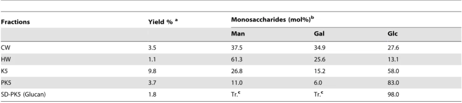
latter was used for the following extractions. The cold (CW) and hot (HW) water extracts were kept for the experiments on THP-1. The remaining residue III, after being extracted with 5% KOH solution, presented an extract of 9.8% yield (K5), which was submitted to freeze-thawing for several times (x 5), and centrifu-gation to separate the soluble polysaccharides from the non-soluble ones. This procedure gave rise to two fractions: the non-soluble one (SK5), that was studied previously and contained a glucogalactomannan [38]; and the insoluble one (PK5) that presented the higher amount of glucose (83%) in its composition (Table 1). The fraction PK5 was treated with Me2SO, which is
able to solubilize mainlyb-glucans, and after centrifugation, the Me2SO-soluble fraction (SD-PK5) was recovered showing
pre-dominantly glucose as monosaccharide component, suggesting the presence of a glucan polysaccharide (Table 1).
In order to characterize the glycosidic linkages of the isolated glucan, methylation analysis was performed, and the major derivative encountered was 2,4,6-Me3-Glcp (99%), which
con-firmed that this polymer is a linear glucan (1R3)-linked. NMR analysis 13C of the fractions PK5 (Figure 2A) and SD-PK5 (Figure 2B) showed that the purification procedure using Me2SO was efficient to remove the contaminant signals (d100.2;
99.8; 72.9; 71.8; 71.4; 70.0; 60.2 ppm). The six main signals in the
13
C-NMR spectra could be attributed to the carbons of the glucose ring by comparison with reported data [14,39,40]. The low-field C1 signal at 102.7 ppm indicates theb-configuration, and the low-field shift of the C3 signal at 85.9 ppm indicates the 3-O-substitution. The non-substituted C6 was confirmed by an inverted CH2 signal at 60.7 ppm on DEPT experiment (data
not shown). The other signals observed were attributed to C2 (d
72.6), C4 (d68.2), and C5 (d76.0). The presence of only six signals on the SD-PK513C-NMR spectrum is characteristic of an hexose homopolysaccharide, presenting a linear chain. These data shows that a linearb-(1R3)-D-glucan can be extracted fromC. militaris and purified by a Me2SO treatment.
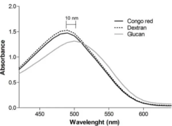
According to Ogawa et al. (1972) [41], polysaccharides existing in an ordered three-dimensional structure, generally triple helical conformation, form a complex with Congo red in dilute NaOH solutions. The complex is stabilized by strong hydrogen bonds and/or hydrophobic interactions between the polysaccharide and the dye molecule. The complex formation of polysaccharides with Congo red is commonly evaluated by means of the shift in the visible absorption maxima (lmax) of the Congo red spectrum [42].
The dye concentration was optimized to give a high and stable absorbance. The concentration of 80mM gave the best result and it was chosen for the analysis [29]. Theb-(1R3)-D-glucan was allowed to complex with Congo red in a 50 mM NaOH solution (Figure 3). Dextran was used as a random-coil control, and its absorbance was similar to the Congo red, which shows no complex formation. While theb-(1R3)-D-glucan sample showed a bath-ochromic shift (10 nm), which indicates that this polysaccharide displayed a triple helical structure.
Similar b-(1R3)-D-glucans are common in Ascomycetes and they have been isolated fromSaccharomyces cerevisiae (curdlan) [43], and from Basidiomycete fungi as Poria cocos (pachyman) [14], andLaetiporus sulphureus[39].
Therefore, the extracts were added to the THP-1 macrophages at 50mg/mL and the expression of pro-inflammatory genes (IL-1b, TNF-a, COX-2) was evaluated. For both incubation periods (3 h and 6 h) (Figure 4), the aqueous extracts significantly stimulated the production of IL-1b, TNF-a, and COX-2 mRNAs, while the alkaline extract did not show any stimulation, as well as the negative control (PBS).
The monosaccharide compositions of the aqueous extracts presented greater amounts of mannose and galactose, while the glucose levels were below 30%. On the other hand, the alkaline extract presented a higher amount of glucose and a lower content of the two other monosaccharides (Table 1). It was demonstrated that b-galactofuranosyl glycosides present immunomodulatory effects by the induction of phagocytic activity of macrophages and stimulation of the production of IL-1b, TNF-a, and IL-6 [44]. In our previous work [38] we isolated a glucogalactomannan from C. militaris, which contains 8.5% of b-galactofuranosyl in its structure. This monosaccharide may be the responsible for the
stimulation of the expression of pro-inflammatory genes observed for the cells treated with CW and HW. Moreover, the galactose content of K5 is the lowest (15.2%) in comparison with the aqueous extracts, which includes both galactofuranose and galactopyranose units (Table 1). It is known that galactopyranose form does not stimulate the macrophages, therefore its presence in the alkaline extract may be the majority.
All of the extracts were tested about a possible LPS contam-ination, and the LPS content of CW and K5 extracts was 13.3 ng/ mg and 4.0 ng/mg of dry extract, respectively. Considering that 50mg/mL of extract was added to the cells, less than 0.7 ng/mL of LPS was present as contaminant, which is too low to interfere in the results [32]. The HW extract was negative for LPS contamination.
In order to test the ability of the polysaccharide extracts to reduce the effects caused by LPS stimulation, 1mg/mL of LPS plus 50mg/mL of extracts (CW, HW, K5) were added to the cells concomitantly. After 3 h of incubation, the cells that received the
Table 1.Monosaccharide composition of the fractions obtained fromC. militaris.
Fractions Yield %a Monosaccharides (mol%)b
Man Gal Glc
CW 3.5 37.5 34.9 27.6
HW 1.1 61.3 25.6 13.1
K5 9.8 26.8 15.2 58.0
PK5 3.7 11.0 6.0 83.0
SD-PK5 (Glucan) 1.8 Tr.c Tr.c 98.0
Footnote:
aCalculated based on the initial dry mushroom weight.
bAlditol acetates obtained on successive hydrolysis, NaBH4reduction, and acetylation, followed by GC-MS analysis.
doi:10.1371/journal.pone.0110266.t001
Figure 2.13C-NMR spectra of fractions PK5 (A), and SD-PK5 (B), in Me2SO-d6at 70uC (chemical shifts are expressed indppm).
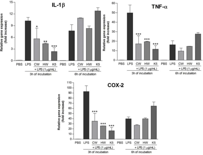
extracts+LPS, showed a significant lower expression of IL-1b, TNF-a, and COX-2 mRNAs than the cells that received LPS (Figure 5). These results suggest that theC. militaris polysaccha-ride extracts exhibit an anti-inflammatory effectin vitro.
It is important to notice that the monosaccharide composition of CW, HW, and K5 present differences and the glucose content is the highest (58.0%) in the alkaline extract, which can be the
responsible for the different stimulation of the THP-1 cells. The inhibitory effect of the LPS stimulation was more evident for the alkaline (K5) extract, inhibiting the expression of the three genes by over than 75%, as table 2 shows. The inhibition observed for CW and HW was statistically significant, although it was not stronger as for K5, which can be observed at table 2.
In a previous study, a glucogalactomannan was isolated from this mushroom and chemically characterized [38]. Although it was isolated from a soluble fraction of an alkaline extract, the aqueous extracts also showed a large amount of this heteropolysaccharide due to its high solubility in water. Nevertheless, the b-glucan, which is part of the fungal cell wall, requires a stronger method to be extracted, being present usually in higher concentrations in the alkaline extracts [45]. Therefore, we decided to proceed with an alkaline extraction, followed by a careful purification procedure with the aim to obtain high yields of the purifiedb-D-glucan and test its properties on the THP-1 cells.
The purified b-(1R3)-D-glucan (SD-PK5) was firstly tested about its cytotoxicity and none negative effect was observed on macrophages (Figure S1). Then, theb-(1R3)-D-glucan was added to the cells and incubated for 3 h and 6 h. There was no stimulation of pro-inflammatory gene expression (data not shown). However, when theb-(1R3)-D-glucan was added concomitantly with LPS (1mg/mL), and incubated for 3 h, it was observed an inhibition of the mRNA expression of 75.560.37% (IL-1b), 81.062.12% (TNF-a), and 81.660.56% (COX-2) at a concen-Figure 3. Absortion spectra of Congo Red (control), Congo Red
with dextran (random coil control), and Congo Red withb -D-Glucan fromC. militaris.
doi:10.1371/journal.pone.0110266.g003
Figure 4. mRNA expression level of pro-inflammatory genes, after treatment with polysaccharide extracts for 3 h and 6 h. Footnote:Negative control (PBS) and positive control (LPS; 1mg/mL). CW (cold water extract; 50mg/mL), HW (hot water extract; 50mg/mL), K5
(alkaline extract; 50mg/mL). Statistical analyses were performed by means of one-way analysis of variance (ANOVA) followed by Bonferronis’ test,
selected pairs. The results represent the mean6SD of duplicate cultures of three representative experiments. *p,0.05; **p,0.01; ***p,0.001 versus negative control.
tration of 50mg/mL (Figure 6). This inhibition was higher than the aqueous extracts, and similar to the alkaline extract (Table 2), which suggests that the isolated b-(1R3)-D-glucan is the most potent anti-inflammatory compound present in the polysaccharide extracts ofC. militaris.
Researchers have observed that pattern recognition receptors (PRRs) of immune cells, such as dectin-1, complement receptor 3 (CR3), scavenger receptors, lactosylceramide (LacCer), and
toll-like receptors (TLRs), recognize theb-glucans and initiate immune responses [15,16]. It was demonstrated that heteropolysaccharides from Polyporus umbellatus and Cordyceps militaris activated macrophages and dendritic cells, respectively, via TLR-4 signaling pathways [46,47]. Besides, it is well known that TLR-4 is the primary signal transducer for LPS, while TLR-2 is either a low affinity receptor for this bacterial endotoxin [48]. Taking this information into account, it is possible that theb-(1R3)-D-glucan Figure 5. mRNA expression level of pro-inflammatory genes, after treatment with LPS+polysaccharide extracts for 3 h and 6 h. Footnote:Negative control (PBS) and positive control (LPS; 1mg/mL). CW (cold water extract; 50mg/mL), HW (hot water extract; 50mg/mL), K5
(alkaline extract; 50mg/mL). Statistical analyses were performed by means of one-way analysis of variance (ANOVA) followed by Bonferronis’ test,
selected pairs. The results represent the mean6SD of duplicate cultures of three representative experiments. *p,0.05; **p,0.01; ***p,0.001 versus positive control.
doi:10.1371/journal.pone.0110266.g005
Table 2.Inhibitory effect of the extracts/glucan on the mRNA expression of pro-inflammatory genes by the THP-1 macrophages, after 3 h of incubation.
Extracts/Glucan (50mg/mL) Inhibitory effect on the mRNA expression (%)a
IL-1b TNF-a COX-2
CW 44.863.85 65.4611.95 62.2621.37
HW 57.160.23 60.860.42 72.060.22
K5 76.861.40 76.665.18 82.369.29
SD-PK5 (Glucan) 75.560.37 81.062.12 81.660.56
Footnote:
is competing with LPS for binding the TLR-4, thus inhibiting the LPS-stimulation of pro-inflammatory genes, after 3 h of incuba-tion. However, in the present study, the inhibitory effect of theb -(1R3)-D-glucan did not last for 6 h of incubation, which may be explained by the LPS-stimulation via TLR-2. This could be observed also for the K5 treatment (Figure 5), which is consistent because the main component of K5 is theb-(1R3)-D-glucan. The molecular mechanisms of LPS-induced macrophage activation and desensitization have been extensively investigated. These studies suggested the involvement of various kinases and that the cooperation of MyD88-dependent and MyD88-independent (TRIF-dependent) signaling is required [49]. The inhibition of any phase of these pathways leads to a negative effect of the LPS-stimulation, with no expression of pro-inflammatory genes [48,49]. Most of theb-(1R3),(1R6)-D-glucans isolated from mushrooms are able to activate macrophages and stimulate the production of pro-inflammatory cytokines in vitro [15], although these effects were not observed on this study. One possible explanation could be that the activation of macrophages requires a branched polysaccharide structure. However, the linear chain of the b -(1R3)-D-glucan of C. militaris showed to be more efficient in inhibiting inflammation of THP-1 cellsin vitro. Up to now, no conclusions were found in the literature pointing to the most potentb-D-glucan structure or which membrane receptor is the preferable to bind these molecules [40]. Further investigation is
required to explain the anti-inflammatory effect exhibited by the
b-(1R3)-D-glucanin vitro.
Evaluation of the antinociceptive and anti-inflammatory propertiesin vivo
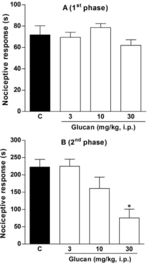
As theb-(1R3)-D-glucan of C. militarisshowed in vitro anti-inflammatory activity, we decided to confirm its biological action in vivo.It is well known that formalin administration causes local tissue injury to the paw of mice and has been used as a model for tonic pain and localized inflammatory pain [50]. This model is constituted by two distinct phases: the neurogenic pain and the inflammatory pain. The early phase (neurogenic pain) results from the direct irritating effect on nociceptors activating primary afferent fibers, and the late phase (inflammatory pain) is related to the release of pro-inflammatory mediators, such as bradykinin, prostaglandins, and cytokines [50,51].
The results depicted in Figures 7A and 7B show that intraperitoneal administration ofb-D-glucan did not inhibit the nociception on the early phase, although it significantly inhibited the nociceptive response on the inflammatory phase by 69611%, at a dose of 30 mg/kg, when compared to control group (C: 242.5625.4 s). Despite of this test is mainly considered a pain model, anti-inflammatory drugs can also be effective on the late phase. Indeed, it was demonstrated that classical non-steroidal anti-inflammatory drugs (NSAIDs), such as acetylsalicylic acid, Figure 6. mRNA expression level of pro-inflammatory genes, after treatment with LPS+b-(1R3)-D-glucan (Glucan) for 3 h and 6 h. Footnote:Negative control (PBS) and positive control (LPS; 1mg/mL).b-(1R3)-D-glucan was added at concentrations of 25, 50, and 100mg/mL. Statistical analyses were performed by means of one-way analysis of variance (ANOVA) followed by Bonferronis’ test, selected pairs. The results represent the mean6SD of duplicate cultures of three representative experiments. *p,0.05; **p,0.01; ***p,0.001 versus positive control.#p
,
0.05;##
indomethacin, paracetamol, and diclofenac can only attenuate the inflammatory phase of formalin-induced nociceptive response [50,52,53]. Thus, our results suggest thatb-D-glucan isolated from C. militaris was effective against the inflammatory pain, with a similar anti-inflammatory effect of NSAIDs.
Then, to confirm thein vivoanti-inflammatory activity ofb -D-glucan, we performed an acute inflammation model, the peritonitis induced by LPS in mice. Intraperitoneal administration of LPS initiates an inflammatory response with recruitment and activation of leukocytes (both mononuclear cells and neutrophils), and subsequent release of pro-inflammatory mediators [54]. In the present study, we observed that intraperitoneal administration of LPS (2mg/kg) produced an increase of leukocyte migration when compared to animals treated only with saline (S: 2.9960.446106 cells) (Figure 8A). However, the treatment of mice with b -D-glucan, at dose of 30 mg/kg, inhibited the migration of total leukocytes to the peritoneal cavity by 70615% compared to control group (C: 6.9060.626106 cells). Dexamethasone, a positive control of the test, also reduced the number of total
leukocytes by 100% (Figure 8A). Additionally, we also evaluated the MPO levels, an indirect marker of neutrophils, and it was observed thatb-D-glucan (30 mg/kg, i.p.) did not alter the MPO levels but dexamethasone reduced them by 9464% compared do control group (C: 0.2960.07 O.D./mL) (Figure 8B). These results confirm the anti-inflammatory activity ofb-D-glucan, reducing the migration of total leukocytes but indirectly indicating no alteration of neutrophils recruitment.
Some authors have showed that cordycepin, which is easily extracted with ethanol fromCordycepsspecies, is partly responsible for the anti-inflammatory effects of these mushrooms [23]. Although, our results demonstrated that other compounds, as theb-(1R3)-D-glucan can also exhibit such effect. The necessity of finding anti-inflammatory drugs with less side effects still remains unsolved [53], and the possibility of using a natural product as
Figure 7. Effect of b-(1R3)-D-glucan on neurogenic (A) and inflammatory phase (B) of nociception induced by formalin in mice. Footnote:Mice received vehicle (saline plus 5% Me2SO, 10 mL/ kg, i.p.) orb-(1R3)-D-glucan (3, 10 and 30 mg/kg, i.p.) 30 min before formalin administration. Statistical analyses were performed by means of one-way analysis of variance (ANOVA) followed by Newman–Keuls’ test. The results represent the mean6SEM of 10–12 animals. *p,0.05 versus control group.
doi:10.1371/journal.pone.0110266.g007
Figure 8. Effect of b-(1R3)-D-glucan on number of total leukocytes (A) and myeloperoxidase levels (B) induced by LPS in mice. Footnote:Mice received vehicle (saline plus 5% Me2SO, 10 mL/kg, i.p.), dexamethasone (DEXA, 0.5 mg/kg, i.p.), orb-(1R 3)-D-glucan (30 mg/kg, i.p.) 30 min before LPS administration. Statistical analyses were performed by means of one-way analysis of variance (ANOVA) followed by Newman–Keuls’ test. The results represent the mean6SEM of 6–8 animals.#
p,0.05 versus saline group; *p,0.05 versus control group.
inhibitor of inflammation open a new research field based on medicinal usage of mushrooms and their compounds.
Conclusions
The medicinal mushroomC. militariswas evaluated for its anti-inflammatory properties. The polysaccharide extracts from this mushroom exhibited different effects related to their monosac-charide composition. The alkaline extract, from which a linearb -(1R3)-D-glucan was isolated, showed the higher anti-inflamma-tory effect by the inhibition of IL-1b, TNF-a, and COX-2 expression. Theb-(1R3)-D-glucan showed the same effect as well, indicating that this polymer is the most potent anti-inflammatory compound present in the polysaccharide extracts ofC. militaris.In addition, we also observed that the isolated b-(1R3)-D-glucan presented antinociceptive and anti-inflammatory activities against formalin-induced nociception and LPS-induced peritonitis in mice.
Supporting Information
Figure S1 Viability of THP-1 macrophages after
incu-bation with the extracts (CW, HW, K5) or theb-D-glucan
(Glucan), for 24 h and 48 h. Footnote:The cells were treated with 10, 50 or 250mg/mL of extracts or glucan. Saponin was added to C+to lysate the cells. C- received only PBS and it was set as 1.0 (100% of viable cells).
(TIF)
Acknowledgments
The authors would like to thank Dr. J. M. Sung of Kangwon National University (Chuncheon, Korea), for the fungal material.
Author Contributions
Conceived and designed the experiments: FRS CHB DGB APSF GLS MI LVG. Performed the experiments: FRS CHB DGB APSF. Analyzed the data: FRS CHB DGB APSF GLS MI LVG. Contributed reagents/ materials/analysis tools: FRS CHB DGB APSF GLS MI LVG. Contributed to the writing of the manuscript: FRS CHB DGB APSF GLS MI LVG.
References
1. Daba AS, Ezeronye OU (2003) Anti-cancer effect of polysaccharides isolated from higher basidiomycetes mushrooms. Afr J Biotechnol 2(12): 672–678. 2. Jo WL, Choi YJ, Kim HJ, Lee JY, Nam BH, et al. (2010) The anti-inflammatory
effects of water extract from Cordyceps militaris in murine macrophage. Mycobiology 38(1): 46–51.
3. Lindequist U, Niedermeyer THJ, Ju¨lich WT (2005) The pharmacological potential of mushrooms. Evid Based Complement Alternat Med 2(3): 285–299. 4. Chanput W, Reitsma M, Kleinjans L, Mes JJ, Savelkoul HF, et al. (2012)b -Glucans are involved in immune-modulation of THP-1 macrophages. Mol Nutr Food Res 56(5): 822–33, doi: 10.1002/mnfr.201100715.
5. Lin JG, Fan MJ, Tang NY, Yang JS, Hsia TC, et al. (2012) An extract of
Agaricus blazei Murill administered orally promotes immune responses in murine leukemia BALB/c mice in vivo. Integr Cancer Ther 11: 29, doi: 10.1177/1534735411400314.
6. Smiderle FR, Ruthes AC, van Arkel J, Chanput W, Iacomini M, et al. (2011) Polysaccharides from Agaricus bisporus and Agaricus brasiliensis show similarities in their structures and their immunomodulatory effects on human monocytic THP-1 cells. BMC Complement Altern Med 11: 58.
7. Chen W, Zhang W, Shen W, Wang K (2010) Effects of the acid polysaccharide fraction isolated from a cultivatedCordyceps sinensison macrophagesin vitro. Cell Immunol 262: 69–74.
8. Das SK, Masuda M, Sakurai A, Sakakibara M (2010) Medicinal uses of the mushroomCordyceps militaris: Current state and prospects. Fitoterapia 81: 961– 968.
9. Wang M, Meng XY, Yang RL, Qin T, Wang XY, et al. (2012)Cordyceps militarispolysaccharides can enhance the immunity and antioxidation activity in immunesuppressed mice. Carbohydr Polym 89: 461–466.
10. Yu R, Yin Y, Yang W, Ma W, Yang L, et al. (2009) Structural elucidation and biological activity of a novel polysaccharide by alkaline extraction from cultured
Cordyceps militaris. Carbohydr Polym 75: 166–171.
11. Ohta Y, Lee JB, Hayashi K, Fujita A (2007) In vivo anti-influenza virus activity of an immunomodulatory acidic polysaccharide isolated fromCordyceps militaris
grown on germinated soybeans. J Agric Food Chem 55: 10194–10199. 12. Yan JK, Wang WQ, Wu JY (2014) Recent advances inCordyceps sinensis
polysaccharides: Mycelial fermentation, isolation, structure, and bioactivities: A review. J Funct Foods 6: 33–47.
13. Holliday J, Cleaver M (2008) Medicinal value of the caterpillar fungi species of the genusCordyceps(Fr.) Link (Ascomycetes). A review. Int J Med Mushrooms 10(3): 219–134.
14. Synytsya A, Nova´k M (2013) Structural diversity of fungal glucans. Review. Carbohydr Polym 92: 792–809.
15. Chen J, Seviour R (2007) Medicinal importance of fungal b-(1R3),(1R
6)-glucans. Mycol Res 111: 635–652.
16. Moradali MF, Mostafavi H, Ghods S, Hedjaroude GA (2007) Immunomodu-lating and anticancer agents in the realm of macromycetes fungi (macrofungi). Int Immunopharmacol 7: 701–724.
17. Brown GD, Gordon S (2005) Immune recognition of fungalb-glucans. Cell Microbiol 7(4): 471–479.
18. Smiderle FR, Olsen LM, Carbonero ER, Baggio CH, Freitas CS, et al. (2008) Anti-inflammatory and analgesic properties in a rodent model of a (1R3),(1R 6)-linkedb-glucan isolated fromPleurotus pulmonarius. Eur J Pharmacol 597: 86– 91.
19. Komura DL, Carbonero ER, Gracher AHP, Baggio CH, Freitas CS, et al. (2010) Structure ofAgaricusspp. fucogalactans and their anti-inflammatory and antinociceptive properties. Bioresour Technol 101: 6192–6199.
20. Dinarello CA (2000) Proinflammatory cytokines. Chest 118: 503–508. 21. Wu Y, Antony S, Meitzler JL, Doroshow JH (2013) Molecular mechanisms
underlying chronic inflammation-associated cancers. Cancer Lett, 345(2): 164– 173.
22. Li Y, Zhang J, Ma H (2013) Chronic inflammation and gallbladder cancer. Cancer Lett, 345(2): 242–248.
23. Won SY, Park EH (2005) Anti-inflammatory and related pharmacological activities of cultured mycelia and fruiting bodies of Cordyceps militaris. J Ethnopharmacol 96: 555–561.
24. Gorin PAJ, Iacomini M (1984) Polysaccharides of the lichensCetraria islandica
andRamalina usnea. Carbohydr Res 128: 119–132.
25. Sassaki GL, Souza LM, Serrato RV, Cipriani TR, Gorin PAJ, et al. (2008) Application of acetates derivatives for gas chromatography-mass spectrometry: Novel approaches on carbohydrates, lipids and amino acids analysis. J Chromatogr A 1208: 215–222.
26. Sassaki GL, Gorin PAJ, Souza LM, Czelusniak PA, Iacomini M (2005) Rapid synthesis of partiallyO-methylated alditol acetate standards for GC-MS: some relative activities of hydroxyl groups of methyl glycopyranosides on Purdie methylation. Carbohydr Res 340: 731–739.
27. Haworth WN (1915) A new method of preparing alkylated sugars. J Chem Soc 107: 8–16.
28. Ciucanu I, Kerek F (1984) A simple and rapid method for the permethylation of carbohydrates. Carbohydr Res 131: 209–217.
29. Palacios I, Garcı´a-Lafuente A, Guillamo´n E, Villares A (2012) Novel isolation of water-soluble polysaccharides from the fruiting bodies of Pleurotus ostreatus
mushrooms. Carbohydr Res 358: 72–77.
30. Santana-Filho AP, Noleto GR, Gorin PAJ, Souza LM, Iacomini M, et al. (2012) GC–MS detection and quantification of lipopolysaccharides in polysaccharides through 3-O-acetyl fatty acid methyl esters. Carbohydr Polym 87: 2730–2734. 31. Reilly TP, Bellevue FH, Woster PM, Svensson CK (1998) Comparison of theIn
vitro cytotoxicity of hydroxylamine metabolites of sulfamethoxazole and dapsone. Biochem Pharmacol 55: 803–810.
32. Chanput W, Mes J, Vreeburg RAM, Savelkoul HFJ, Wichers HJ (2010) Transcription profiles of LPS-stimulated THP-1 monocytes and macrophages: a tool to study inflammation modulating effects of food-derived compounds. Food Funct 1: 254–261.
33. Livak KJ, Schmittgen TD (2001) Analysis of relative gene expression data using real-time quantitative PCR and the 22DDC
Tmethod. Methods 25: 402–408. 34. Zimmermann M (1983) Ethical guidelines for investigations of experimental
pain in conscious animals. Pain 16: 109–110.
35. Hunskaar S, Fasmer OB, Hole K (1985) Formalin test in mice, a useful technique for evaluating mild analgesics. J Neurosci Methods 14: 69–76. 36. Borges FR, Silva MD, Co´rdova MM, Schambach TR, Pizzolatti MG, et al.
(2014) Anti-inflammatory action of hydroalcoholic extract, dichloromethane fraction and steroid a-spinasterol from Polygala sabulosa in LPS-induced peritonitis in mice. J Ethnopharmacol 151(1): 144–50.
38. Smiderle FR, Sassaki GL, van Griensven LJLD, Iacomini M (2013) Isolation and chemical characterization of a glucogalactomannan of the medicinal mushroomCordyceps militaris. Carbohydr Polym 97: 74–80.
39. Alquini G, Carbonero ER, Rosado FR, Cosentino C, Iacomini M (2004) Polysaccharides from the fruit bodies of the basidiomyceteLaetiporus sulphureus
(Bull.: Fr.) Murr. FEMS Microbiol Lett 230: 47–52.
40. Lehtovaara BC, Gu FX (2011) Pharmacological structural, and drug delivery properties and applications of 1,3-b-glucans. J Agric Food Chem 59: 6813– 6828.
41. Ogawa K, Tsurugi J, Watanabe T (1972) Complex of gel-formingb -1,3-D-glucan with congored in alkaline solution. Chem Lett 689–692.
42. Ogawa K, Dohmaru T, Yui T (1994) Dependence of complex formation of (1R3)-b-D-Glucan with Congo red on temperature in alkaline solutions. Biosci, Biotechnol Biochem 58: 1870–1872.
43. Ramberg JE, Nelson ED, Sinnott RA (2010) Immunomodulatory dietary polysaccharides: a systematic review of the literature. Nutr J 9(54): 1–22. 44. Sassaki GL, Rattmann YD, Santana-Filho AP, Riter DS, Iagher F, et al. (2013)
Galactofuranosyl glycosides: Immunomodulatory effects on macrophages and
in vivoenhancement of lethality on sepsis. Chem Biol Interact 205: 29–37. 45. Netea MG, Brown GD, Kullberg BJ, Gow NAR (2008) An integrated model of
the recognition ofCandida albicans by the innate immune system. N Rev Microbiol 6: 67–78.
46. Li X, Xu W (2011) TLR4-mediated activation of macrophages by the polysaccharide fraction fromPolyporus umbellatus (pers.) Fries. J Ethnophar-macol 135: 1–6.
47. Kim HS, Kim JY, Kang JS, Kim HM, Kim YO, et al. (2010) Cordlan polysaccharide isolated from mushroomCordyceps militarisinduces dendritic cell maturation through toll-like receptor 4 signalings. Food Chem Toxicol 48: 1926–1933.
48. Zhang G, Ghosh S (2001) Toll-like receptor–mediated NF-kB activation: a phylogenetically conserved paradigm in innate immunity. J Clin Investig 107: 13–19.
49. Fujihara M, Muroi M, Tanamoto K, Suzuki T, Azuma H, et al. (2003) Molecular mechanisms of macrophage activation and deactivation by lipopolysaccharide: roles of the receptor complex. Pharmacol Therap 100: 171–194.
50. Hunskaar S, Hole K (1987) The formalin test in mice: dissociation between inflammatory and non-inflammatory pain. Pain 30: 103–114.
51. Tjølsen A, Berge OG, Hunskaar S, Rosland JH, Hole K (1992) The formalin test: an evaluation of the method. Pain 51: 5–17.
52. Malmberg AB, Yaksh TL (1995) Cyclooxygenase inhibition and the spinal release of prostaglandin E2 and amino acids evoked by paw formalin injection: A microdialysis study in unanesthetized rats. J Neurosci, 15: 2768–2776. 53. Santos ARS, Vedana EMA, De Freitas GAG (1998) Antinociceptive effect of
meloxicam, in neurogenic and inflammatory nociceptive models in mice. Inflamm Res, 47: 302–307.