Iranian Journal of Basic Medical Sciences
ijbms.mums.ac.ir
The preventive and therapeutic roles of phytoestrogen
α
-Zearalanol on osteoporetic rats due to ovariectomization
Shaochun Li
1, 2, Weiwei Zhang
1, Fei Duan
1, Weihua Liu
1, Xiaofang Sun
1, Xuefeng Pan
1, 3*
1 School of Basic Medical Sciences, Hebei University, Baoding 071002, China 2 Xiyuan Hospital, China Academy of Chinese Medical Sciences, Beijing 100091, China 3 School of Life Science, Beijing Institute of Technology, Beijing 10081, China
A R T I C L E I N F O A B S T R A C T
Article type:
Original article Objective(s): The aim of this study was to observe the influence of phytoestrogen α-Zearalanol on
ovariectomy-induced postmenopausal osteoporosis in rats.
Materials and Methods:40 SD female rats were randomly divided into four groups: Sham group, OVX
group ovariectomized and fed estrogen , α-Zearalanol group ovariectomized and fed α-Zearalanol) and untreated group ovariectomized . Three weeks later after surgery, α-Zearalanol and estradiol
valerate were administered by oral gavage for weeks to the α-Zearalanol group and the OVX group, respectively. In contrast, the sham and untreated controls were treated with distilled water in a daily basis. After the treatments, uterus histomorphometry, bone mechanical strength, bone histomorphometry, bone mineral density (BMD) of femur, and serum biochemical indicators, such as serum E2, CT and PTH, as well as the levels of TNF and IL-1 were examined.
Results: The BMD was overall declined rigorously in the OVX rats, and that could be mitigated through feeding on either estrogen or α-Zearalanol. Estrogen or α-Zearalanol was found to decrease the levels of serum ALP and BGP in OVX rats, while, α-Zearalanol was found to increase the levels of serum E2
and CT, the thickness of the endometrium, and decrease the levels of PTH, TNF and IL-1 in serum in OVX rats. Feeding the OVX rats on α-Zearalanol improved the bone histomorphometric parameters impaired due to estrogen deficiency and enhanced the bone mechanical properties in the ovariectomized rats.
Conclusion:α-Zearalanol treated rats reduced the resorption of bone, and showed a preventive and therapeutic effect of α-Zearalanol on postmenopausal osteoporosis.
Article history: Received: Oct 25, 2015 Accepted: Jun 30, 2016
Keywords:
Bone histomorphometric Bone mechanical -properties
Bone mineral density Osteoporosis Ovariectomized rats
α-Zearalanol
►
Please cite this article as:Li SH, Zhang W, Duan F, Liu W, Sun X, Pan X. The preventive and therapeutic roles of phytoestrogen α-Zearalanol on osteoporetic rats due to ovariectomization. Iran J Basic Med Sci 2016; 19:1216-1221.
Introduction
Osteoporosis is often characterized by reduced bone mass and structural deterioration, and increased bone fragility and susceptibility of bone fracture, which is believed to be increasing as the extension of the average life expectancy in the worldwide, and which thereby turns to be a main cause of increased morbidity and mortality in human beings (1).
The common treatment for postmenopausal osteoporo- sis is hormone replacement therapy (HRT), which reduces the rate of postmenopausal bone loss effectively (2), while risking the occurrence of endometrial or breast cancer as side effects. It is therefore needed for finding new substitutes owning estrogenic effects in osteoporosis treatment but showing less undesirable side effects on endometrium or breast.
Recently, plant-derived phytoestrogen have been reported to be capable of reducing the side effects of estrogen on the endometrium or breast, while retaining thebenefits in osteoporosisprevention (3). α-Zearalanol
α-ZAL) is a reductive product of the Gibberella zeae
metabolite zearalenone, which owns estrogenic
property, and can potentially be useful in the treatments of estrogen-related human disorders like postmenopausal osteoporosis (4).
Several investigations have demonstrated that α-ZAL
retards the development of atherosclerosis with fewer side effects on the growth of mammary gland and uterine (5). However, the mechanism underlined has not been fully elucidated. In this work, we have investigated the
effects of α-ZAL on the estrogen-deficiency caused bone
loss in the ovariectomized (OVX) rats, and compared the similarities and differences with the effects of estrogen physiologically and biochemically.
Materials and Methods
Experimental animals and preparations of
experimental materials
All animal experiments were undertaken with the approval of Animal Ethics Committee of Hebei
*Corresponding author: Xuefeng Pan. School of Basic Medical Sciences, Hebei University, Baoding 071002, China, School of Life Science, Beijing Institute of
University. Forty adult virgin female (6-month-old) Sprague-Dawley rats were obtained from Laboratory Animal Center of Hebei Province. The animal house is
maintained at temperature 22±2 ᵒC with relative
humidity 50±15% and 12 hr dark/light cycle throughout the study. Rats had free access to food and water ad libitum.
The rats were randomly divided into four groups, each group comprised of ten rats. After 1 week acclimation, three groups were ovariectomized bilaterally and one group underwent a sham operation as described before (6) . After the surgery, the rats were housed individually for one week, and then housed in a group of five each for subsequent treatments: Drug administrations were performed three weeks later after the surgery. Both sham group and an untreated OVX group were used as controls, and were administered orally with appropriate amount of distilled water. The other groups were treated orally with either estradiol valerate (Bayer Inc., Germany) or
α-ZAL (Sigma Inc., Germany) by 10 mg/kg BW per day.
After 12 weeks treatments, blood samples were collected from abdominal aorta, and blood serum was prepared and preserved at -20 ᵒC till further analysis. Femur and tibia of all rats were excised and their soft tissues were removed. The femurs were parceled with saline gauze and preserved at -20 ᵒC till further analysis. And the tibias were preserved in 70% ethanol till further analysis. Uterus were isolated, and the absolute weight of uterine tissue was recorded and normalized with body weight (relative weight of uterus, i.e., weight of uterus per 1 kg of BW) of animals, then fixed in 10% phosphate buffered formalin for further analysis.
Histomorphometry of uterus
The uterus samples fixed by using 10% phosphate buffered formalin were further processed with paraffin-embedding and sectioned by 5 µM. The biopsies were stained with hematoxylin and eosin for histological examination. The stained sections were observed under a microscope and subjected to morphometric analysis using the eye piece scale and the stage micrometer.
Biochemical analysis on bone markers
Serum alkaline phosphatase (ALP), serum calcium (Ca) and phosphorus (P) were quantified by using an automatic biochemical analyzer (OLYMPUS, Japan).
Serum estradiol, calcitonin (CT), bone glaprotein
(BGP), TNFα and IL-1
Radioimmunoassay (RIA) was carried out by using a kit (Huaying biotechnology institute, Beijing, China) for estimating the circulating levels of estradiol, CT, BGP,
TNFα and )L-1.
Measurements of the bone density
Bone density of the left femurs was measured using dual-energy X-ray absorptiometry (Lunar, USA). During which, the femurs were carefully brought to the room temperature in a saline bath before measurements.
Measurements of the bone mechanical strength
Mechanical strength of right femurs was measured by three point bending test (TPBT) (7) using mechanical testing machine. Before the testing, the femurs were taken out from freezer and thawed at room temperature for 12 hrs. During the TPBT testing, the femurs were positioned on 2 supports 15 mm apart. Load was applied to midshaft, with a loading speed of 15 mm/min. The bone was compressed in the middle of the femur shaft until fracture occurred. During testing, data on displacement and loading were displayed and recorded by using a computer connecting to the interface of the machine. Indexes of structural mechanics of bone, including maximal load, maximal deflection, elastic load, and elastic deformation were observed on the computer. The inner and outer diameters of cross section were measured with slide gauge, and the indexes of mechanics of materials (maximal stress and maximal strain) of bone were calculated.
The histomorphometry of bone
The proximal tibias of the left limb were fixed with 10% formaldehyde for 18 hrs, followed by decalcification with 15% EDTA, and then dehydrated in 95% (v/v) ethanol and embedded in paraffin. 7 μm-thick sections were cut from the proximal tibias and stained with Haematoxylum and eosin. The sections were then examined under light microscopy, and analyzed by using an image analyzing system (NIS-Elements D 3.1 image analyzing system, Nikon corp., Japan). The following indexes were recorded (8): 1) Total trabecular bone volume (BV/TV; %) of mineralized and unmineralized, which are presented as a percentage of the total medullar volume. 2) Mean trabecular plate thickness (MTPT; µM), the mean thickness of trabecular plates in ten visual fields. 3) Mean trabecular plate separation (MTPS; µM), an index of the mean distance between trabecular plates in ten visual fields. 4) Mean trabecular plate density (MTPD; /mm), the mean number of trabeculaer plates in ten visual fields.
Statistical analysis of the data
All data obtained in this study were presented as mean±SD. The means between groups were compared using analysis of variance (ANOVA). Two group means
were tested using Fisher’s protected least-significant
difference (PLSD) post hoc tests. For all tests, differences were considered significant when P<0.05.
Results
The effects of α-ZAL on uterus index and
histomorphometry
The final body weights of the rats in the treated groups and the control groups were unable to find differences of significance (Table 1). However, the final uterine indexes were found to be decreased in all the rats in the OVX group compared to the rats in the sham group. Through treatments of estradiol valerate and
Table 1.The effect of estradiol and α-ZAL on uterus index and endometrial thickness (ET) in ovariectomized rats
Group Uterus index Body weight (g) ET (μM)
Sham 2.54±0.919 a 308.40±38.52 538.12±122.19 a
OVX 0.35±0.061 b 312.33±37.46 235.94±47.47 b
E2 0.84±0.029 a b 301.00±17.34 444.58±70.40 a b α-ZAL 0.61±0.305 a b c 312.74±32.36 351.00±28.63 a b c
Values are means±SD and values with different letters are significantly different P<0.05). Sham, sham-operated group; OVX, ovariectomied group; E2, ovariectomied fed estradiol valerate; α-ZAL, ovariectomied fed α-ZAL; ET, Endometrial thickness
however, the uterine indexes of the α-ZAL treated rats were found to be lower than those rats treated by using E2 (Table1).
The endometrial thickness (ET) were measured at the time of death, and the efficacy of OVX, E2and α-ZAL
replacement were assessed and characterized to see the uterine response to these interventions in the groups. Compared with sham group, endometrial atrophy was observed in OVX rats, but those rats
treated with estradiol valerate and α-ZAL experienced
increases in endometrial thickness. Similar to these, the degrees of the endometrium in the α-ZAL treated rats were smaller than those rats treated with E2 (Table 1).
The effects of α-ZAL on bone biochemical markers
After the treatments for 12 weeks by feeding α-ZAL, the levels of Ca and P in the serum of the rats were measured, however no significant differences between groups were found (Table 2), while the levels of serum ALP and BGP were found to be higher significantly in the OVX rats compared to the rats in the sham group (Table2), indicating the treatments of estradiol valerate to the OVX rats elevated the levels of serum ALP and BGP, respectively. Interestingly, a similar effect of α-ZAL and estrogen on the levels of serum ALP and BGP in the
E2group and α-ZAL group was observed (Table2).
The effects of α-ZAL on serum E2, CT and PTH
As expected that OVX rats did show a significant
reduction in serum E2 compared to the rats in the sham
group, we then observed that E2 could be significantly
enhanced through feeding the OVX rats on either E2or α
-ZAL in a similar way. Further, the serum level of CT was also low in the OVX rats compare to the sham rats, but it could be enhanced by feeding on either E2 or α-ZAL. In
contrast, the level of PTH in serum were found to be higher in OVX rats than it was in the rats in the sham group, while it can be reduced by feeding on E2 or α-ZAL
(Table 3).
The effects of α-ZAL on serum IL-1 and TNFα
The mean concentrations of the serum IL- and TNFα
were high in the OVX rats than in the sham rats, indicating that concentrations of serum IL- and TNFα were significantly increased by feeding on estradiol valerate (Table 4). By contrast, rats fed on α-ZAL were also found to decrease the concentration of the serum
TNFα and IL-1 (Table 4), suggesting that α-zearalanol worked similarly with the estradiol valerate by regulating the production of cytokine.
The effects of α-ZAL on bone density
The bone of OVX rats was found less condensed than those rats in the sham group, while it could be increased through the treatments of estradiol valerate and α-ZAL ( Table 5), indicating that both estradiol valerate and α -ZAL increased the bone densities significantly.
Table 2.The effects of ovariectomy, E2and α-ZAL treatments on serum Ca, P, ALP and BGP levels in rats
Group Ca P ALP BGP
Sham 2.451±0.326 1.889±0.346 45.92±4.83 a 3.233±0.543*
OVX 2.427±0.563 1.721±0.414 91.55±11.74 b 4.740±0.327a
E2 2.464±0.624 1.855±0.260 68.59±6.97 a b 3.843±0.220 a b α-ZAL 2.405±0.568 1.863±0.262 66.15±11.90 a b 3.642±0.379 a b
Values are means±SD and values with different letters are significantly different P<0.05). Sham, sham-operated group; OVX, ovariectomied group; E2, ovariectomied fed estradiol valerate; α-ZAL, ovariectomied fed α-ZAL; ALP, alkaline phosphatase; BGP, Osteocalcin
Table 3.The effects of ovariectomy, E and α-ZAL treatments on serum E2, CT, PTH in rats
Group E2 CT PTH
Sham 76.93±12.03 a 312.85±28.99 a 56.32±8.22 a
OVX 43.77±6.95 b 229.68±32.78 b 68.05±6.14 b
E2 78.26±10.14 a 320.92±42.80 a 55.66±6.56 a α-ZAL 72.55±10.13 a 311.07±44.48 a 54.27±11.06 b
Values are means±SD and values with different letters are significantly different (P<0.05). Sham, sham-operated group; OVX, ovariectomied group;
Table 4.The effects of ovariectomy, E2and α-ZAL treatments on
serum IL- and TNFα in rats
Group IL-1 TNFα
Sham 0.194±0.048 a 0.308±0.059 a
OVX 0.301±0.030 b 0.471±0.043 b
E2 0.203±0.030 a 0.306±0.054 a α-ZAL 0.210±0.035 b 0.348±0.087 a
Values are means±SD and values with different letters are shown, and
they are significantly different P<0.05). Sham, sham-operated group; OVX, ovariectomied group; E2, ovariectomied fed estradiol valerate; α-ZAL, ovariectomied fed α-ZAL
Table 5.The effects of ovariectomy, E2and α-ZAL treatments on left
femur density in rats
Group BMD(g/cm2)
Sham 0.188±0.020a
OVX 0.110±0.030b E2 0.176±0.010a α-ZAL 0.164±0.037a
Values are means±SD and values with different letters are significantly
different (P<0.05). Sham, sham-operated group; OVX, ovariectomied group; E2, ovariectomied fed estradiol valerate; α-ZAL, ovariectomied
fed α-ZAL. BMD, Bone mineral density
The effects of α-ZAL on bone mechanical strength
The mean maximal load, maximal deflection, elastic load, and elastic deformation were found to be reduced significantly in the OVX rats (Table 6). Treatments with either estradiol valerate or α-ZAL were found to be capable of improving the maximal load, maximal deflection, elastic load, and elastic deformation of femurs without big differences (Table 6). Similarly, the maximal stress and maximal strain in the OVX rats were found to be elevated significantly by the
treatments of estradiol valerate and α-ZAL, compared
to those of rats in the sham group (Table 6).
The effects of α-ZAL on bone histomorphometry
parameters
Total trabecular bone volume was found to be significantly decreased in the OVX rats compare to those in the sham group, indicating that estradiol
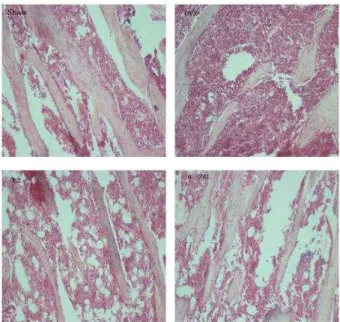
Figure 1. Histomorphologies of bone trabeculae by ovariectomy and by treatments with estrogen and α-ZAL (Haematoxylin-Eosin Staining, 100×). In sham group, the cancellous bone showed intervening trabecular bone with connectivity of the trabecular elements. While in ovariectomized rats, there was significant thinning and disconnection
of trabeculae. When the rats were fed estrogen and α-ZAL for three months, the trabeculation in the cancellous bone was significantly higher than that of ovariectomized rats
valerate and α-ZAL could improve the total volume of
trabecular bone. Similarly, the mean thickness of the trabecular plate (MTPT) was decreased significantly in OVX rats, while it could be increased by the treatments
of estradiol valerate and α-ZAL. The mean trabecular
plate separation (MTPS) was found to be enlarged in OVX rats, but it became narrowed in the rats of E2 group and α-ZAL group by the treatments of estradiol
valerate and α-ZAL. The mean trabecular plate density
(MTPD) was seen to be decreased in OVX rats, but it was enhanced in the rats of E2 and α-ZAL groups by the
treatments of estradiol valerate and α-ZAL, respectively
(Table 7 and Figure 1).
Table 6.The effects of ovariectomy, E2and α-ZAL treatments on mechanical properties of left femur
Parameters Sham OVX E2 α-ZAL
maximal load 84.4±7.59 a 65.27±8.60 b 83.37±4.48 a 82.71±6.55 a
maximal deflection 1.04±0.13 a 0.71±0.12 b 1.04±0.10 a 0.95±0.11 a
elastic load 63.10±5.84 a 44.92±6.91 b 64.28±4.88 a 58.42±7.62 a
elastic deformation 0.82±0.12 a 0.39±0.21 b 0.90±0.11 a 0.74±0.11 a
Maximal stress 1339.26±253.86 a 817.07±241.34 b 1213.04±313.09 a 1338.32±479.50 a
maximal strain 0.19±0.02 a 0.15±0.01 b 0.18±0.01 a 0.18±0.01 a Values are means±SD and values with different letters are significantly different P<0.05). Sham, sham-operated group; OVX, ovariectomied group; E2, ovariectomied fed estradiol valerate; α-ZAL, ovariectomied fed α-ZAL
Table 7.The effects of ovariectomy, E2and α-ZAL treatments on mechanical properties of left femur
Group n TBV/TTV (%) MTPT(µM) MTPD(/mm2) MTPS(µM)
Sham 10 44.13±6.61a 81.31±9.65 a 2.35±0.56 a 545.63±10.23 a
OVX 9 27.09±6.98b 58.37±16.09 b 1.02±0.33 b 965.52±35.34 b
E2 8 40.13±4.63a 76.56±11.20 a 1.96±0.97 a 873.49±28.54 a α-ZAL 9 35.43±4.35a 66.27±15.81 a 1.67±0.18 a 719.37±43.66 a
Values are means±SD and values with different letters are significantly different P<0.05). Sham, sham-operated group; OVX, ovariectomied grou E2,
Discussion
Deficiency in estrogen due to post-menopause is responsible for postmenopausal osteoporosis, for which hormone replacement therapy has been designed and utilized as alternative treatments (7, 8). However, the therapy shows serious side effects on the uterus and also risk breast cancer occurrence when performed for a long-term.
In this study, we have investigated the possible roles of α-zearalanol, a novel phytoestrogen, in the prevention of bone loss in the experimentally established rat models. Our results showed that α-zearalanol was capable of improving the bone density, the bone biomechanics properties and the bone histomorphometry parameters with fewer side effects to uterus, while giving less effect on uterus. By contrast, treatments using estradiol increased uterus index and endometrium thickness significantly.
In clinic, changes in bone density is often seen as one of the main indicators of osteoporosis, decreased bone density increases the incidence of bone fracture (9). Therefore, roles of increasing bone density by α -zearalanol in OVX rats was meaningful to demonstrate its potential in bone protection, which in this regard, was found to be similar to the roles of estradiol valerate. These can further be supported by the bone-sparing
effects of α-ZAL as indicated by the improvements
of bone mechanical strength test and bone
histomorphometry parameters.
Biomechanics is a straight forward test for evaluating the risk of bone fractures due to osteoporosis (10). We found the biomechanics properties were indeed decreased in OVX rats when compare with the rats in the sham group. Interestingly the bone losses can be prevented by treating the rats with estradiol valerate or α-zearalanol. Consistence with this, the improvements on bone histomorphmetry parameters also indicated the bone protective effects of
α-zearalanol on OVX rats.
The positive effect of α-zearalanol on bone may be
associated with its role of inhibiting the bone turnover, which behaves somehow similar to estrogen. Indeed, the osteoporotic rats when treated with α-zearalanol are clearly demonstrating an increase in bone turnover as associated with elevated rates of bone formation and bone resorption. It has been reported that estrogen blunts the bone turnover of osteoporosis and decreases the markers of bone formation and resorption (11).
Similarly α-zearalanol regulates the bone metabolic
processes like estrogen (12). We found that, as indicated by the variations of serum biochemical
markers, α-zearalanol decreased the alkaline
phosphatase and osteocalcin in serum, which served as
markers of bone formation, but didn’t affect the blood
calcium and phosphorus, suggesting that α-zearalanol
behaves similarly to the estrogen that inhibits the bone formation and reduces the level of bone turnover. It was known that hormones, such as PTH (13-15), CT (16, 17), are also involved in the regulations of bone
formation and bone resorption. PTH and CT up-regulate the activity of the bone-resorbig cells by regulating the blood calcium. Interestingly, in this study, we found that estradiol valerate showed an up-regulating effect on CT and a down-up-regulating effect on
PT(, while α-zearalanol can only up-regulate the level
of serum CT, but didn’t affect the level of serum PTH,
suggesting that α-zearalanol may regulate bone
turnover through regulation to endocrinium.
It has been argued that bone-protective effects of estrogen are due partially to its capacities of suppressing the productions of osteoclastogenic cytokines from osteoblasts, bone marrow stromal cells and T-cells (18), which otherwise increase the number of osteoclasts. The decrease of the level of estrogen at menopause is often associated with increases of interleukins, such as IL-1, IL-6, IL-7 and tumor necrosis
factor α TNFα (19-22). Estrogen replacement therapy,
predominantly exert bone-protective effects through regulating the expression of cytokine such as IL-1, IL-6,
TNFα. Consistence with this, in this research, our data
showed that serum IL- and TNFα levels were
significantly elevated in OVX rats and all the pharmacological interventions including both estradiol valerate and α-zearalanol treatments significantly decreased the concentration of TNFα in serum, while α
-zearalanol can only increase the level of serum TNFα ,
but cannot increase the IL-1, suggesting that
regulations on the expression of cytokine by α
-zearalanol may indeed be involved in its bone-protective effects.
Conclusion
Our study clearly showed that α-zearalanol can effectively abate bone loss due to ovariectomy in a
manner similar to that of estrogen. However, α
-zearalanol shows anti-atherosclerotic activity that presumably through binding to estrogen receptors (23), and an antioxidant ion effect through inhibiting the homocysteine-induced endothelin-1 expression and oxidative stress (24, 25). Bsed upon this idea, we
conclude that α-zearalanol can be a new substitute of
estrogen in preventing postmenopausal osteoporosis. However, further investigation on any possible adverse actions, and more detailed mechanism of α-zearalanol actions should also be addressed.
Acknowledgment
This work was jointly supported by grants from Medical Commission of Hebei Province, China (20130048), Population and Family Planning Committee of Hebei Province, China (2010-A03), Advance Research Foundation of Hebei University (2007JY04) and Youth Foundation of Hebei University (2010Q37).
Conflict of interest
References
1. Hohenhaus MH, McGarry KA, Col NF. Hormone therapy for the prevention of bone loss in menopausal women with osteopenia: is it a viable
option? Drugs 2007; 67:2311–2321.
2. Lorraine AF. Estrogen therapy for postmenopausal osteoporosis. Arq Bras Endocrinol Metabol 2006; 50: 705-719.
3. Landete JM, Arqués J, Medina M, Gaya P, de Las Rivas B, Muñoz R.Bioactivation of Phytoestrogens: Intestinal bacteria and health. Crit Rev Food Sci Nutr 2015; 56:1826-1843.
4.Liu T, Hou DD, Zhao Q, Liu W, Zhen PP, Xu JP.
Phyto-estrogen α-Zearalanol Attenuates Homocysteine-Induced Apoptosis in Human Umbilical Vein Endothelial Cells. BioMed Res Int 2013; 2013:813450.
5. Dai S, Duan J, Lu Y, Cheng J, Ren J, Deng W, et al. α
-Zearalanol inhibits the progression of atherosclerosis
and improves lipid profile in ovariectomized
cholesterol-fed rabbits. Endocrine 2004; 25:121–130.
6. Celotto AC, Fukada SY, Laurindo FR, Haddad R,
Eberlin MN, De Oliveira AM et al. Chronic
hyperhomocysteinemia impairs vascular function in ovariectomized rat carotid arteries. Amino Acids 2010; 38:1515-1522.
7. Peng Z, Tuukkanen J, Zhang H. The mechanical strength of bone in different rat models of
experimental osteoporosis. Bone 1994;15:523–532.
8. Stellon AJ, Webb, A, Compston JE. Bone histomorphometry and structure in corticosteroid treated chronic active hepatitis. Gut 1988; 29:378-384.
9. Lee, Il Jae, Lee, Jong Joo, Bae, Joon-Ho, et al.
Significance of Osteoporosis in Facial Bone Density Using Computed Tomography. J Craniofac Surg 2013; 24:428-431.
10. Routh RH, Rumancik S, Pathak RD, Burshell AL,
Nauman EA, et al. The relationship between bone
mineral density and biomechanics in patients with osteoporosis and scoliosis. Osteoporos Int 2005; 16:1857-1863.
11. Weitzmann MN, Pacifici R. Estrogen deficiency and bone loss: an inflammatory tale. J Clin Invest
2006; 116:1186–1194.
12. Dai S, Duan J, Lu Y, Cheng J, Ren J, Deng W, et al.
α-zearalanol, a phytoestrogen for cardiovascular
therapy. Endocrine 2004; 25:117-119.
13. Takeuchi Y. Treatment of osteoporosis with PTH. Clin Calcium 2014; 24:893-902.
14. McNeilly T, McNally C, Finch M, Beringer T. Effects of PTH (1-84) on bone quality in a validated model of osteoporosis due to androgenic deprivation. Aging Male 2014; 17:42-50.
15. Martín-Fernández M, Martínez E, Díaz-Curiel M,
Guede D, Caeiro JR, De la Piedra C, et al. Effects of
PTH (1-84) on bone quality in a validated model of osteoporosis due to androgenic deprivation. Aging Male 2014; 17:42-50.
16. Kuo YJ, Tsuang FY, Sun JS, Lin CH, Chen CH, Li JY,
et al. Calcitonin Inhibits SDCP-Induced Osteoclast
Apoptosis and Increases Its Efficacy in a Rat Model of Osteoporosis. PLoS One 2012; 7:e40272.
17. Rodríguez Rodríguez LP. Etidronate and calcitonin to PTH (1-84) in postmenopausal osteoporosis. An R Acad Nac Med (Madr) 2011; 128:169-198.
18. Ophoff VK, Callewaert F, Boonen S, Bouillon R, Vanderschueren D. Sex steroids during bone growth: a comparative study between mouse models for hypogonadal and senile osteoporosis. Osteoporos Int 2009; 20:1749-1757.
19. Pacifici R, Rifas L, McCracken R, Vered I,
McMurtry C, Avioli LV, et al. Ovarian steroid
treatment blocks a postmenopausal increase in blood monocyte interleukin 1 release. Proc Natl Acad Sci USA 1989; 86:2398-2402.
20. Jilka RL, Hangoc G, Girasole G, Passeri G, Williams
DC, Abrams JS, et al. Increased osteoclast
development after estrogen loss: mediation by interleukin-6. Science 1992; 257:88-91.
21. Weitzmann MN, Roggia C, Toraldo G, Weitzmann L, Pacifici R. Increased production of IL-7 uncouples bone formation from bone resorption during estrogen deficiency. J Clin Invest 2002; 110:1643-1650.
22. Pacifici R, Brown C, Puscheck E, Friedrich E,
Slatopolsky E, Maggio D, et al. Effect of surgical
menopause and estrogen replacement on cytokine release from human blood mononuclear cells. Proc Natl Acad Sci USA 1991; 88:5134-5138.
23. Dai S, Duan J,Lu Y, Dai S, Wu Y, Sun R, et al.
Phytoestrogen alpha-zearalanol inhibits
atherogenesis and improves lipid profile in ovariectomized cholesterol-fed rabbits. Endocrine 2004; 25:121-129.
24. Duan J, Xu H, Dai S, Wang X, Wu Y, Zhang Y, et al.
Phytoestrogen alpha-zearalanol inhibits
homocysteine-induced endothelin-1 expression and oxidative stress in human umbilical vein endothelial cells. Atherosclerosis 2008; 197:549-555.
25. Xu H, Duan J, Sun R, Wang X, Xu X, Wu Y, et al.