Synchronous Bilateral Warthin Tumors:
A Case Report
Luiz Augusto Nascimento
1Julia Alessandra Santos Ferreira
2Raquel Baptista Pio
2Gustavo Henrique Soares Takano
3Hélcio Luiz Miziara
41Department of Otolaryngology Head and Neck Surgery, Hospital
Universitário de Brasília (UnB), Brasília/DF, Brazil
2Student, Department of Medicine, Universidade Católica de Brasília,
Brasília/DF, Brazil
3Master Degree, Department of Medical Pathology, Hospital
Universitário de Brasília, Brasília/DF, Brazil
4Doctor Degree, Department of Medical Pathology, Universidade
Católica de Brasília, Brasília/DF, Brazil
Int Arch Otorhinolaryngol 2014;18:217–220.
Address for correspondence Luiz Augusto Nascimento, MD, PhD, Hospital Universitário de Brasília, SGAN 605, Av. L2 Norte Brasilia/DF, CEP 70840901, Brazil (e-mail: [email protected]).
Keywords
►
cystadenoma
►
papillary
►
salivary glands
►
neoplasms
►
multiple primary
Abstract
Introduction
Warthin tumor is described as papillary cystadenoma lymphomatosum
and is the second most common tumor of the parotid glands. Bilateral synchronous
incidence is rare, occurring in 7 to 10% of the cases. It is more common in males between
60 and 70 years of age and is closely related to smoking. There is slow growth and the
condition is a delimited nodule of regular outlines; it has low rates of malignant
progression and recurrence.
Objective
Report a case of synchronous bilateral Warthin tumor occurring in an
elderly patient, and review incidence and peculiarities of this tumor.
Resumed Report
A 78-year-old man who used to smoke had a history of mild pain in
the topography of right parotid three weeks ago. Patient with hypertension, diabetes
and a longtime smoker (smoking a pack per day for 32 years) noticed a progressive
bulging in the right parotid region for about 2.5 years ago, and noticed another
progressive bulging (althought in the left parotid region), for about one year ago.
Patient denied fever, redness, skin lesions and pain during this period until last three
weeks, when he sought medical attention for a mild pain in the right facial region. The
patient underwent cervical magnetic resonance imaging that showed tumor lesions in
both parotids. Fine needle aspiration revealed a typical lesion of epithelial oxyphilic cells
associated with reactive lymphoid proliferation, suggesting Warthin tumor. The patient
underwent two super
fi
cial parotidectomies, and the histopathologic result from both
tumors of parotid glands showed papillary cystadenoma lymphomatosum.
Conclusion
The occurrence of synchronous bilateral Warthin tumor is extremely rare,
and anamnesis and physical examination, as well as some complementary
examina-tions, are important means for diagnostic evaluation. Con
fi
rmation of the diagnosis can
only be obtained through a histopathologic study. A super
fi
cial or total parotidectomy is
the recommended treatment for the disease.
received October 24, 2012 accepted February 18, 2013
DOI http://dx.doi.org/ 10.1055/s-0033-1351676. ISSN 1809-9777.
Copyright © 2014 by Thieme Publicações Ltda, Rio de Janeiro, Brazil
THIEME
Introduction
Salivary gland tumors represent3% of all head and neck tumors.1Warthin tumor (papillary cystadenoma lymphoma-tosum) is the second most common benign tumor of the parotid gland, representing 5 to 12% of all tumors of the salivary glands.2–5This tumor is more prevalent in men (10: 1) in the sixth to seventh decades of life and is described predominantly in white subjects, less frequently in Oriental subjects, and rarely in black subjects.6Its pathogenesis is not yet completely understood, but there is an intrinsic relation-ship with smoking.5–8Generally, the patient starts the clinical picture with a complaint of a painless nodule that is slow-growing, but progressive, in the region of the angle of the mandible.
Warthin tumor may present as unilateral or bilateral, synchronous or metachronous. Bilateral tumors are more infrequent and occur in 7 to 10% of the cases, and multifocal tumors and recurrences occur in2% of the cases,2with rare malignant transformation. Most of the bilateral tumors are metachronous, with a few cases of synchronous bilateral tumors reported in the literature. The following case report describes the case of an individual of 78 years with a synchronous bilateral Warthin tumor.
Case Report
A 78-year-old man who used to smoke had a history of mild pain in the topography of right parotid three weeks ago; however, a painless bulge was noticed in the right parotid region 2.5 years previously and another painless bulge was noticed in left parotid region almost a year ago. The patient denied fever, redness, and skin lesions but complained of mild pain in the right facial region that started about 3 weeks ago. Physical examination revealed an2-cm mass near the right parotid and an2.5-cm mass near the left parotid, both of them smooth, mobile, slightly painful on palpation, not adhering to deep planes, well defined, and without associated cervical lymph nodes. The patient did not show facial nerve deficit or any other motor deficit. He was then subjected to fine needle aspiration (FNA) of the tumor of the right parotid gland, which indicated a cytopathologic result of lesion of typical oxyphilic epithelial cells, associated with reactive lymphoid proliferation (and being able to correspond to Warthin tumor).
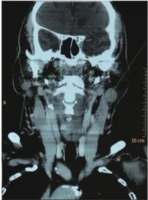
Cervical magnetic resonance image (MRI) showed tumors in the right and left parotids measuring 2.0 cm and 2.5 cm, respectively, in their largest diameters (►Figs. 1,2).
With an intention of carrying out a superficial parotidec-tomy, this procedure was performedfirst on the side with the larger tumor (left) and, after 4 months, a tumor resection was performed on the right side. Both surgical procedures were performed as superficial parotidectomies with preservation of the facial nerve and intraoperative neural monitoring (Viasys Endeavor CR monitor, with 16 channels).
In the immediate postoperative period of thefirst surgical procedure, there were no complications or facial nerve pare-sis in any of its branches. In the second postoperative surgical
period (right side), the patient developed temporary paralysis grade II ocular and grade III marginal mandibular, according to the Blackman-House scale. The individual underwent motor rehabilitation exercises with total motor recovery in 6 months.
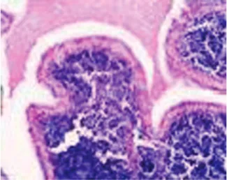
The histopathologic results showed papillary cystade-noma lymphomatosum in both parotid tumors (►Figs. 3,4).
Discussion
Warthin tumor, today also known aspapillary cystadenoma lymphomatosum, was first observed in 1895 by Hildebran, Fig. 1 Magnetic resonance image.
Fig. 2 Magnetic resonance image.
International Archives of Otorhinolaryngology Vol. 18 No. 2/2014
who considered it a variant of a congenital cyst of the neck.2In 1929, Alfred Scott Warthin systematically reviewed all parot-id tumors and brachial cervical cysts recorded over a period of 35 years at the University of Michigan. In two cases, which were called papillary cystadenoma lymphomatosums, the condition was believed to be a rare form of parotid tumor, not only for their low incidence among the other tumors found in the review but also because there were no reports in the literature with similar characteristics. However, Warthin stated that these papilliferous cystadenomas with lymphoid stroma represent a grotesque development of the eustachian tube, which had been modified and grown slowly over the years and taken a late cancer trend. In 1944, Martin and Ehrlich14reported 22 cases of papillary cystadenoma lym-phomatosum of the parotid gland and suggested the eponym of Warthin’s tumor in honor of the first researcher who described it.3
Warthin tumor is the second most common cancer of the parotid glands—the most common is pleomorphic
adeno-ma1,5—and represents2 to 15% of all epithelial neoplasms of the parotid, being bilateral in 7 to 10% and multifocal in 2%. Both recurrences as well as malignancies are rare.4,9Warthin tumor can occur unilaterally or bilaterally, metachronously or synchronously. Most salivary gland neoplasms have unilater-al presentation unilater-alone, and simultaneous and bilaterunilater-alfindings are unusual.10There are a few reports of synchronous bilat-eral tumors in the literature. In a systematic review of Tveterås and Kristensen, only seven synchronous bilateral tumors were identified in the 71 cases reviewed. It is even rarer to find multiple synchronous bilateral Warthin tumors.11
Most tumors occur in men (man-to-woman ratio of 10:1), although recent studies show a fall in this proportion, with a smaller distribution between the sexes.8,9 The condition usually occurs between sixth and seventh decades of life and is described predominantly in white subjects, less fre-quently in Oriental subjects, and rarely in black subjects.6
Smoking is an important risk factor for the development of Warthin tumor. Some authors report that the risk is closely related to the amount smoked and that smokers are eight times more likely to develop the tumor than nonsmokers.1,4,8 It is believed that this relationship is justified by the retro-gradeflow of substances in cigarette smoke to the salivary ducts or the excretion by the cigarette of noxious substances in the ducts.12The association with cigarettes is independent of sex and age.
Although there are some theories to explain the origin and development of the tumor, there is still controversy in the scientific world. Among the most well-accepted theo-ries, one hypothesis relates that the tumor is an adenoma with concomitant lymphocytic infiltration; another hy-pothesis refers to the neoplastic proliferation of salivary ductal cells in its course of development near the lymph nodes and peri- and intraparotid, associated with the major salivary glands, especially the parotid, because this is encapsulated later in the submandibular and the sublingual glands.7Some authors suggest that the tumor has a lym-phocytic component with an immunologic reaction to the epithelial component, rather than the tumor being completely made up of lymphatic tissue.9There are still countless theories against Warthin tumor being a true neoplasm. Honda et al16have shown that the epithelial tumor components are polyclonal cellular populations— which exclude a pattern of malignant growth—and accord-ing to the World Health Organization, these lesions should be classified as tumorlike lesions.2Some features of War-thin tumor speak in favor of benignity, such as its slow growth, its well-defined nodule format with regular out-lines, the unusual involvement of the facial nerve, and the low rates of malignant progression and recurrence.
The clinical history is usually a patient seeking medical attention for a nodular lesion that is painless or a bit painful, with slow growth, top of the parotid gland, near the angle of the mandible. The patient may also report buzzes and even deafness, although rare.5 The average size of the tumor at presentation varies from a few millimeters to centimeters, averaging 2 to 4 cm.5,7,9A sudden increase of the tumor can Fig. 3 Left parotid tumor. Papilla with lymphomatoid center and
oncocytic coating (20, hematoxylin and eosin).
Fig. 4 Right parotid tumor. Papillary projections coated by oncocytic cells upon a lymphocytic stroma. These projections penetrate in cystic areas (20, hematoxylin and eosin).
indicate secondary infection in the location or the need to add another diagnostic hypothesis.
A detailed clinical history and thorough physical examina-tion can direct the diagnostic hypothesis toward Warthin tumor. Additional tests such as ultrasonography may be useful in elucidating the content of the lesion. Computed tomography and magnetic resonance imaging may be re-quested in special situations, such as multiple tumors, suspi-cion of other concomitant neoplasms, and cases of relapse.7 Although quite controversial in the scientific world, some studies have shown high sensitivity and specificity of Warthin tumor infine FNA. Other authors report that the main goal of FNA in parotid nodes is the distinction between a malignant and a benign neoplasm, whereas the definitive diagnosis can only be given with histopathology after tumor excision.13,15 Macroscopically, the tumor is cystic and filled with a brown gelatinous liquid. Cytologicfindings of the tumor in FNA are epithelial parenchyma and lymphoid stroma, with amorphous cell groups and a mixture of lymphocytes, epi-thelial cells, oncocytes, and mast cells.7The parenchymatous tissue is composed of tubules and dilated cystic spaces within the lumen from which thin papillary processes protrude like ribbons.3
The treatment recommended in the literature for Warthin tumor is superficial parotidectomy when the tumor occurs in a superficial lobe—90% of the cases—or total parotidectomy when the tumor is in a deep lobe or for recurrent tumors, with both procedures involving the identification and preserva-tion of the facial nerve.1,5,7
Postsurgical complications are uncommon, with low inci-dence and morbidity. The most important are facial nerve lesions, Frey syndrome, wound infections, dehiscence, hema-tomas, and seromas. Some studies report incidence of tem-porary facial paralysis in 16 to 47% and permanent facial paralysis in 0 to 9% of parotidectomies.5
Conclusion
This article reports an extremely rare case of bilateral and synchronous Warthin tumor as well as presents a literary review on the incidence and etiology of this lesion in the population. Recent studies imply that this is not a true neoplasm tumor and therefore can be reclassified as a tu-morlike lesion. The standard recommended treatment is
either a superficial or total parotidectomy, depending on the extent and location of the tumor in the gland. We believe that more clinical and histopathologic studies are needed to clarify its synchronous occurrence.
References
1 Ascani G, Pieramici T, Rubini C, Messi M, Balercia P. Synchronous bilateral Warthin’s tumours of the parotid glands: a case report. Acta Otorhinolaryngol Ital 2010;30:310–312
2 Naujoks C, Sproll C, Singh DD, et al. Bilateral multifocal Warthin’s tumors in upper neck lymph nodes. Report of a case and brief review of the literature. Head Face Med 2012;8:11
3 Viveros ALM, Sánchez MJF. Tumor de Warthin. Anales médicos de la Asociacion del Hospital ABC 2001;46(2):88–91
4 Alwan MH, Kirkwood S. Periparotid multifocal Warthin’s tumour. ANZ J Surg 2011;81:183–184
5 Chedid HM, et al. Tumor de Warthin da glândula parótida: estudo de 70 casos. Rev Col Bras Cir 2011;38(2):090–094
6 Pinkston JA, Cole P. Cigarette smoking and Warthin’s tumor. Am J Epidemiol 1996;144:183–187
7 Castro GA, et al. Tumor de Warthin de Glândula Parótida. Int Arch Otorhinolaryngol 2004;8(2):3–5
8 Lamelas J, Terry JH Jr, Alfonso AE. Warthin’s tumor: multicentricity and increasing incidence in women. Am J Surg 1987;154:347–351 9 Hilton JM, Phillips JS, Hellquist HB, Premachandra DJ. Multifocal multi-site Warthin tumour. Eur Arch Otorhinolaryngol 2008;265: 1573–1575
10 Gnepp DR, Schroeder W, Heffner D. Synchronous tumors arising in a single major salivary gland. Cancer 1989;63:1219–1224 11 Tveterås K, Kristensen S. Warthin’s tumour with bilateral
synchro-nous presentation. Survey of the literature and a new case. J Laryngol Otol 1986;100:487–492
12 Gallo O, Bocciolini C. Warthin’s tumour associated with autoim-mune diseases and tobacco use. Acta Otolaryngol 1997;117: 623–627
13 Azua-Romeo J, et al. Synchronous salivary gland tumors with regard to two cases. Rev Esp Cir Oral y Maxilofac 2005;27 (3):145–149
14 Martin H, Ehrlich HE. Papillary cystadenoma lymphomatosum (Warthin’s tumor) of the parotid gland. Surg Gynecol Obstet 1944;79:611–623
15 Brennan PA, Davies B, Poller D, et al. Fine needle aspiration cytology (FNAC) of salivary gland tumours: repeat aspiration provides further information in cases with an unclear initial cytological diagnosis. Br J Oral Maxillofac Surg 2010;48:26–29 16 Honda K, Kashima K, Daa T, Yokoyama S. Nakayama I: Clonal
analysis of the epithelial component of Warthin’s tumor. Hum Pathol 2000;31:1377–1380
International Archives of Otorhinolaryngology Vol. 18 No. 2/2014