Recebido em 30.01.2002. / Received in January, 30th of 2002.
Aprovado pelo Conselho Consultivo e aceito para publicação em 30.07.2002. / Approved by the Consultive Council and accepted for publication in July, 30th of 2002.
* Trabalho realizado com bolsa de pós-doutorado da Capes e da Fundação Alexander von Humboldt, no Laboratório de Diagnóstico Genético, Universidade de Colônia, Alemanha (Serviço do Prof. Thomas Krieg) /Work done with a post-doctorate grant from Capes and the Alexander von Humbold Foundation, in the Genetic Diagnosis Laboratory, University of Cologne, Germany (Service of Prof. Thomas Krieg)
1Professor Adjunto de Dermatologia, Universidade Federal de Pelotas. / Adjunct Professor of Dermatology, Federal University of Pelotas. ©2002by Anais Brasileiros de Dermatologia
Genética Molecular das Epidermólises Bolhosas
*Molecular Genetics of Epidermolysis Bullosa
*Hiram Larangeira de Almeida Jr.
Educação Médica Continuada /
Continuing Medical Education
Resumo:O estudo das alterações moleculares das epidermólises bolhosas tem contribuído para que se
com-preenda melhor essas enfermidades. Na epidermólise bolhosa simples a maioria dos casos está associada com alteração nas citoqueratinas basais 5 (gen KRT5) e 14 (gen KRT14), o que modifica o citoesqueleto na camada basal da epiderme, levando à degeneração dessa camada, formando bolha intra-epidérmica. Mutações na plecti-na (gen PLEC1), componente da placa interplecti-na do hemidesmossoma, levam também à clivagem intra-epidérmi-ca. Na epidermólise bolhosa juncional vários gens estão envolvidos, em decorrência da complexidade da zona da membrana basal, todos levando ao descolamento dos queratinócitos basais na lâmina lúcida, pela disfunção da aderência entre esses e a lâmina densa. Alterações na laminina 5 (gens LAMA3, LAMB3e LAMC2), integrina
α6β4 (gens ITGA6e ITGB4) e colágeno XVII (gen COL17A1) foram descritas. Por fim, na epidermólise bolhosa distrófica apenas um gen está mutado, alterando o colágeno VII (gen COL7A1), principal componente das fibri-las ancorantes, produzindo clivagem abaixo da lâmina densa, variando fenotipicamente de acordo com a con-seqüência da mutação. Outra aplicação importante dessas informações refere-se ao diagnóstico pré-natal, com a perspectiva no futuro da terapia gênica.
Palavras-chave: Diagnóstico pré-natal; epidermólise bolhosa; genética bioquímica; mutação; reação em cadeia da polimerase.
Summary: New data regarding the molecular aspects of the heterogeneous group of epidermolysis bullosa has brought some important information about its pathogenesis. In epidermolysis bullosa simplex the majority of mutations are localized in the genes of the basal cytokeratin 5 (gene KRT5) and 14 (gene KRT14), cytolysis at this layer with intraepidermal blister is seen under light microscopy. Mutations of plectin (gene
PLEC1), a protein found in the inner hemidesmosomal plaque, leads also to intraepidermal blisters. In junc-tional epidermolysis bullosa many proteins from the basal membrane zone are involved, such as laminin 5 (genes LAMA3, LAMB3and LAMC2), integrin α6β4(genes ITGA6and ITGB4) and collagen XVII (gene COL17A1), the dysfunction which leads to a subepidermal blister, at the level of the lamina lucida. In the third group, epidermolysis bullosa dystrophica, the mutations are localized in only one gene (gene COL7A1), where they alter the structure of collagen VII, the principal compound of anchoring fibrils, splitting the skin under the lamina densa. This information can also be used in the prenatal diagnosis of epidermolysis bullosa, with future perspectives of gene therapy.
Key words: prenatal diagnosis; epidermolysis bullosa; genetics, biochemical; mutation; polymerase chain reaction.
INTRODUÇÃO
Antes da descoberta e padronização da reação em cadeia da polimerase (PCR) o seqüenciamento gênico era tarefa lenta, levando-se muito tempo para analisar peque-nos segmentos. A PCR permite a amplificação rápida de segmentos de DNA, os quais são posteriormente
seqüencia-INTRODUCTION
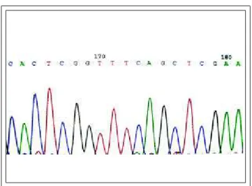
enor-Figure 1: Example of gene sequencing
Figura 1: Exemplo de seqüenciamento gênico
dos, tendo trazido enorme evolução nessa área. Por ocasião dessa revisão, ao se utilizar PCR como palavra-chave no Medline, estavam disponíveis mais de 150.000 publicações com essa técnica, num período de pouco mais de 12 anos, ilustrando a importância da mesma na pesquisa médica.
O princípio da PCR é bastante simples: o primeiro passo é o isolamento de DNA, por exemplo a partir de san-gue, fazendo uso de sua insolubilidade e precipitação em alguns solventes e de sua hidrossolubilidade, havendo co-mercialmente vários kits para essa função.
Posteriormente uma parte do DNAobtido é incubado com uma polimerase termorresistente (já que o mesmo é aquecido para que a cadeia dupla se desfaça) juntamente com seqüências conhecidas de DNA, os chamados primers (inicia-dores). Havendo no DNAem questão seqüência igual à do primer, a polimerase amplificará esse segmento de DNA. Os nucleotídeos (adenina, timidina, citosina e guanina) fazem parte da reação, para que obviamente a enzima tenha a maté-ria-prima necessária à polimerização. Ciclos de aquecimento e resfriamento são repetidos inúmeras vezes, aumentando cada vez mais o produto da PCR. Posteriormente é feita ele-troforese para identificar a presença de uma banda de DNA, mostrando a positividade ou não da reação.
Numa etapa posterior o produto obtido pela PCR é seqüenciado, o que é feito com uma variante da PCR. O seqüenciamento é atualmente automatizado, sendo feita uma leitura a laser, obtendo-se gráficos policromáticos, representando a cor azul, citosina; a cor vermelha, timidina; o preto, guanina; e o verde, adenina (Figura 1). A compara-ção do resultado obtido no paciente investigado e em seus genitores com a seqüência normal do gen pode demonstrar mutação e o padrão da herança.
Cada conjunto de três bases do DNA codifica um aminoácido para a síntese protéica no ribossoma; havendo uma mutação, ou seja, a troca de uma base, haverá durante a síntese protéica a inserção de outro aminoácido, alterando a estrutura da proteína, com as conseqüências que isso pode acarretar.
Inúmeros gens já foram seqüenciados, estando sua composição disponível em bancos de dados digitais. O mais utilizado é o Genbank, do
Centro Nacional de Infor-mação em Biotecnologia dos Estados Unidos, disponível no endereço eletrônico: www.ncbi.nlm.nih.gov.
As mutações são des-critas citando-se os aminoáci-dos ou as bases envolvidas. Primeiro é citado o
aminoáci-mous progress in this field. To date, inputting PCRas a key word in the Medline database, lists over 150,000 publica-tions using this technique, in a period of little over 12 years, thus illustrating the importance of PCRto medical research. The principle of PCRis quite simple: the first step is to isolate the DNA, for instance taking blood and making use of its insolubility and precipitation in certain solvents and its water solubility, several commercial kits are availa-ble for this function.
Then, part of the DNAobtained is incubated with a thermoresistant polymerase (since the DNA is heated in order to separate the double chain) together with known sequences of DNA, the so-called primers. If the DNA in question has the same sequence as that of the primer, the polymerase will amplify this segment of DNA. The nucleoti-des (adenine, thymidine, cytosine and guanine) are part of the reaction, such that obviously the enzyme has the neces-sary raw material for the polymerization. Heating and coo-ling cycles are repeated innumerous times, thereby increa-sing the product of PCRmore and more. After which, elec-trophoresis is performed to identify the presence of a band of DNA, demonstrating the positivity or otherwise of the reaction.
In a subsequent stage the product obtained by PCRis sequenced using a variant of PCR. The sequencing is now automated with laser readings providing polychromatic graphs, with blue representing cytosine, red thymidine, black guanine and green adenine (Figure 1). Comparison of the result obtained in the investigated patient and their progenitors with the normal gene sequence can demonstra-te mutation and an inheridemonstra-ted patdemonstra-tern.
Each set of three bases of DNA codifies an amino acid for the protein synthesis in the ribosome; leading to a mutation, or in other words, the change of a base, during the protein synthesis there will be an insertion of another amino acid, thus altering the structure of the protein and resultant consequences.
Countless genes have already been sequenced and their composition is available in digital databases. These can be accessed at the Genbank of the National Center of Biotechnology Information of the United States: www.ncbi.nlm.nih.gov.
corresponds to the location of the same in the protein/gene under investigation and finally the aminoacid/base inserted in the place of the first, such as, for instance, Glu20Arg, in other words, the twentieth amino acid should be a glutami-ne, but the mutation leads to an insertion in the protein of an arginine, which alters its structure. Some sequences of bases codify the end of the protein synthesis; a mutation can lead to this interruption, the so-called premature termina-tion codon, which is described in the following manner:
Lys472Stop, in other words, instead of the insertion of a lysi-ne, at the 472position, the protein synthesis was interrup-ted. Abbreviated descriptions with only a single letter can be found, for example such an interruption of this synthesis is denoted by an X; in the above example this would then be
L472X.
The bullous dermatoses make a fascinating chapter in dermatology, they comprise acquired or congenital defects of the intraepidermal or dermoepidermal adhesion, leading to blisters which can be spontaneous or provoked by minimal trauma.
In epidermolysis bullosa (EB) these defects are con-genital and can be identified by gene sequencing, which affords a greater understanding of their molecular base,1,2
complements the clinicohistological diagnosis and maybe in the medium term will partly modify the classification of these dermatoses.
Three subgroups of EBare recognized:2
epidermoly-sis bullosa simplex (EBS), in the which the cleaving occurs inside the epidermis; junctional epidermolysis bullosa (EBJ), with subepidermal cleaving in the lamina lucida; and epidermolysis bullosa dystrophica (EBD), also subepider-mal, but below the lamina densa. EBJand EBDcannot be differentiated by optical microscopy alone. There are seve-ral classifications, but in this work the second international consensus has been adopted regarding the diagnosis and classification of epidermolysis bullosa.3
Epidermolysis bullosa simplex
The group of EBShas several subtypes, according to the intensity and location of the blisters, all of which with autosomal dominant inheritance;4
these are also called epi-dermolytic EB, since the defect is intraepidermal.3
The his-tological aspect most commonly found is degeneration of the basal layer, in the absence of any inflammatory infiltra-tion and without deposit of antibodies in the tissue.
The most serious form of EBS is Dowling-Meara syndrome (EBS-DM),5
in which disseminated blisters that also involve the mucous membranes, are accompanied by palmoplantar hyperkeratosis. The mildest form is Weber-Cockayne syndrome (EBS-WC) with lesions restricted to the palmar and plantar regions.5
An intermediate form exists, again with disseminated blisters, but with a less intense pic-ture than that of EBS-DM, denominated EBS-Koebner ( EBS-K), although certain authors consider this to be a mild variant of EBS-DM.6
do/base que deveria constituir a proteína/gen, seguido por um número, o qual corresponde à localização do mesmo na proteína/gen investigado, e por fim o aminoácido/base inse-rido no lugar do primeiro, como, por exemplo, Glu20Arg, ou seja, o vigésimo aminoácido deveria ser uma glutamina, mas a mutação leva à inserção na proteína de uma arginina, o que altera sua estrutura. Algumas seqüências de bases codificam o final da síntese protéica; uma mutação pode levar a essa interrupção, o chamado premature termination codon (códon finalizador prematuro), a qual é descrita da seguinte forma: Lys472Stop, ou seja, em vez da inserção de uma lisina, na posição 472, foi interrompida a síntese pro-téica. Encontram-se também descrições abreviadas com apenas uma letra, sendo a interrupção da síntese descrita com X; o exemplo acima ficaria L472X.
As dermatoses bolhosas são capítulo fascinante da dermatologia, constituídas por defeitos adquiridos ou natos da adesão intra-epidérmica ou dermoepidérmica, levando a bolhas espontâneas ou provocadas por trauma mínimo, decorrentes do defeito dessa adesão.
Nas epidermólises bolhosas (EB) esses defeitos são natos e podem ser identificados por seqüenciamento gêni-co, o que traz maior compreensão à base molecular das mesmas,1,2complementa o diagnóstico clínico-histológico e talvez a médio prazo modifique em parte a classificação dessas dermatoses.
Três subgrupos de EB são reconhecidos:2 a epider-mólise bolhosa simples (EBS), na qual a clivagem ocorre dentro da epiderme; a epidermólise bolhosa juncional (EBJ), cuja clivagem é subepidérmica, na lâmina lúcida; e a epidermólise bolhosa distrófica (EBD), sendo também sube-pidérmica, mas abaixo da lâmina densa. A EBJe a EBDnão podem ser diferenciadas apenas por microscopia ótica. Existem diversas classificações, sendo adotada aqui a do segundo consenso international sobre diagnóstico e classifi-cação das epidermólises bolhosas.3
Epidermólise Bolhosa Simples
O grupo da EBStem vários subtipos de acordo com a intensidade e localização das bolhas, sendo todos de herança autossômica dominante;4 são também chamados de
EB epidermolítica, pois o defeito é intra-epidérmico.3 O aspecto histológico mais comumente encontrado é a dege-neração da camada basal, na ausência de infiltrado inflama-tório e sem depósito de anticorpos no tecido.
A forma mais grave da EBS é a Dowling-Meara (EBS-DM),5na qual bolhas disseminadas, envolvendo tam-bém as mucosas, são acompanhadas de hiperqueratose pal-moplantar. A forma mais leve é a Weber-Cockayne ( EBS-WC) com lesões restritas às palmas e plantas.5Existe uma forma intermediária com bolhas disseminadas, mas com quadro menos intenso do que o da EBS-DM, denominado
EBS - Koebner (EBS-K), que certos autores consideram variante leve da EBS-DM.6
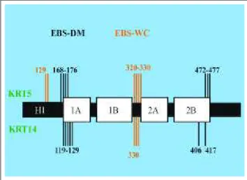
Figure 2: Schematic represen-tation of the molecule of cytokeratin 5 and 14. The mutations of cytokeratin 5 are represented above the scheme and those of cytokeratin 14 below, in that each line could represent more than one mutation. It can be observed that the mutations of epider-molysis bullosa simplex Dowling-Meara (EBS-DM) are located in the extremities of the molecule, while most of those of EBS Weber-Cockayne (EBS-WC), in the non-helical segment between 1B and 2A.
Figura 2: Representação esquemática da molécula das citoqueratinas 5 e 14. As mutações da citoqueratina 5 estão representadas acima do esquema e as da citoqueratina 14 abaixo, sendo que cada traço pode representar mais de uma mutação. Note-se que as mutações da epidermólise bol-hosa simples Dowling-Meara (EBS-DM) localizam-se nas extremidades da molécula, e a maioria das da EBS Weber-Cockayne (EBS-WC), no segmen-to não helicoidal entre 1B e 2A
epiteliais aos pares,7 os quais formam heterodímeros, ou seja, a união das duas moléculas, configurando o citosque-leto dos epitélios, havendo especificidade de acordo com o epitélio envolvido.7A camada basal diferencia-se de outros epitélios e dos segmentos suprabasais da epiderme pela expressão das citoqueratinas 5 e 14.
As citoqueratinas 5 e 14 são reguladas pelos gens
KRT5e KRT14, localizados nos cromossomas 17 e 12, res-pectivamente. É interessante observar que defeitos genéti-cos distintos na EBS, um afetando a citoqueratina 5, outro, a 14,6,8,9 levam à mesma alteração histológica, pois todos esses defeitos produzem alterações estruturais de uma ou outra citoqueratina,10 impedindo a função estrutural das mesmas no citoesqueleto11– a formação dos heterodímeros, responsáveis pela configuração tridimensional da célula. Essa alteração é vista facilmente na histologia e culmina com a formação das bolhas, sendo esse o único subgrupo das EBdecorrente de citólise e não de defeito de adesão.
As citoqueratinas são constituídas por quatro seg-mentos helicoidais, 1A, 1B, 2Ae 2B,12tendo sido a maioria das mutações da EBS-DMdescrita no início do segmento 1A
e no final do segmento 2B(Figura 2)13,14 das citoqueratinas basais. As mutações da EBS-Ktêm localização semelhan-te,15reforçando a hipótese de ser variante da EBS-DM. Na
EBS-WC a maioria das mutações localiza-se no segmento não-helicoidal entre 1B e 2A das mesmas citoqueratinas, sem que com isso se explique a localização palmoplantar das lesões.
Um quarto tipo de EBSé descrito, no qual não ocor-re citólise na camada basal. É a EBScom distrofia muscular tardia, decorrente de alteração da plectina, presente na placa interna do hemidesmossoma (Figura 3). Como a clivagem ocorre dentro da epiderme, é incluída nesse grupo. A plec-tina é regulada pelo gen PLEC11e está também envolvida no
citoesqueleto da musculatura lisa,16daí a miopatia associa-da.1,17Outro componente da placa interna do hemidesmos-soma é o antígeno do penfigóide bolhoso de 230 KDde peso molecular, não havendo até o momento descrição de muta-ção no gen que o regula.1
Some cytokeratins are expressed in the epithelial cells in pairs,7
which form heterodimers, in other words, the union of two molecules, configuring the cytoskeleton of the epithelia, with specificity according to the epithelium invol-ved.7
The basal layer differs from other epithelia and supra-basal segments of the epidermis by the expression of cyto-keratins 5 and 14.
Cytokeratins 5 and 14 are regulated by the genes
KRT5and KRT14, located in chromosomes 17 and 12, res-pectively. It is interesting to note that different genetic defects in EBS, one affecting cytokeratin 5 and the other 14,6,8,9
lead to the same histological alteration, because all these defects produce structural alterations in one or ano-ther cytokeratin,10
impeding their structural function in the cytoskeleton11
– i.e. the formation of the heterodimers, res-ponsible for the three-dimensional configuration of the cell. This alteration is easily seen in the histology and culmina-tes with the formation of blisters, making this the only sub-group of EB due to cytolysis and not to an adhesion defect. The cytokeratins are constituted by four helical seg-ments, 1A, 1B, 2Aand 2B,12
the majority of the mutations of
EBS-DMare described in the beginning of segment 1Aand at the end of segment 2B(Figure 2)13,14
of the basal cytokeratins. The mutations of EBS-Khave a similar location,15
reinforcing the hypothesis that it is a variant of EBS-DM. In EBS-WC
most of the mutations are located in the non-helical segment between 1B and 2A of the same cytokeratins, though this does not explain the palmoplantar location of the lesions.
A fourth type of EBSis described, in which cytolysis in the basal layer does not occur. It is EBSwith tardive muscular dystrophy, due to alteration of the plectin, present in the inter-nal plaque of the hemidesmosome (Figure 3). Since the clea-ving occurs within the epidermis, it is included in this group. The plectin is regulated by the gene PLEC11and is also invol-ved in the cytoskeleton of the smooth musculature,16
hence the associated myopathy.1,17
Junctional Epidermolysis Bullosa
Given the complexity of the basal membrane zone, alterations in several proteins involved in the dermoepider-mal adhesion can lead to the various clinical pictures of
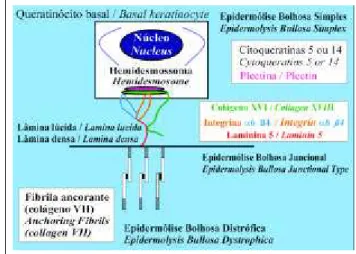
EBJ; for these molecular alterations to be understood, it is important to be familiar with the substances responsible for the adhesion between the basal keratinocytes and the colla-gen IV– the lamina densa (Figure 3).
The antigen of bullous pemphigoid (180 KD) and integrin α6β4, which are transmembranous proteins, are found in the external plaque.18
The antigen of bullous pemphigoid (180 KD) is in reality a transmembranous collagen, denominated collagen
XVII, and is regulated by the gene COL17A1.1
Each segment of the integrin a6ß4 is regulated by two different genes,
ITGA6and ITGB4, which are also expressed in the skin and digestive tract.1,18
Finally, some substances present in the lamina luci-da complement this molecular net,1
of which the most important is laminin 5. The laminins are heterotrimers, or that is, they are constituted by three distinct classes of poly-peptides α, β e γ,18-20
and hence regulated by three genes. Laminin 5 is composed of one α3, one β3 and one γ2, regu-lated by the genes LAMA3, LAMB3and LAMC2, respectively. An absence or alteration of these substances produ-ces a rupture of this adhesion net, with the formation of blis-ters.2
Some mutations occur due to the so-called premature termination codon (PTC), which provokes an interruption of the protein synthesis and consequently absence of protein in the tissue, resulting in a more serious clinical picture.
Several genophenotype correlations have already been made. Such as integrin α6β4 is expressed in the skin and intestine, mutations of which lead to forms of EBJwith atresia pilori, the clinical picture varies according to whe-ther or not it is associated to PTC.
Regarding generalized, benign and atrophic EBJ, cha-racterized by disseminated blisters with nail dystrophy, in which the immunohistochemistry with antibody against colla-gen XVII is negative, PTChas been demonstrated in the gene
Epidermólise Bolhosa Juncional
Dada a complexidade da zona da membrana basal, alterações de várias proteínas envolvidas na adesão dermoe-pidérmica podem levar aos diversos quadros clínicos da
EBJ; para que se compreendam essas alterações molecula-res, é importante conhecer as substâncias responsáveis pela adesão entre os queratinócitos basais e o colágeno IV– a lâmina densa (Figura 3).
Na placa externa do hemidesmossoma encontram-se o antígeno do penfigóide bolhoso de 180 KDe a integrina
α6β4, os quais são proteínas transmembranosas.18
O antígeno do penfigóide bolhoso de 180 KD é na realidade um colágeno transmembranoso, sendo denomina-do colágeno XVII, e é reguladenomina-do pelo gen COL17A1.1Cada segmento da integrina α6ß4 é regulado por dois gens dis-tintos, ITGA6e ITGB4, sendo a mesma expressa na pele e no tubo digestivo.1,18
Finalmente algumas substâncias presentes na lâmina lúcida complementam essa rede molecular,1 sendo a mais importante a laminina 5. As lamininas são heterotrímeros, ou seja, são constituídas por três classes distintas de polipep-tídeos α, βe γ,18-20e daí reguladas por três gens. A laminina 5 é composta por uma classe α3, uma β3 e uma γ2, regula-das pelos gens LAMA3, LAMB3e LAMC2, respectivamente. A ausência ou alteração dessas substâncias produz a ruptura dessa rede de adesão, com a formação das bolhas.2 De algumas mutações decorre o chamado “premature termi-nation codon” (PTC), o que provoca a interrupção da sínte-se protéica e consínte-seqüentemente a ausência da proteína no tecido, com quadro clínico mais grave.
Várias correlações genofenotípicas já foram feitas. Como a integrina α6β4 é expressa na pele e no intestino, suas mutações levam a formas de EBJcom atresia pilórica, sendo o quadro clínico variável de acordo com sua associa-ção com PTCou não.
Na EBJgeneralizada atrófica benigna, caracterizada por bolhas disseminadas com distrofia ungueal, na qual a imuno-histoquímica com anticorpo contra o colágeno XVII é negativa, foi demonstrada PTCno gen COL17A,1,21o que
Figure 3: The adherence between the basal keratinocyte and the lamina densa is made by the transmembranous proteins based on the external plaque of the hemi-desmosome (collagen XVII and integrin α6β4) and by laminin 5, present in the lamina lucida. The adherence between the lamina densa and the dermis is promoted by anchorage fibrils (collagen VII). The plectin is present in the inter-nal plaque of the hemidesmosome, and its alterations lead to the intraepidermal separation, belon-ging to the group of the epidermoly-sis bullosa simplex, as well as the alterations of the basal cytokeratins.
Figura 3: A aderência entre o queratinócito basal e a lâmina densa é feita pelas proteínas trans-membranosas a partir da placa externa do hemidesmossoma
(colágeno XVII e integrina α6β4) e
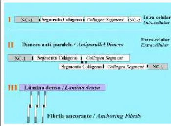
Figure 4: Diagram of the synthesis of collagen VII and anchoring fibrils. After the loss of segment NC2 the formation of antiparallel dimers occurs, which group together and form anchorage fibrils.
Figura 4: Esquema da síntese do colágeno VII e das fibrilas ancorantes. Após a perda do segmento NC2 ocorre a
formação dos dímeros antiparalelos, os quais se
agrupam e formam as fibrilas ancorantes.
correlaciona com a ausência tecidual do colágeno XVII. Alguns autores denominam essa forma, por ter curso bran-do e expectativa de vida normal, de EBJnão-Herlitz.21,22
Semelhante às mutações dos componentes do hemidesmossoma descritas acima, as mutações da lamini-na também provocam o descolamento da epiderme. A maior parte das mutações dos gens da laminina 5 leva à
PTC, provocando ausência da proteína e quadro clínico intenso,23,24caracterizado por lesões disseminadas afetando também as mucosas,23com sobrevida curta em função das complicações bacterianas, denominadas EBJ tipo Herlitz ou EBJletal.
As alterações já foram demonstradas no três gens que codificam a laminina 5,25não havendo diferença fenotí-pica de acordo com o segmento envolvido,26sugerindo que todos são importantes para a função adesiva da mesma.26,27 Oitenta por cento das mutações residem no gen LAMB3,22, 24,28-30havendo duas delas recorrentes (R635Xe R42X),26,28as quais perfazem metade das mutações no LAMB3.1 Nesse gen já foram relatadas 35 mutações diferentes.22
Existem relatos de mutação da laminina 5 em pacien-tes nos quais a imuno-histoquímica consegue demonstrar diminuição da laminina 5, e não ausência, como na EBJ -Herlitz, e, em decorrência disso, o quadro clínico não é tão grave.31,32Essas formas têm sido também denominadas EBJ não-Herlitz,33as quais clinicamente são semelhantes às for-mas decorrentes da mutação do colágeno XVII.33
Todas as formas de EBJsão autossômicas recessivas.23
Epidermólise Bolhosa Distrófica
A característica clínica principal das EBD são as cicatrizes decorrentes da perda tecidual, pois a clivagem ocorre abaixo da lâmina densa.34Como nos outros grupos, há variantes de acordo com o quadro clínico; apesar dessas variantes, o defeito genético foi localizado em um único gen, o COL7A1.35 Esse gen é responsável pela codificação do colágeno VII, principal constituinte das fibrilas ancoran-tes,35as quais participam da aderência da lâmina densa com a derme (Figura 3), daí também a denominação EB dermo-lítica. Nesse grupo existem formas com herança autossômi-ca dominante e recessiva.
Para que se entenda a correlação entre genótipo e fenótipo na EBDé necessário que se entenda também o papel do colágeno VIIna
ade-COL17A,1,21
which correlates with the tissular absence of colla-gen XVII. Some authors denominate this form non-Herlitz EBJ, as it presents a mild course and normal life expectancy.21,22
Similar to the mutations in components of the hemides-mosome described above, mutations in the laminin also pro-voke dislocation of the epidermis. Most of the mutations of the genes of laminin 5 lead to PTC, provoking absence of the pro-tein and intense clinical picture,23,24
characterized by dissemi-nated lesions also affecting the mucous membranes,23
with low survival in function of bacterial complications, denominated Herlitz syndrome or epidermolysis bullosa lethalis.
The alterations have already been demonstrated in the three genes that codify laminin 5,25
without a phenotype difference according to the segment involved,26
suggesting that all are important for its adhesion function.26,27
Eighty percent of the mutations reside in the gene LAMB3,22,24,28-30
two of these are recurrent (R635X and R42X),26,28
which amount to half of the mutations in LAMB3.1
In this gene alone 35 different mutations have already been reported.22
There are reports of mutation in laminin 5 among patients in which the immunohistochemistry demonstrated a reduction in laminin 5, but not a complete absence, as in Herlitz Syndrome, and consequently the clinical picture was not so serious.31,32
These forms have also been denominated non-Herlitz EBJ,33
which are clinically similar to the forms arising from mutation of collagen XVII.33
All forms of EBJ are inherited as an autosomal recessive trait.23
Epidermolysis bullosa dystrophica
The main clinical characteristic of EBDis scarring after tissue loss, since the separation occurs below the lamina densa.34
As in the other groups, there are variants reflecting the clinical picture; in spite of these variants, the genetic defect is located in a single gene, COL7A1.35
This gene is responsible for codifying collagen VII, the main representative of the anchoring fibrils,35
which participate in the adherence of the lamina densa to the dermis (Figure 3), hence it is also denominated dermolytic EB. In this group there are forms inherited as autosomal dominant and
recessive traits.
In order to unders-tand the correlation between genotype and phenotype in
Figura 5: Representação esquemática da molécula do colágeno VII. A maioria das mutações da epidermólise bol-hosa distrófica recessiva Halloupeau-Siemens (EBD-RHS) é premature termination codon (PTC) levando à ausência da molécula no tecido. Na forma recessiva Mitis (EBD-RM) os defeitos localizam-se no final do segmento colágeno e no NC-2. Na forma dominante (EBD-D) as substituições de glicina – repre-sentadas embaixo – ocorrem geralmente no segundo segmen-to colágeno. Cada traço pode rep-resentar mais de uma mutação.
Figure 5: Schematic representa-tion of the molecule of collagen VII. The majority of mutations in epidermolysis bullosa dys-trophica recessive Halloupeau-Siemens (EBD-RHS) are prema-ture termination codon (PTC) leading to the absence of the molecule in the tissue. In the recessive mitis form (EBD-RM) the defects are located at the end of the collagen segment and in NC-2. In the dominant form (EBD-D) the substitutions of glycine – shown below – usually occur in the second collagen segment. Each line can repre-sent more than one mutation.
understand the role of collagen VIIin the dermoepidermal adhesion.
This is produced by the keratinocytes and has a tri-ple helix configuration of collagen, preceded and followed by non-collagen segments (NC-1and NC-2, respectively).36, 37
In the center of the triple helix there is small non-collagen segment, which probably provides flexibility to the protein. Later, at the extracellular level, a fusion occurs between two of these molecules with loss of the NC-2segment, for-ming antiparallel dimers. The union of several dimers forms the anchoring fibrils34,36,37
(Figure 4).
Three subtypes of EBDare well characterized: reces-sive EBDHalloupeau-Siemens (EBD-RHS), with an intense clinical picture, producing acral retractions with synechiae of the digits and involvement of the digestive tract;37
recessi-ve EBDmitis (EBD-RM), in which the clinical picture is much less intense in comparison with that of EBD-RHS, with loca-lized lesions in the areas of greatest trauma, such as the knees and extremities; and the dominant form (EBD-D), with a similar picture to that of EBD-RM,37
associated to nail dystrophy and, in some cases, with white papular lesions.
Electron microscopy and immunohistochemical cha-racterization with antibodies against collagen VII show alteration in the anchoring fibrils to the extent of their absence in EBD-RHS and reduction in the milder forms,
EBD-RM and EBD-D.38
In some cases the immunohistoche-mistry is positive but without anchoring fibrils revealed by electron microscopy, which demonstrates the presence of part of the molecule, but with structural alteration.38
The identification of the mutations responsible for EBD
has brought a greater understanding of this spectrum (Figure 5). In EBD-RHSthe genetic alteration is a PTC, with con-sequent interruption in the synthesis of collagen VII,39
which correlates with the intensity of the clinical picture and with the findings of electron microscopy and immunohistoche-mistry, in which the anchoring fibrils are not detected.34,37
In EBD-RMthe greater part of the mutations occur at the end of the collagen segment and in NC-2, interfering in the formation of the antiparallel dimers and altering the são dermoepidérmica.
O mesmo é produzido pelos queratinócitos e possui uma tripla hélice de colágeno, precedida e seguida por seg-mentos não colágenos (NC-1 e NC-2, respectivamente).36,37 No centro da tripla hélice há pequeno segmento não coláge-no, o qual provavelmente dá flexibilidade à proteína. Posteriormente, no nível extracelular, ocorrerá com duas dessas moléculas uma fusão com perda do segmento NC-2, formando dímeros antiparalelos. A união de vários dímeros forma as fibrilas ancorantes34,36,37(Figura 4).
Três subtipos da EBD estão bem caracterizados: a
EBD recessiva Halloupeau-Siemens (EBD-RHS), com quadro clínico intenso, produzindo retrações acrais com sinéquia dos dígitos e acometimento do tubo digestivo;37 a
EBD recessiva Mitis (EBD-RM), na qual a intensidade do quadro clínico é bem menor em comparação ao da EBD-RHS, com lesões localizadas nas áreas de maior trauma, como joelhos e extremidades; e a forma dominante ( EBD-D), com quadro semelhante ao da EBD-RM,37 associada com distrofia ungueal e, em alguns casos, com lesões albo-papulóides.
A microscopia eletrônica e a caracterização imuno-histoquímica com anticorpos contra colágeno VIImostram alteração nas fibrilas ancorantes, indo da ausência das mes-mas, na EBD-RHS, à diminuição nas formas mais leves,
EBD-RMe EBD-D.38Em alguns casos a imuno-histoquímica é positiva, sem que se observe as fibrilas ancorantes na microscopia eletrônica, o que demonstra a presença de parte da molécula, mas com alteração de sua estrutura.38
A identificação das mutações responsáveis pela EBD
trouxe maior compreensão a esse espectro (Figura 5). Na EBD-RHSa alteração genética é uma PTC, com conseqüente interrupção na síntese do colágeno VII,39o que correlaciona com a intensidade do quadro clínico e com os achados de microscopia eletrônica e imuno-histoquímica, com os quais não se detectam as fibrilas ancorantes.34,37
da proteína, estando, porém, presente, decorrendo um qua-dro clínico mais leve e a presença de fibrilas ancorantes na microscopia eletrônica.37,40
Na EBD-Da alteração característica é a substituição de uma glicina no segmento colágeno,41,42 alterando sua estabilidade e talvez propiciando sua degradação.36,37,43 Como na EBD-RM, as fibrilas ancorantes estão presentes, mas com sua função comprometida. A maior parte das mutações localiza-se logo depois do segmento não coláge-no do centro da tripla hélice;42 a mutação G2043R é a mais comumente descrita.36,41Também já foi demonstrado que a alteração funcional da fibrila ancorante depende da locali-zação da substituição da glicina,34,44o que, por sua vez, con-tribui para a variabilidade clínica. Não existe explicação convincente a respeito de por que a substituição de glicina é herdada de forma dominante.
A maioria dos casos da forma pré-tibial da EBDé autossômica dominante, tendo sido descrita também a subs-tituição de glicina.45 Casos recessivos foram igualmente publicados,45 podendo ser considerados variantes das for-mas leves de EBD, não se sabendo o porquê da ocorrência localizada das lesões.
Cerca de 100 mutações diferentes já foram descritas na EBD,34 sendo encontradas em 80% dos casos examina-dos.37Assim como nas outras formas de EB, algumas muta-ções não se enquadram no esquema acima exposto, pois, por exemplo, algumas substituições de glicina foram encon-tradas na EBD-RM;37,40,41razão de, nesses casos, os genitores que apresentam essa substituição de glicina serem normais, ou seja, a mutação não ser dominante, só se expressando de forma recessiva, com a herança de dois alelos mutados, e até o momento não pôde ser esclarecida.41
Alguns quadros clínicos intermediários, de difícil clas-sificação clínica, já foram também descritos com essas muta-ções incomuns, como, por exemplo, EBDrecessiva com uma
PTCem um alelo e uma substituição de glicina em outro.46
Discussão
Os novos aspectos moleculares, tanto gênico quanto protéico, mostram o quão variado é o espectro da EB
(Tabela 1). Na EBSos defeitos genéticos das citoqueratinas basais produzem alteração histológica em função da modi-ficação do citoesqueleto na camada basal da epiderme, sendo que a alteração da plectina, componente da placa interna do hemidesmossoma, também leva à clivagem intra-epidérmica. Na EBJvários gens estão envolvidos, devido à complexidade da zona da membrana basal, mas todos levam ao descolamento dos queratinócitos basais da lâmina densa, ou seja, a clivagem ocorre na lâmina lúcida. Por fim, na EBDapenas um gen está mutado, alterando o colágeno
VII, sendo a clivagem abaixo da lâmina densa, mas, mesmo, assim variando fenotipicamente, de acordo com a conse-qüência da mutação.
Apesar de contribuir com importantes avanços na compreensão dessas enfermidades, o seqüenciamento
gêni-compliance of the protein, though these continue present, giving rise to a milder clinical picture and the presence of anchoring fibrils in the electron microscopy.37,40
In EBD-D, the characteristic alteration is the substi-tution of a glycine in the collagen segment,41,42
altering its stability and maybe propitiating its degradation.36,37,43
As in
EBD-RM, the anchorage fibrils are present, but their func-tion is impaired. Most of the mutafunc-tions are located imme-diately after the non-collagen segment of the center of the triple helix;42
the G2043R mutation is the most commonly described.36,41
Likewise it has already been demonstrated that the functional alteration of the anchoring fibrils depends on the location in which the glycine is substitu-ted,34,44
which in turn contributes to the clinical variability. As yet, there is no convincing explanation as to why the glycine substitution is an inherited dominant trait.
The majority of cases involving the pretibial form of
EBDare autosomal dominant and the substitution of glyci-ne has also been described.45
Recessive cases have been published,45
these could equally be considered variants of the mild forms of EBD, the reason behind the localized occurrence of the lesions is not known.
About 100 different mutations have already been described in EBD,34
and are found in 80% of the cases exa-mined.37
As in other forms of EB, some mutations are not defined within the above described outline, because, for ins-tance, some substitutions of glycine have been found in
EBD-RM;37,40,41
to date, it has yet to be clarified why in these cases the progenitors that present such glycine substitution may be normal, or in other words, the mutation is not domi-nant and is only expressed in a recessive manner, with the inheritance of two changed alleles.41
Various intermediate clinical pictures, presenting diffi-cult clinical classification, have already been described with such uncommon mutations, for instance, recessive EBDwith
PTCin one allele and a glycine substitution in the other.46
Discussion
The new molecular aspects, involving both genes and proteins, demonstrate just how varied the spectrum of EB can be (Table 1). In EBSthe genetic defects of the basal cytokeratins produce a histological alteration due to the modification of the cytoskeleton in the basal layer of the epidermis, in that alteration of the plectin, a component of the internal plaque of the hemidesmosome, also leads to the intraepidermal separation. In EBJseveral genes are invol-ved, due to the complexity of the basal membrane zone, but all lead to the dislocation of the basal keratinocytes of the lamina densa, in other words, the cleaving occurs in the lamina lucida. Finally, in EBDonly one gene is modified, altering the collagen VII, cleaving below the lamina densa, but even so with phenotype variation, according to the con-sequence of the mutation.
used together with clinical, histological, electron microscopy and immunohistochemical findings in the diagnosis of EB.47
Another important application for molecular gene-tics is in prenatal diagnosis (PND),2,48
examining fetal DNA
obtained from the chorion rather than the fetal skin. PND
performed on the basis of the lesions requires the collection of a skin specimen, which should be representative of the ill-ness, in order to avoid a false-negative result, one should wait until the eighteenth or twentieth week.43
Sequencing has the advantage that it can be performed around the tenth week, which means that a more precocious decision to ter-minate the gestation can be made in those countries in which this procedure is permitted. Furthermore, complica-tions arising from fetoscopy with biopsy occur in between four to 7% of cases compared to 1% in chorionic biopsy.43
Genetic sequencing has already been used in PND
for all forms of EB,23,24,43,49-51
and has already been performed before implantation, based on a cell obtained from an embr-yo with a number of cells varying from five to eight.52
Genetic counseling is another important application of this new information, since it helps to explain the inheri-tance pattern, especially when dealing with frequent and well-known mutations. Also in the case of de novo muta-tions, when the DNAexam of the progenitors is normal and the mutation is only found in the patient, it can be affirmed that the risk factor for the next gestation is very low. co deve ser utilizado em conjunto com a clínica, histologia,
microscopia eletrônica e a imuno-histoquímica no diagnós-tico da EB.47
Outra aplicação importante da genética molecular ocorre no diagnóstico pré-natal (DPN),2,48examinando-se o
DNAfetal obtido do córion e não a pele fetal. O DPNfeito a partir das lesões necessita da obtenção de fragmento da pele, o qual deve ser representativo da enfermidade, para evitar resultado falso-negativo, devendo-se esperar até a décima oitava ou vigésima semana.43 O seqüenciamento tem a vantagem de poder ser feito em torno da décima semana, o que permite, nos países em que a gestação pode ser interrompida nessas situações, decisão mais precoce. Além disso as complicações da fetoscopia com biópsia ocorrem entre quatro e 7% dos casos, e na biópsia coriôni-ca em 1%.43
O seqüenciamento genético já foi utilizado no DPN
de todas as formas de EB,23,24,43,49-51já tendo sido também rea-lizado antes da implantação, a partir de célula obtida de embrião com número de células variável de cinco a oito.52 Aconselhamento genético é outra aplicação importante des-sas novas informações, pois ajuda a esclarecer o padrão de herança, principalmente tratando-se de mutações freqüentes e bem conhecidas. Também no caso de mutações de novo, quando o exame do DNAdos genitores é normal e a
muta-EBS
EBJ
EBD
Dowling-Meara
Dowling-Meara
Weber-Cockaine
Weber-Cockaine
EBS - distrofia muscular
EBS - muscular dystrophy
Herlitz
Herlitz
Não-Herlitz
Non-Herlitz
EBJ - atresia pilórica
EBJ - atresia pilori
Halloupeau-Siemens
Halloupeau-Siemens
EBD-Recessiva Mitis
EBD-Recessive Mitis
EBD-Dominante
EBD-Dominant form
Citoqueratina 5 ou 14
Cytokeratin 5 or 14
Citoqueratina 5 ou 14
Cytokeratin 5 or 14
Plectina
Plectin
Laminina 5
Laminin 5
Colágeno XVII
Collagen XVII
Integrina alpha6 beta4
Integrin alpha6 beta4
Colágeno VII
Collagen VII
Colágeno VII
Collagen VII
Colágeno VII
Collagen VII
KRT5 ou KRT14
KRT5 or KRT14
KRT5 ou KRT14
KRT5 or KRT14
PLEC1
PLEC1
LAMA3, LAMB3 ou LAMC2
LAMA3, LAMB3 or LAMC2
COL17A1
COL17A1
ITGA6 ou ITGB4
ITGA6 or ITGB4
COL7A1
COL7A1
COL7A1
COL7A1
COL7A1
COL7A1
17 ou 12
17 or 12
17 ou 12
17 or 12
8
18, 1 ou 1
18. 1 or 1
10
2 ou 17
2 or 17
3
3
3 Proteína alterada
Altered protein
Gen envolvido
Gene involved
Cromosoma
Chromosome
Subtipo
Subtype
Tabela 1: Principais tipos de EB, com as proteínas, gens e cromossomas envolvidos.
ção só é encontrada no paciente, pode-se afirmar que o risco para uma próxima gestação é muito baixo. Com relação à prole do paciente, dependerá do tipo da mutação encontra-da, dominante ou recessiva,42ou seja, presente em um só alelo ou em dois.
Em função dessas recentes informações sobre a expres-são gênica,53novas perspectivas terapêuticas existem para as
EB, embora ainda em fase experimental. Já há relatos da mani-pulação ex vivo de queratinócitos de portadores de EBJ, inca-pazes de produzir a cadeia ß3 da laminina 5, os quais, após transferência gênica, se mostraram capazes – ainda que transi-toriamente – de sintetizá-la, abrindo novas perspectivas tera-pêuticas para esse grupo de genodermatoses.54Modelo animal com ratos transgênicos, simulando a doença humana, tem acrescentado informações relevantes na pesquisa das EB.10,12
Alguns autores consideram que as correlações entre genótipo e fenótipo estejam apenas começando34 e que a expansão dos bancos de dados sobre as alterações gênicas seja de extrema importância, pois permitirá, cada vez mais, que se melhore essa correlação e talvez até se reclassifique, com base em aspectos moleculares, parte das genodermatoses.38
q
Regarding the patient's offspring, this will depend on whe-ther the type of mutation found, is dominant or recessive,42
present in only one allele or both.
In function of this recent information regarding gene expression,53
there are new therapeutic perspectives for EB, although these are still in an experimental phase. There have already been reports of ex vivo manipulation of kera-tinocytes from patients with EBJ, unable to produce the ß3 chain of laminin 5, which, after gene transfer, were demons-trated to be capable – albeit transitorily – of synthesizing it, thereby opening new therapeutic perspectives for this group of genodermatoses.54
Animal models using transgenic mice to simulate human disease, has been contributing informa-tion relevant to the research of EB.10,12
Some authors consider that research into the corre-lation between genotype and phenotype is just at the begin-ning34
and that the expansion of the databases on gene alte-rations is of extreme importance, since it will enable an ever increasingly improved correlation and perhaps even a reclassification of some genodermatoses based on molecu-lar aspects.38
q
REFERÊNCIAS / REFERENCES
1. Pulkkinen L, Uitto J. Mutation analysis and molecular genetics of epidermolysis bullosa. Matrix Biol 1999;18:29-42.
2. Uitto J, Pulkkinen L. Molecular genetics of heritable blistering disorders. Arch Dermatol 2001; 137: 1458-61.
3. Fine JD, Eady RAJ, Bauer EA, et al. Revised classification system for inherited epidermolysis bullosa: report of the second international consensus meeting on diagnosis ans classification of epidermolysis bullosa. J Am Acad 2000;42:1051-66.
4. Müller FB, Küster W, Tuderman LB, Korge BP. Novel K5 and K14 mutations in German patients with the Weber-Cockayne variant of epidermolysis bullosa simplex. J Invest Dermatol 1998; 111:900-2.
5. Horn HM, Tidman MJ. The clinical spectrum of epidermolysis bullosa simplex. Br J Dermatol 2000; 142: 468-72.
6. Shemanko CS, Mellerio JE, Tidman MJ, Lane EB, Eady RAJ. Severe palmo-plantar hyperkeratosis in Dowling-Meara epider-molysis bullosa simplex caused by a mutation in the keratin 14 gene (KRT14). J Invest Dermatol 1998; 111:893-5.
7. Irvine AD, Mclean WHI. Human keratin diseases: the increa-sing spectrum of disease and sublety of the phenotype-genotype correlation. Br J Dermatol 1999; 140: 815-28.
8. Sasaki Y, Shimizu H, Akiyama M, et al. A recurrent keratin 14 mutation in Dowling-Meara epidermolysis bullosa simplex . Br J Dermatol 1999; 141: 747-8.
9. Livingston RJ, Sybert VP, Smith LT, Dale BA, Presland RB, Stephens K. Expression of a truncated keratin 5 may contribute to severe palmo-plantar hyperkeratosis in epidermolysis bullosa simplex patients. J Invest Dermatol 2001; 116:970-4.
10. Peters B, Kirfel J, Büssow H, Vidal M, Magin TM. Complete cytolysis and neonatal lethality in keratin 5 knockout mice reveal its fundamental role in skin integrity and in epidermolysis bullosa simplex. Mol Biol Cell 2001; 12: 1775-89.
11. Ma L, Yamada S, Wirtz D, Coulombe PA. A hot-spot mutation alters the mechanical properties of keratin filament networks. Nat Cell Biol 2001; 3: 503-6.
12. Cao T, Longley MA, Wang XJ, Roop DR. An inducible mouse model for epidermolysis bullosa simplex: implications for gene therapy. J Cell Biol 2001; 152: 651-6.
13. Batta K, Rugg EL, Wilson NJ, et al. A keratin 14 knockout mutation in recessive epidermolysis bullosa simplex resulting in less severe disease. Br J Dermatol 2000; 143: 621-7.
14. Müller FB, Almeida Jr. HL, Schumann H, et al. An update on keratin mutations in epidermolysis bullosa simplex Dowling-Meara (in press) .
15. Liovic M, Stojan J, Bowden PE, et al. A novel keratin 5 muta-tion (K5V186L) in a family with EBS-K: a conservative substitu-tion can lead to development of different disease phenotypes. J Invest Dermatol 2001; 116: 964-9.
16. Bauer JW, Rouan F, Kofler B, et al. A compound heterozy-gous one amino-acid insertion/nonsense mutation in the plectin gene causes epidermolysis bullosa simplex with plectin defi-ciency. Am J Pathol 2001; 158: 617-25.
17. Kurose K, Mori O, Hashisuka H, Shimizu H, Owaribe K, Hashimoto T. Cultured keratinocytes from plectin/HD1-deficient epidermolysis bullosa simplex showed altered ability of adhesion to the matrix. J Dermatol Sci 2000; 24: 184-9.
18. Nievers MG, Schaapveld RQJ, Sonnenberg A. Biology and function of hemidesmossomes. Matrix Biol 1999; 18:5-17. 19. Aumailley M, Krieg T. Laminins: a family of diverse multi-functional molecules of basement membranes. J Invest Dermatol 1996;106:209-14.
20. Aumailley M, Rousselle P. Laminins of the dermo-epidermal junction. Matrix Biol 1999; 18:19-28.
composed of more exons than any previously characterized gene. Genomics 1994; 21: 169-79.
36. Mellerio JE, Alanis JCS, Talamantes ML, et al. A recurrent glycine substitution mutation, G2043R, in the type VII collagen gene (COL7A1) in dominant dystrophic epidermolysis bullosa. Br J Dermatol 1998; 139: 730-7.
37. Järvikallio A, Pulkkinen L, Uitto J. Molecular basis of dystro-phic epidermolysis bullosa: mutations in the type VII collagen gene (COL7A1). Human Mut 1997; 10: 338-47.
38. Kon A, Pulkkinen L, Yamamoto AI, Hashimoto I, Uitto J. Novel COL7A1 mutations in dystrophic forms of epidermolysis bullosa. J Invest Dermatol 1998; 111:534-7.
39. Christiano AM, Anhalt G, Gibbons S, Bauer EA, Uitto J. Premature termination codons in the type VII collagen gene (COL7A1) underlie severe, mutilating recessive dystrophic epi-dermolysis bullosa. Genomics 1994; 21: 160-8.
40. Ryoo YW, Kim BC, Lee KS. Characterization of mutations of the type VII collagen gene (COL7A1) in recessive dystrophic epi-dermolysis bullosa mitis (M-RDEB) from three Korean patients. J Dermatol Sci 2001; 26: 125-32.
41. Rouan F, Pulkkinen L, Jonkman MF, et al. Novel and de novo glycine substitution mutations in the type VII collagen gene (COL7A1) in dystrophic epidermolysis bullosa: implications for genetic counseling. J Invest Dermatol 1998; 111:1210-13. 42. Lee YY, Li C, Chao SC, Pulkkinen L, Uitto J. A de novo glyci-ne substitution mutation in the collagenous domain of COL7A1 in dominant dystrophic epidermolysis bullosa. Arch Dermatol Res 2000; 292: 159-63.
43. Klingberg S, Mortimore R, Parkes J, et al. Prenatal diagnosis of dominant dystrophic epidermolysis bullosa, by COL7A1 mole-cular analysis. Prenat Diagn 2000; 20: 618-22.
44. Murata T, Masunaga T, Shimizu H, et al. Glycine substitution mutations by different amino acids in the same codon of COL7A1 lead to heteregeneous clinical phenotypes of dominant dystrophic epidermolysis bullosa. Arch Dermatol Res 2000; 292: 477-81. 45. Betts CM, Posteraro P, Costa AM, et al. Pretibial dystrophic epidermolysis bullosa: a recessively inherited COL7A1 splice site mutation affecting procollagen VII processing. Br J Dermatol 1999; 141: 833-39.
46. Nordal EJ, Mecklenbeck S, Hausser I, Skranes J, Tuderman LB, Dahl TG . Generalized dystrophic epidermolysis bullosa: identification of a novel, homozygous glycine substitution, G2031S, in exon 73 of COL7A1 in monozygous triplets.Br J Dermatol 2001; 144: 151-7.
47. Mcgrath JA, Ashton GHS, Mellerio JE, et al. Moderation of phenotypic severity in dystrophic and junctional forms of epider-molysis bullosa through in-frame skipping of exons containing non-sense or frameshift mutations. J Invest Dermatol 1999; 113:314-21.
48. Klausegger A, Pulkkinen L, Gubo GP, et al. Is screening of the candidate gene necessary in unrelated partners of members of families with Herlitz junctional epidermolysis bullosa? J Invest Dermatol 2001; 116:474-5.
49. Christiano AM, Pulkkinen L, Mcgrath JA, Uitto J. Mutation-based prenatal diagnosis of Herlitz junctional epidermolysis bullo-sa. Prenat Diagn 1997; 17: 343-54.
50. Hovnonian A, Hilal L, Bardon CB, et al. DNA-based prenatal diagnosis of generalized recessive dystrophic epidermolysis bullo-sa in six pregnancies at risk for recurrence. J Invest Dermatol 1995; 104:456-61.
mutation in type XVII collagen gene (COL17A1) uncovers an alternatively spliced mRNA accounting for an unusually mild form of non-Herlitz junctional epidermolysis bullosa. J Invest Dermatol 2001; 116:182-7.
22. Nakano A, Pfendner E, Pulkkinen L, Hashimoto I, Uitto J. Herlitz junctional epidermolysis bullosa: novel and recurrent mutations in the LAMB3 gene and the population carrier fre-quency. J Invest Dermatol 2000; 115:493-8.
23. Vailly J, Pulkkinen L, Miguel C, et al. Identification of a homozygous one-basepair deletion in exon 14 of the LAMB3 gene in a patient with Herlitz junctional epidermolysis bullosa and prenatal diagnosis in a family at risk for recurrence. J Invest Dermatol 1995; 104:462-6.
24. Takizawa Y, Shimizu H, Pulkkinen L, et al. Novel mutations in the LAMB3 gene shared by two japanese unrelated families with Herlitz junctional epidermolysis bullosa, and their applica-tion for prenatal testing. J Invest Dermatol 1998; 110:174-8. 25. Takizawa Y, Shimizu H, Pulkkinen L, et al. Novel premature termination codon mutations in the laminin _2-chain gene (LAMC2) in Herlitz junctional epidermolysis bullosa. J Invest Dermatol 1998; 111:1233-4.
26. Pulkkinen L, Meneguzzi G, Mcgrath JA, et al. Predominance ot the recurrent mutation R635X in the LAMB3 gene in european patients with Herlitz junctional epidermolysis bullosa has impli-cations for mutation detection strategy. J Invest Dermatol 1997; 109:232-7.
27. Mcgrath JA, Kivirikko S, Ciatti S, Moss C, Christiano AM, Uitto J. A recurrent homozygous nonsense mutation within the LAMA3 gene as a cause of Herlitz junctional epidermolysis bul-losa in patients of pakistani ancestry: evidence for a founder effect. J Invest Dermatol 1996; 106:781-4.
28. Cserhalmi PB, Horvath A, Boros V, et al. Identification ot the LAMB3 hotspot mutation R635X in a hungarian case of Herlitz junctional epidermolysis bullosa. Exp Dermatol 1997;6:70-4. 29. Takizawa Y, Shimizu H, Pulkkinen L, et al. Combination of a novel frameshift mutation (1929delCA) and a recurrent nonsense mutation (W610X) of the LAMB3 gene in a japanese patient with Herlitz junctional epidermolysis bullosa, and their application for prenatal testing. J Invest Dermatol 1998; 111:1239-40.
30. Hauschild R, Wollina U, Tuderman LB. Junctional epider-molysis bullosa gravis (Herlitz): diagnostic and genetic aspects. J Eu Acad Dermatol Venereol 2001; 15: 73-6.
31. Christiano AM, Pulkkinen L, Eady RAJ, Uitto J. Compound heterozygosity for nonsense and missense mutations in the LAMB3 gene in nonlethal junctional epidermolysis bullosa. J Invest Dermatol 1996; 106:775-7.
32. Mcgrath JA, Pulkkinen L, Christiano AM, Leigh IM, Eady RAJ, Uitto J. Altered laminin 5 expression due to mutations in the gene encoding the ß3 chain (LAMB3) in generalized atrophic benign epidermolysis bullosa. J Invest Dermatol 1995; 104:467-74. 33. Inoue M, Tamai K, Shimizu H. A homozygous missense mutation in the cytoplasmatic tail of ß4 integrin, G931D, that dis-rupts hemidesmossome assembly and underlies non- Herlitz junc-tional epidermolysis bullosa without pyoric atresia? J Invest Dermatol 2000; 114:1061-3.
34. Tuderman LB, Höpfner B, Hauasli NH. Biology of anchoring fibrils: lessons from dystrophic epidermolysis bullosa. Matrix Biol 1999; 18: 43-54.
51. Rugg EL, Baty D, Shemanko CS, et al. DNA based prenatal testing for the skin blistering disorder epidermolysis bullosa sim-plex. Prenat Diagn 2000;20:371-7.
52. Friedman PBC, Tang Y, Adler A, Krey L, Grifo JA, Christiano AM. Preimplantation genetic diagnosis in two families at risk for recurrence of Herlitz junctional epidermolysis bullosa. Exp Dermatol 2000; 9:290-7.
53. Khavari PA. Gene therapy for genetic skin disease. J Invest Dermatol 1998; 110:462-6.
54. Vailly J, Palacios LG, Dell’Ambra E, et al. Corrective gene transfer of keratinocytes from patients with junctional epidermoly-sis bullosa restores assembly of hemidesmossomes in reconstruc-ted epithelia. Gene Ther 1998; 5: 1322-32.
ENDEREÇO PARA CORRESPONDÊNCIA: / MAILINGADDRESS:
Prof. Dr. Hiram Larangeira de Almeida Jr. Departamento de Medicina Especializada Faculdade de Medicina da UFPEL
1. Qual a herança genética no grupo da EBS? a) autossômica dominante
b) autossômica recessiva c) ligada a X dominante d) ligada a X recessiva e) poligênica
2. Com respeito à EBS qual dos achados abaixo NÃO é encontrado?
a) degeneração da camada basal b) clivagem sub-epidérmica
c) ausência de infiltrado inflamatório d) ausência de anticorpos no tecido e) citólise
3. Quais das proteínas abaixo está alterada no grupo da EBS?
a) citoqueratina 14 b) citoqueratina 10 c) laminina 5 d) colágeno XVII e) colágeno VII
4. Qual manifestação extra-cutânea está descrita na EBS? a) atresia de piloro
b) porfíria cutânea tarda c) paralisia espástica d) distrofia muscular tardia e) estenose de esôfago
5. Em que parte do gen das citoqueratinas basais localiza-se a maioria das mutações da EBS- Weber-Cockayne?
a) no início do segmento 1A b) no final do segmento 2B
c) no segmento não-helicoidal entre 1B e 2A d) no início do segmento 2B
e) no final do segmento 1A
6. Qual a herança genética no grupo da EBJ? a) autossômica dominante
b) autossômica recessiva c) ligada a X dominante d) ligada a X recessiva e) poligênica
7. Em qual das proteínas abaixo NÃO foi ainda descrita mutação?
a) plectina
b) antígeno do penfigóide bolhoso de 180 KD c) antígeno do penfigóide bolhoso de 230 KD d) laminina 5
e) integrina α6β4
8. Qual manifestação extra-cutânea está descrita na EBJ? a) atresia de piloro
b) megacólon
c) estenose de esôfago d) distrofia muscular tardia e) acloridria
9. Assinale a alternativa falsa.
a) As mutações tipo premature termination codon (PTC), levam à interrupção da síntese proteica com ausência da proteína no tecido e quadro clínico mais grave
b) As mutações já foram demonstradas nos três gens que codificam a laminina 5, não havendo diferença fenotípica de acordo com o segmento envolvido
c) O antígeno do penfigóide bolhoso de 180 KD é um colágeno transmembranoso
d) No grupo da EBJ não ocorrem mutações recurrentes e) A integrina α6β4 é uma proteína transmembranosa
10. Qual proteína está envolvida na EBJ tipo Herlitz ou EBJ Letal?
a) integrina α6β4 b) colágeno XVII c) laminina 5 d) plectina e) colágeno VII
11. Em qual das formas abaixo da EB apenas um gen está mutado?
a) Epidermólise Bolhosa Simples b) Epidermólise Bolhosa Juncional c) Epidermólise Bolhosa Epidermolítica d) Epidermólise Bolhosa Distrófica e) Epidermólise Bolhosa Adquirida
12. Qual alteração foi demostrada na EBJ generalizada atró-fica benigna?
a) PTC no gen COL17A1 b) mutação no gen LAMB 3
c) mutação nos gens ITGA6 e ITGB4 d) PTC no gen LAMC2
e) PTC no gen COL7A1
13. A laminina 5 é um: a) heterodímero b) dímero antiparalelo c) colágeno transmembranoso d) heterotrímero
e) componente da placa interna do hemidesmossoma.
d) Na EBD-Dominante a alteração encontrada é uma substituição de glicina no segmento colágeno
e) A localização da substituição de glicina não contribui para a variabilidade clínica na EBD-Dominante
18. Qual das formas de EB não é decorrente do defeito de adesão e sim por citólise?
a) Epidermólise Bolhosa Simples b) Epidermólise Bolhosa Juncional c) Epidermólise Bolhosa Distrófica d) Epedermólise Bolhosa Dermolítica e) Epidermólise Bolhosa Adquirida
19. Com relação à utilização do sequenciamento gênico a partir do corion no diagnóstico pré-natal, assinale a alterna-tiva falsa.
a) Não necessita de biópsia da pele
b) As complicações ocorrem em 1 % dos casos c) Pode ser realizado ainda antes da implantação d) Pode ser utilizado em todas as formas de EB e) Deve ser feito na 20asemana
20. Assinale a alternativa correta.
a) Nas mutações de novo o risco para a prole do paciente é muito baixo
b) A manipulação ex vivo de células de portadores de EB não é possível
c) Modelo animal com ratos transgênicos tem ajudado a esclarecer a patogenia das diversas formas de EB d) Na EB não ocorrem mutações de novo
e) O sequenciamento gênico substitui outros exames no diagnóstico da EB
14. Na EBD a clivagem ocorre: a) intraepidérmica
b) na lâmina lúcida c) acima da lâmina densa
d) na placa interna do hemidesmossoma e) abaixo da lâmina densa
15. Assinale a alternativa correta com relação à EBD. a) Várias proteínas encontra-se mutadas
b) Apenas uma proteína está mutada, o que leva somente a um quadro clínico
c) A herança pode ser autossômica dominante ou recessiva d) As fibrilas ancorantes estão sempre ausentes
e) A mutação leva sempre a PTC
16. Com relação EBD recessiva Halloupeau-Siemens qual alternativa é falsa?
a) A microscopia eletrônica mostra ausência das fibrilas ancorantes
b) A imuno-histoquímica com anticorpos contra o colágeno 7 é negativa
c) Ocorre uma PTC no gen COL7A1 com conseqüente interrupção na síntese do colágeno VII
d) O quadro clínico é leve com lesões nos joelhos e cotovelos
e) Ocorre sinéquia nas extremidades
17. Assinale a alternativa correta
a) Na EBD-D não são encontradas mutações recorrentes b) Na EBD-Dominante a substituição de glicina esclarece a herança autossômica dominante
c) Nas diferentes formas de EB as mutações do tipo PTC levam a quadros clínicos mais leves
Gabarito:
Eritema Nodoso Hansênico: atualização clínica e terapêutica.
2002;77 (4):389-407
1.e 2.e 3.a 4.d 5.b 6.c 7.d 8.e 9.b 10.e