768
Srp Arh Celok Lek. 2012 Nov-Dec;140(11-12):768-771 DOI: 10.2298/SARH1212768S
ПРИКАЗ БОЛЕСНИКА / CASE REPORT UDC: 616-002.7
Correspondence to:
Vesna ŠKODRIĆ-TRIFUNOVIĆ Clinic for Lung Diseases Clinical Center of Serbia Višegradska 26, 11000 Belgrade Serbia
vesna.skodric@kcs.ac.rs; vesnaskodric@eunet.rs
SUMMARY
Introduction This is a presentation of a 61-year-old female patient. Since 44 years have passed from the onset of her first symptoms until the final diagnosis of sarcoidosis, this was the reason of our decision to publish the case.
Case Outline During the follow-up period of 44 years the patient had ocassional polymorphic complains, such as adynamia, nausea, abdominal pains, myalgia, arthralgia, body weight loss (8-10 kg) etc. The clinical course was predominated by splenomegaly, hepatitis and arthralgia, and later chronic renal failure also developed. Laboratory findings showed elevated markers of acute inflammation and autoantibodies. The patient was hospitalized in different university internal hospitals (gastroenterology, allergology, rheumatology, nephrology and pulmology). Liver biopsy was performed three times, rectum and kidney biopsy once each and finally bronchoscopy and pulmonary biopsy was done. At last, about 40 years from the onset of the first symptoms, in 2006 the diagnosis of lung sarcoidosis was established.
Conclusion The final diagnosis of spleen sarcoidosis was confirmed by pathologically verified sarcoidosis of the lungs. This case is particularly interesting because of the presence of familial sarcoidosis (the patient’s son also had recurrent pulmonary sarcoidosis).
Keywords: sarcoidosis; liver; spleen; lung; kidney
Mystery Called Sarcoidosis: Forty-Four Years
Follow-Up of Chronic Systemic Disease
Vesna Škodrić-Trifunović1,2, Violeta Vučinić1,2, Sanja Simić-Ogrizović1,3, Ruža Stević1,4, Mihailo Stjepanović2, Katarina Ilić5, Živorad Savić4
1School of Medicine, University of Belgrade, Belgrade, Serbia; 2Clinic for Lung Diseases, Clinical Center of Serbia, Belgrade, Serbia; 3Clinic for Nephrology, Clinical Center of Serbia, Belgrade, Serbia; 4Center for Radiology and MRI, Clinical Center of Serbia, Belgrade, Serbia;
5Department of Pharmacology, School of Pharmacy, University of Belgrade, Belgrade, Serbia
INTRODUCTION
Sarcoidosis is a chronic granulomatous disease of unknown origin, with pulmonary involve-ment in more than 90% of patients. Sometimes sarcoidosis can be an enigmatic disease with extremely variable manifestations in pattern, severity and course [1, 2, 3]. Abdominal sar-coidosis is a rare manifestation with the in-volvement of the liver, spleen or kidney. It can be also commonly underestimated thus causing unnecessary diagnostic dilemma. The diagnosis of spleen sarcoidosis may become a complex clinical problem, because differential diagnosis varies between hematological dis-orders or disseminated infectious or tropical diseases and other multisystemic diseases with spleen involvement [4, 5, 6].The final diagnosis of abdominal sarcoidosis can be passed when there is a pathological diagnosis of other organ involvement and clinical or radiological symp-toms or signs consistent with liver or spleen involvement [7].
CASE REPORT
In 1967, when aged 17 years, our patient ex-perienced sudden abdominal pains, followed by general feeling of weakness and malaise. Clinical and abdominal examination at that
769
Srp Arh Celok Lek. 2012 Nov-Dec;140(11-12):768-771
www.srp-arh.rs
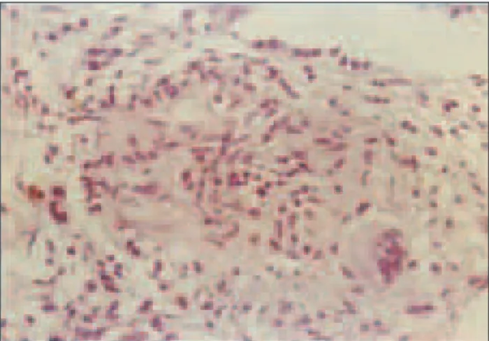
disease of the connective tissue. Therefore the patient was referred to the Department of Rheumatology for further investigation. In 1991 splenomegaly and chronic hepatitis were detected again. During 1993 the patient developed arterial hypertension and kidney failure (glomerular fil-tration rate – GFR 50-60 ml/min) and proteinuria (0.5-1 g/24 h). In 1997 clinical and laboratory signs of different systemic disease persisted. The criteria for systemic lu-pus erythematosus were not met, however non-specific markers of inflammation and autoantibodies were de-tected (FITC ANA 0, anti-DNK antibodies 0). Kidney biopsy was performed (pathologic analysis showed a few glomeruli only, with a thickened basal membrane, but with predominant tubulointerstitial fibrosis and sclerosis with vascular sclerosis). During the next two years, the patient was treated with azathioprine (75 mg) and corticoids (5-20 mg/day). In 2000, she was on corticosteroid therapy due to renal failure. First detected changes of the chest were interpreted as interstitial fibrosis, but it was not further investigated. The spleen and hepatogram were normal and only gamma globins were slightly elevated. In 2006, wors-ening of kidney function was detected (rising serum cre-atinine level up to 170 µmol/L and declining GFR of less than 40 ml/min). Kidney ultrasound revealed small sized kidneys with thin and bright renal parenchyma (features of chronic kidney disease). Since she had fever, chest CT was done revealing bilateral lymph node enlargement with interstitial changes (Figure 1). This time a pulmologist was consulted and bronchoscopy was indicated. Bronhoscopy revealed hyperemic mucosa covered with white nodules. Pathological analysis discovered epithelioid noncaseating granuloma verisimilar sarcoidosis (Figure 2). Also, the val-ue of angiotensin-converting enzyme (60.2 U/L) and cal-cium serum level (2.85 mmol/l) were elevated. Meanwhile, hepatogram and ultrasound liver and spleen findings were normal. The diagnosis of pulmonary sarcoidosis grade I/ II was established. In the meantime, the patient’s son aged 28 years was prior to our patient diagnosed pulmonary sarcoidosis. Since 2006 corticoid therapy (at initial dose of 30 mg/day) was decreased and excluded during 2008.
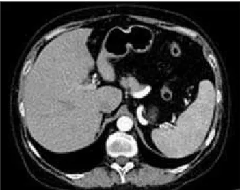
Throughout the first 6 months in 2009, the patient was feeling well. Corticosteroids were withdrawn because chest and abdominal computerized tomography (CT) findings and ACE level were normal. However, in the second part of 2009 she started to loose on weight (over 10 kg) and had nausea, and abdominal pains. Due to these complaints and renal failure progression the patient was admitted at Nephrology Clinic in January 2010. Abdominal CT re-vealed enlarged spleen with diffuse hypodense nodules (Figure 3) and chest CT showed a bilateral pulmonary re-ticulonodular pattern with mediastinal lymphadenopathy. For the first time ACE level was increased (up to 99 U/L) and polyclonal hypergammaglobulinemia was revealed (particularly IgG and IgA). Bone marrow biopsy was performed and findings were negative for neoplasia/lym-phoma. Tumor markers were also negative and infections were excluded. During this time dermatology examina-tion of the skin revealed changes typical of skin sarcoidosis (papular erythema with minimal whitish squamosis of the hands and legs). Thus, according to chest and abdominal CT, high ACE level, skin changes and other examinations, the diagnosis of pulmonary and extrapulmonary sarcoid-osis recurrence was established. Prednisolone (30 mg/day) therapy was started again in March 2010. Follow-up of the patient in June and September 2010 revealed improve-ment of subjective feeling, without prior symptoms during
Figure 1. Chest CT scan shows thickened interstitium of both lungs and enlarged hilar lymph nodes
Figure 3. Coronary reformatted abdominal CT revealed enlarged sple-en with numerous hypodsple-ense nodular lesions (arrows)
770
doi: 10.2298/SARH1212768S
prednisolone in dose of 10-20 mg/day and renal insuffi-ciency which stabilized (creatinine clearance 40 ml/min). Chest and abdominal CT showed absence of lymphade-nopathy and hepatosplenomegaly (Figure 4). Laboratory analysis showed that ACE level was within normal limits. On the last follow-up the patient was without pulmonary and spleen sarcoidosis.
DISCUSSION
In patients with splenic sarcoidosis the disease is usually severe, persistent and chronic, and tends to affect extra-thoracic organs [8, 9, 10]. At the time when our patient ex-perienced first signs and symptoms of splenic sarcoidosis almost 40 years ago, diagnostic procedures for sarcoidosis were developing. This case report confirms the rule that the diagnosis of sarcoidosis is more accurate when it is suspected by the doctor. Extrapulmonary presentation as the initial manifestation of the disease is often unrecog-nized and may mimic other etiologies. Therefore, in this patient the diagnosis of sarcoidosis remained an unsolved puzzle for over 40 years. The acute onset of the disease, with clinical manifestations of splenomegaly and hepati-tis, were significant prognostic parameters of the chronic nature of the disease in our case, as well as the indicated results by other investigators [5, 11]. At that time, during the 70-ies similar examples were found in the literature when splenectomy was performed because of suspected lymphoma but later pathological finding revealed sarcoid-osis [12, 13, 14]. It is well known that corticosteroids are indicated as a very effective therapeutic agent at the clini-cal course of “disease with no name”. Possibly, temporary doses of prednisolone in the dose of 5-20 mg were not sufficient to stop this chronic inflammatory disease and besides, there were permanently appearing new problems. Thus, in our case splenomegaly was established for the first time in 1976 during laparoscopy, but also in 1987, 1991 and 2010 by ultrasound or CT examination and at that time, according to the medical data, corticosteroid therapy was started many times with different durations
(1-2 years). Spontaneous remissions of sarcoidosis occur in nearly two thirds of patients, but chronic, progressive disease may result in severe sequelae [4, 5, 10]. This could probably be the explanation for the undefined liver find-ings. Three times repeated liver biopsies pointed to fibro-sis in a systemic disease or suspected liver TBC because of granuloma detection. Laboratory findings suggested spreading of the disease to the kidneys, furthermore bi-opsy of kidneys confirmed interstitial nephritis without granulomas, which was also found by Mahévas M. et al. [15] in 10 of 43 examined patients. Therapy with azathio-prine and corticosteroids during the next two years did not stop the progression of chronic renal failure but the progression was relatively slow during the next 10 years, with proteinuria as can be expected in chronic mainly tubulointerstitial processes. During 2006, when cortico-steroid therapy was stopped, radiological, bronchoscopy and pulmonary changes appeared which was typical of sarcoidosis. For the first time, after a lymph node biopsy epithelioid non-caseinating granuloma verisimilar sar-coidosis was established. On corticoid therapy a complete remission was achieved. After long time (1991-2009) the patient was without splenomegaly but in 2010, the disease reactivated with enlarged spleen and diffuse hypodense nodules; a rare form of spleen sarcoidosis seen on abdomi-nal CT that was previously reported in the literature [16]. Chest CT and high level of ACE confirmed reactivation of sarcoidosis.
At the end it is very important to note that during a long-term follow-up period there was no development of hypersplenism, anemia or elevated transaminases. Only once, hypercalcemia was detected, but calciuria was not measured because GFR was less than 60 ml/min. All the time the high sedimentation rate as well as hypergama-globulinemia was presented as signs of nonspecific inflam-mation. Finally, after 5 months of corticosteroid therapy the patient was again in a complete remission. Also, this case is interesting because of the presence of sarcoidosis in the patient’s family (patient’s son also had recurrent pul-monary sarcoidosis). It is possible that genetic factors and environmental agents of infection may result in autoim-mune response that is manifested as sarcoidosis [17]. But it is also likely that genetic factors may be important in defining the pattern of disease presentation and progres-sion as well as its overall prognosis. In concluprogres-sion, clinical manifestations of sarcoidosis, especially abdominal local-ization are extremely heterogeneous and overlap with a wide spectrum of diseases, and due to this fact biopsy and pathologic confirmation of sarcoidosis is a necessity whenever possible. Final diagnosis of spleen sarcoidosis can be confirmed in the presence of pathologically verified sarcoidosis of the lungs as in our case.
ACKNOWLEDGEMENTS
This work was supported by the Ministry of Science and Technological Development of Serbia, contract No. 175046, 2011–2014.
Škodrić-Trifunović V. et al. Mystery Called Sarcoidosis: Forty-Four Years Follow-Up of Chronic Systemic Disease
Figure 4. Contrast enhanced abdominal CT after treatment
771
Srp Arh Celok Lek. 2012 Nov-Dec;140(11-12):768-771
www.srp-arh.rs REFERENCES
1. James DG, Zumla A. The Granulomatous Disorders. Cambridge: Cambridge University Press; 1999.
2. Milovanović A, Milovanović J, Torbica N. Ocena radne sposobnosti kod neurosarkoidoze. Srp Arh Celok Lek. 2006; 134(5-6):238-40.
3. Morgenthau SA, Iannuzzi MC. Recent advances in sarcoidosis. Chest. 2011; 139(1):174-82.
4. Sestini P, Marchi B, Marri D. Liver disorders in pulmonary sarcoidosis. Sarcoidosis. 1993; 10:177-9.
5. James DG, Sherlock S. Sarcoidosis of the liver. Sarcoidosis. 1994; 11:2-6.
6. Čolović N, Čemerikić V, Čolović R, Zogović S, Stojković M. Hamartom slezine. Srp Arh Celok Lek. 2000; 128(3-4):331-4. 7. Hunninghake GW, Costabel U, Ando M, Baughman RP, Cordier
JF, du Bois R, et al. ATS/ERS/WASOG statement on sarcoidosis. Sarcoidosis Vasc Diff Lung Dis. 1999; 16:149-73.
8. Salazar A, Mañá J, Corbella X, Albareda JM, Pujol R.
Splenomegaly in sarcoidosis: a report of 16 cases. Sarcoidosis. 1995; 12:131-4.
9. Fordice J, Katras T, Jackson RE, Cagle PT, Jackson D, Zaleski H, et al. Massive splenomegaly in sarcoidosis. Sou Med J. 1992; 85:774-8.
10. Judson M. Hepatic and splenic sarcoidosis in Baughmans sarcoidosis. Lung Biology in Health and Diseases. 2006; 26:571-92.
11. Sharma OP, Vucinic V, James DG. Splenectomy in sarcoidosis: indications, complications, and long-term follow-up. Sarcoidosis Vasc Diff Lung Dis. 2002; 19(1):66-70.
12. Longcope WT, Freiman DG. A study of sarcoidosis based on a combined investigation of 160 cases including 30 autopsies from The Johns Hopkins Hospital and Massachusetts General Hospital. Medicine (Baltimore). 1952; 31:1-132.
13. Sharma OP. Splenic rupture in sarcoidosis: Report of unusual case. Am Rev Respir Dis. 1967; 96:101-2.
14. Di Sabatino A, Carsetti R, Corazza GR. Posplenectomy and hyposplenic states. Lancet. 2011; 378(9785):86-97. 15. Mahévas M, Lescure FX, Boffa JJ, Delastour V, Belenfant X,
Chapelon C, et al. Renal sarcoidosis: clinical, laboratory, and histological presentation and outcome in 47 patients. Medicine (Baltimore). 2009; 88(2):98-106.
16. Warshauer DM, Dumbleton SA, Molina PL, Yankaskas BC, Parker LA, Woosley JT. Abdominal CT findings in sarcoidosis: radiologic and clinical correlation. Radiology. 1994; 192:93-8.
17. Schurmann M, Lympany PA, Philipp R, Muller-Myhsok B, Wurm K, Schlaak M, et al. Familial sarcoidosis is linked to the major histocompatibility complex region. Am J Respir Crit Care Med. 2000; 162(3):861-4.
КРАТАК САДРЖАЈ
Увод Ово је при каз бо ле сни це ста ре 61 го ди ну. С об зи ром на то да су од пр вих симп то ма бо ле сти до ко нач не ди јаг но-зе сар ко и до но-зе про шле 44 го ди не, то је био раз лог да овај слу чај об ја ви мо.
При каз бо ле сни ка То ком на ве де ног пе ри о да с вре ме на на вре ме су се ја вља ле по ли морф не те го бе, као што су ади на-ми ја, муч ни на, бол у тр бу ху, ар трал ги ја, на-ми ал ги ја, гу би так те ле сне те жи не (8–10 kg) итд. У кли нич кој сли ци до ми ни-ра ли су спле но ме га ли ја, хе па ти тис и ар тни-рал ги ја, а ка сни је се раз ви ла и хро нич на ин су фи ци јен ци ја бу бре га. У ла бо ра-то риј ским ана ли за ма мар ке ри ин фла ма ци је и аура-то ан ти те ла би ли су по ви ше ни. Бо ле сни ца је би ла хо спи та ли зо ва на у
раз ли чи тим ин тер ни стич ким кли ни ка ма (га стро ен те ро ги ја, алер го ло ги ја, ре у ма то ло ги ја, не фро ло ги ја, пул мо ло-ги ја). Би оп си ја је тре је ура ђе на три пу та, би оп си ја бу бре га и рек ту ма јед ном и на кра ју су ура ђе не брон хо ско пи ја и би оп си ја плу ћа. Ко нач но, че тр де сет го ди на на кон по чет ка пр вих симп то ма бо ле сти, 2006. го ди не је по твр ђе на ди јаг-но за сар ко и до зе плу ћа.
За кљу чак Ко нач на ди јаг но за сар ко и до зе сле зи не по ста-вље на је по сле па то хи сто ло шке по твр де сар ко и до зе плу ћа. Овај слу чај је по себ но за ни мљив због по сто ја ња фа ми ли јар-не сар ко и до зе (син бо ле сни це је та ко ђе бо ло вао од ре ци-ди ви ра ју ће сар ко и до зе плу ћа).
Кључ не ре чи: сар ко и до за; је тра; сле зи на; плу ћа; бу брег
Мистерија звана саркоидоза: четрдесет и четири године клиничког праћења
хроничне системске болести
Весна Шкодрић-Трифуновић1,2, Виолета Вучинић1,2, Сања Симић-Огризовић1,3, Ружа Стевић1,4, Михаило Стјепановић2,
Катарина Илић5, Живорад Савић4
1Медицински факултет, Универзитет у Београду, Београд, Србија; 2Клиника за пулмологију, Клинички центар Србије, Београд, Србија; 3Клиника за нефрологију, Клинички центар Србије, Београд, Србија;
4Центар за радиологију и магнетну резонанцу, Клинички центар Србије, Београд, Србија; 5Катедра за фармакологију, Фармацеутски факултет, Универзитет у Београду, Београд, Србија