Rev. Bras. Cir. Plást. 2011; 26(3): 546-9
546
Batista KT et al.
Plexiform neuroibroma of the upper limb
Neuroibroma plexiforme de membro superior
ABSTRACT
The authors present an unusual case of plexiform neuroibroma affecting the upper limb in a patient diagnosed with type 1 neuroibromatosis. Tumor resection was performed on the median nerve. The patient showed maintenance of limb function and remission of symptoms of pain after four years of follow-up.
Keywords: Neuroibromatosis 1. Neuroibroma. Neuroibroma, plexiform. Upper
extre-mity.
RESUMO
Os autores apresentam um caso incomum de neuroibroma plexiforme acometendo o membro superior, com diagnóstico de neuroibromatose do tipo 1. Realizou-se a ressecção do tumor no nervo mediano. A paciente evoluiu com manutenção da função do membro e remissão dos sintomas de dor após seguimento de quatro anos.
Descritores: Neuroibromatose 1. Neuroibroma. Neuroibroma plexiforme. Extremidade
superior. Study conducted at SARAH
Network of Rehabilitation Hospitals, Brasília, DF, Brazil. Submitted to SGP (Sistema de Gestão de Publicações/Manager Publications System) of RBCP (Revista Brasileira de Cirurgia Plástica/Brazilian Journal of Plastic Surgery). Received: October 16, 2009 Accepted: April 6, 2010
1. Member of the Brazilian Society of Plastic Surgery; Physician; Plastic Surgeon of SARAH Network of Rehabilitation Hospitals, Brasília, DF, Brazil. 2. Doctor of Orthopedics; Surgeon-in-Chief of SARAH Network of Rehabilitation Hospitals, Brasília, DF, Brazil.
KÁTIA TORRES BATISTA1
HUGO JOSÉDE ARAÚJO1
ALOYSIO CAMPOS DA PAZ
JÚNIOR2
Franco T et al.
Vendramin FS et al.
CASE REPORT
INTRODUCTION
Neuroibromas, tumors that originate in Schwann cells and normally show the clinical symptoms of type 1 neuroi-bromatosis (NF1), are rarely found in the hands.
A diagnosis of NF1 is considered if two of the following characteristics are present:
• six or more light brown stains with a diameter of >0.5 cm in prepubertal and >1.5 cm in postpubertal individuals; • two or more neuroibromas of any type or one plexiform
neuroibroma (net shaped);
• ephelides in the axillary or groin area; • optic glioma (tumor in the optic nerve); • two or more Lisch nodules in the iris;
• sphenoid dysplasia or thinning of the cortex of long bones, with or without pseudoarthrosis;
• irst-degree relative with NF1; • biopsy indings1-3.
Neuroibromas are benign tumors, usually single and originating within the nerve fascicles. Enucleation is not possible, so total tumor excision is required, leaving some damage to hand function. Surgery is indicated in cases of excessive tumor growth, pain, and when there is suspicion of malignancy4.
This paper aims to describe the evolution of a case of upper limb plexiform neurofibroma in a patient with NF1.
CASE REPORT
A 3-year-old female child was admitted to the unit of SARAH Network of Rehabilitation Hospitals in Brasília (SARAH-Brasília, Brazil) in 1996 with a history of left wrist tumor measuring 2 cm x 3 cm, diagnosed as neuroibroma. NF1 was diagnosed on the basis of the following criteria: family history and presence of light brown stains, neuroi-bromas, and plexiform neuroibroma.
Rev. Bras. Cir. Plást. 2011; 26(3): 546-9 547
Plexiform neuroibroma of th upper limb
with the second and fifth fingers; preserved hook grip; preserved joint mobility and strength; and neurofibromas distributed bilaterally in the brachial plexus and in other peripheral nerves of the upper limbs. Moreover, there was evidence of a bulky mass in the pelvic region; this mass was pushing against the structures of the bladder and recto-sigmoid region and extending along the lower limb and gluteal region, corresponding to the lumbosacral plexus. The patient did not have functional complaints related to these tumors.
The patient underwent electromyography tests (Figure 1) and MRI in sagittal, coronal, and axial sections. These tests revealed elongated cylindrical structures, grouped into a bulky expansive injury, from the anteromedial face of the arm and forearm to the palmar face of the hand and passing through the carpal tunnel.
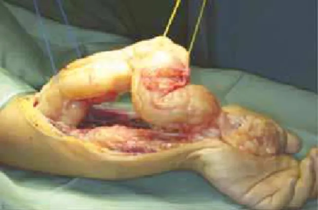
A tumor (size, 23 cm x 4 cm; weight, 172 g) located in the left upper limb was resected (Figures 2 to 5). Histopatho-logical evaluation of frozen tissue sections led to a diagnosis of neuroibroma of the median nerve.
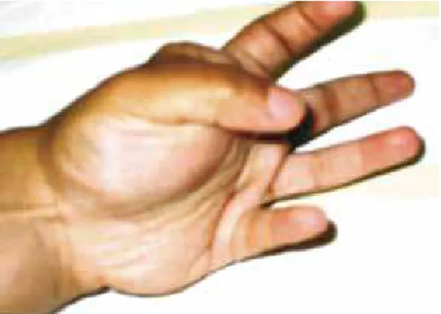
The patient recovered with improvement in hand func-tion and relief from pain symptoms. The patient retained clamping movements of the thumb with the other digits (Figures 6 to 10) and object gripping, and the sensitivity
map, measured using Semmes-Weinstein monofilaments, remained unchanged. Finally, there was no evidence of tumor recurrence in the operated area for four years.
Figure 1 – Results of the electromyographic test, showing marked
reduction in the extent and speed in the motor conduction study.
Figure 2 – Marking held at the immediate preoperative period of
patient with plexiform neuroibroma of the left upper limb.
Figure 3 – Dissection of the tumor on the left median nerve.
Figure 4 – Resection of the tumor on the left median nerve.
Rev. Bras. Cir. Plást. 2011; 26(3): 546-9
548
Batista KT et al.
DISCUSSION
Neuroibromas may occur anywhere in the body and viscera, and rarely occur in the hands. Some authors1,2 have
reported an incidence of 0.8% for neuroibromas in the hands and from 10% to 15% for NF1.
Plexiform neuroibromas show diffuse growth of the endoneurium, affecting all branches of the nerve, as demons-trated in this case of tumor of the median nerve and its branches, with severe constriction of the carpal tunnel3-5.
Plexiform neuroibromas are considered dificult to diagnose because their incidence is very low6, but when the criteria of
neuroibromatosis7 are fulilled, the suspicion of plexiform
neuroibroma increases. However, clinicians should carry out differential diagnoses for other neural tumors, such as schwannoma8 and sarcomatous degeneration2.
The surgical treatment of neuroibromas is controversial, since it is necessary to excise the entire nerve for tumor removal.
According to Seddon4, tumor resection is not necessary,
unless the tumor causes pain, shows exuberant growth, or impairs
function, as in this case. The evolution of neuroibromatosis with neuroibroma as a slow, asymptomatic growth over eight years has been reported. Surgery was indicated because of hand dysfunc-tion and pain due to tumor growth. It was possible to remove the
Figure 6 – Closing of the surgical wound.
Figure 7 – Thumb clamping movement with the second inger,
observed during the preoperative period.
Figure 8 – Maintenance of clamping movement after the sixth
month of tumor resection.
Figure 9 – Failure to perform a clamping movement of the thumb
with the ifth left inger, observed in the preoperative period.
Figure 10 – Recovery of the clamping movement of the thumb with
Rev. Bras. Cir. Plást. 2011; 26(3): 546-9 549
Plexiform neuroibroma of th upper limb
Correspondence to: Kátia Torres Batista
SMHS – Quadra 501 – cj. A – Brasília, DF, Brazil – CEP 70335-901 E-mail: [email protected]
plexiform neuroibroma of the median nerve; the symptoms of pain reduced and hand function improved over a follow-up period of four years, with no signs of recurrence of this tumor.
REFERENCES
1. Basheer H, Rabia F, Basheer H, el-Helw K. Neuroibromas of digital nerves. J Hand Surg Br. 1997;22(1):61-3.
2. Birch R. Peripheral nerve tumors. In: Dyck PJ, Thomas PK, eds. Peri-pheral neuropathy. 3rd ed. Philadelphia: WB Saunders; 1993. p. 1623-39.
3. Nambi GI, Gupta AK, Kumaran S. Plexiform neuroibroma of the inger. J Plast Reconstr Aesthet Surg. 2008;61(11):1402-3.
4. Seddon HJ. Surgical disorders of peripheral nerves. Baltimore: Willia-ms and Wilkins; 1972.
5. Punia RS, Dhingra N, Mohan H. Cutaneous plexiform schwannoma of the inger not associated with neuroibromatosis. Am J Clin Dermatol. 2008;9(2):129-31.
6. Ilyas AM, Nourissat G, Jupiter JB. Segmental neuroibromatosis of the hand and upper extremity: a case report. J Hand Surg Am. 2007;32(10):1538-42.
7. Gajeski BL, Kettner NW, Awwad EE, Boesch RJ. Neuroibromatosis type I: clinical and imaging features of Von Recklinghausen’s disease. J Manipulative Physiol Ther. 2003;26(2):116-27.