Crosstalk between Thyroid Hormone Receptor and Liver
X Receptor in the Regulation of Selective Alzheimer’s
Disease Indicator-1 Gene Expression
Emi Ishida, Koshi Hashimoto*, Shuichi Okada, Tetsurou Satoh, Masanobu Yamada, Masatomo Mori
Department of Medicine and Molecular Science, Gunma University Graduate School of Medicine, Maebashi, Gunma, Japan
Abstract
Selective Alzheimer’s disease (AD) indicator 1 (Seladin-1) has been identified as a gene down-regulated in the degenerated lesions of AD brain. Up-regulation of Seladin-1 reduces the accumulation of b-amyloid and neuronal death. Thyroid hormone (TH) exerts an important effect on the development and maintenance of central nervous systems. In the current study, we demonstrated that Seladin-1 gene and protein expression in the forebrain was increased in thyrotoxic mice compared with that of euthyroid mice. However, unexpectedly, no significant decrease in the gene and protein expression was observed in hypothyroid mice. Interestingly, an agonist of liver X receptor (LXR), TO901317 (TO) administrationin vivo increased Seladin-1 gene and protein expression in the mouse forebrain only in a hypothyroid state and in the presence of mutant TR-b, suggesting that LXR-a would compensate for TR-b function to maintain Seladin-1 gene expression in hypothyroidism and resistance to TH. TH activated the mouse Seladin-1 gene promoter (21936/+21 bp) and site 2
including canonical TH response element (TRE) half-site in the region between2159 and2154 bp is responsible for the positive regulation. RXR-a/TR-b heterodimerization was identified on site 2 by gel-shift assay, and chromatin immunoprecipitation assay revealed the recruitment of TR-b to site 2 and the recruitment was increased upon TH administration. On the other hand, LXR-autilizes a distinct region from site 2 (2120 to2102 bp) to activate the mouse Seladin-1 gene promoter. Taking these findings together, we concluded that TH up-regulates Seladin-1 gene expression at the transcriptional level and LXR-amaintains the gene expression.
Citation:Ishida E, Hashimoto K, Okada S, Satoh T, Yamada M, et al. (2013) Crosstalk between Thyroid Hormone Receptor and Liver X Receptor in the Regulation of Selective Alzheimer’s Disease Indicator-1 Gene Expression. PLoS ONE 8(1): e54901. doi:10.1371/journal.pone.0054901
Editor:Michelina Plateroti, University Claude Bernard Lyon 1, France
ReceivedAugust 13, 2012;AcceptedDecember 17, 2012;PublishedJanuary 24, 2013
Copyright:ß2013 Ishida et al. This is an open-access article distributed under the terms of the Creative Commons Attribution License, which permits unrestricted use, distribution, and reproduction in any medium, provided the original author and source are credited.
Funding:This work was supported by grant from Japan Intractable Disease Research Foundation to KH. The funders had no role in study design, data collection and analysis, decision to publish, or preparation of the manuscript.
Competing Interests:The authors have declared that no competing interests exist.
* E-mail: [email protected]
Introduction
Alzheimer’s disease (AD) is one of the major causes of dementia and a serious concern to the human society [1,2]. However, the pathogenesis of the disease has not yet been revealed.
Thyroid hormone (TH) is well known to play an important role in the development and maintenance of the central nervous system in mammals [3,4]. TH exerts its biological function through thyroid hormone receptors (TRs). TRs are nuclear hormone receptors, to which triiodothyronine (T3) binds at a high-affinity order as a native ligand. TRs possess at least two isoforms, TR-a
and -b (Nr1a1 and Nr1a2), and several isoforms exist as two or three subtypes, respectively (a1,a2, b1,b2, andb3) [5]. It is of note that only TR-a1,b1 andb2 have both a ligand binding and a DNA binding domain [6]. TR-a1 is widely expressed in tissues including heart, muscle, intestine, bone, and brain and plays a key role in regulating postnatal development and cardiac metabolism, whereas TR-b1 is also widely expressed in brain, cochlea, pituitary, kidney, lung, heart and at its highest level in the liver regulating multiple steps in hepatic metabolism as well as thyroid hormone levels [6]. TR-b2 expression is mainly in the pituitary, the hypothalamic TRH neurons, the developing inner ear and retina [7]. Thus, both TR-aand TR-bplay an important role for the development and the maintenance of the central nervous
system even though their expression patterns are spatiotemporally distinct [8–10]. Hypothyroidism sometimes leads to the develop-ment of dedevelop-mentia-like symptoms, especially in elderly patients [11,12]. The TH receptor (TR)b-D337T knock-in (TRKI) mouse demonstrates severe cerebellar ataxia and cognitive dysfunction [13]. As such, although case reports and basic studies support the idea that TH is closely related to AD pathogenesis and could be beneficial to cure AD [14–16], large-scale clinical studies examining the relationship between thyroid function and AD have led to controversial conclusions [17–20].
Among many genes related to AD, we focused on selective AD indicator 1, Seladin-1 gene. Seladin-1 gene expression is down-regulated in the vulnerable region in the brain of AD patients [21]. Up-regulation of Seladin-1 in the neuron leads to the reduction of
some neuronal precursor cell lines induced Seladin-1 gene expression [27]. However, the molecular mechanism by which TH up-regulates Seladin-1 mRNA levels is yet to be elucidated.
Liver X receptors (LXRs) are nuclear oxysterol receptors and play pivotal roles in cholesterol metabolism [28,29]. LXRs comprise two isoforms, LXR-a and -b. Both isoforms are expressed in the brain, although the latter is expressed at significantly higher levels [30]. An artificial agonist of LXR, TO901317 (TO), reduces b-amyloid accumulation and AD-specific pathological changes in the brain of an AD model mouse [31–33]. Since LXR response element (LXRE) is identified in the second intron in human Seladin-1 gene promoter, another group has suggested that Seladin-1 gene promoter may be a target of LXR [34]. However, the molecular mechanism by which LXRs regulate Seladin-1 gene expression has been yet to be elucidated. TRs and LXRs are members of the nuclear receptor superfamily. Although LXRs and TRs belong to two distinct receptor subgroups with respect to ligand-binding affinity [35], the two receptor systems show similarity with respect to molecular mechanism, target genes, and physiological roles [36]. Since both TRs and LXRs play an important role in metabolic regulation, form heterodimers with retinoid X receptors (RXRs), and bind to direct repeat-4 (DR-4) with identical geometry and polarity [28,36–38], crosstalk between the two receptors has been reported, especially on lipid metabolism-related genes [37–43].
In the current study, we demonstrate that TH increased Seladin-1 gene and protein expression in the mouse forebrain. However, no significant difference between euthyroid and hypothyroid mice was observed. Interestingly, TO administration
in vivo increased Seladin-1 gene and protein expression in the mouse forebrain only in a hypothyroid state and in the presence of mutant TR-b. We also show that TH activates mouse Seladin-1 gene promoter and identify a novel positive thyroid hormone response element named site 2 including canonical thyroid hormone response element (TRE) half-site in the mouse promoter. Taking these findings together, we concluded that TH up-regulates Seladin-1 gene expression at the transcriptional level and that LXR-awould compensate for TR-bfunction to maintain Seladin-1 gene expression in hypothyroidism and resistance to TH (RTH). Thus, our data strongly suggests that TR and LXR crosstalk in Seladin-1 gene regulation.
Materials and Methods
Animals
8 to 12-week-old male C57/BL6 (B6) and TRKI mice (generously provided by Prof. Fredric E. Wondisford, Johns Hopkins medical institute, Baltimore) were employed for the study. All aspects of animal care were approved by the Institutional Animal Care and Use Committee of Gunma University Graduate School of Medicine (Maebashi, Gunma, Japan, Protocol number #10-059). Animals were maintained on a 12 h light/12 h dark schedule (lights on at 06:00 h) and fed laboratory chow as indicated and given water ad libitum. Hypothyroidism was induced in the B6 mice by the inclusion of 0.1% methimazole (MMI) in the drinking water and 1% (w/w) propylthiouracil (PTU) in the chow for 21 days [37,41]. To induce a thyrotoxic state, the B6 mice were injected daily with 10mg per 20 g body weight of T3 for 7 days [37]. For TO treatment, 1 mg per 20 g body weight of TO901317 (#71810 Cayman, Ann Arbor, MI) was intraperito-neally administered daily for 7 days [31,33] or vehicle (DMSO) was administered for Sham group. The number of mice receiving each treatment is indicated in the figures. Serum-free T3and T4 levels were measured by SRL (Tokyo, Japan). We anatomically
excised cerebellum, diencephalon, pituitary, hypothalamus, and medulla oblongata from the whole brain of mouse and obtained the rest as forebrain for the analysis as we reported previously [13].
Plasmids
The mouse Seladin-1 gene promoter (21936/+21 bp) plasmid, which contained the region from21936 to+21 bp of the mouse Seladin-1 gene, was generated by genomic PCR using 59
-GTGTGGTACCTGGTGGCAGGAGAGAGCCCC-39 as a
sense primer and 59
-GTGTAGATCTG-TAAGTCGGGCCCGCCGCC-39 as an anti-sense primer. An
Asp718orBglIIrestriction enzyme site was introduced into each primer sequence so that the PCR product could be subcloned between these sites in the pGL4-Luc vector (Promega, Madison, WI). The deletion mutants and a site 1 or site 2 point mutants were generated with PCR site-directed mutagenesis [37,43,44]. All human TR-b1 and its mutants, human TR-a1, RXR-a, murine LXR-a, and LXR-bcDNAs were placed into an SV40 expression construct, pSG5 [44]. All PCR-generated constructs were verified by sequencing the DNA.
Transfections and Luciferase Assay
For the luciferase assay, we employed CV-1 cells. 10mg of the reporter plasmid and 0.25mg of human TR-b1 or TR-a1 or mouse LXR-a1 or LXR-bin pSG5 was transfected per plate of a 6-well format into CV-1 cells using the calcium-phosphate method. Sixteen hours after transfection, cultures were treated with Dulbecco’s modified Eagle’s medium (DMEM) containing 10% resin charcoal double-stripped fetal bovine serum for 8 hours in the absence or presence of 1028M T3 or 1026M TO. All transfections were equalized for the same total amount of expression vector using an empty vector as needed. We performed
b-gal assays to confirm the transfection efficiency of the luciferase assay for each experiment at least once and found no significant difference in transfection efficiency among the plates. Data are presented as fold basal activation over vector (pSG5) in the absence of ligand stimulation6 SEM or as otherwise indicated. Luciferase activity is expressed as arbitrary light units permg of cellular protein. All transfection experiments were repeated at least twice with triplicate determinations.
Western Blotting
For analysis of the protein expression of Seladin-1 (DHCR24), 30mg of whole cell extract from mouse forebrain was subjected to SDS-PAGE. Western blotting was performed using a rabbit anti-DHCR24 polyclonal antibody (#ab40490, Abcam, Cambridge, UK) to detect Sealdin-1 protein levels and anti-glyceraldehyde-3-phosphate dehydrogenase (GAPDH) (#ab8245, Abcam, Cam-bridge, UK) as a control. The Seladin-1 detects a specific band at 55 kDa in tissues and cells. The bands were quantitatively measured using Adobe Photoshop 7.0 (Adobe Systems Corp., San Jose, CA) and ImageJ (Rasband, W.S., ImageJ, US National Institutes of Health, Bethesda, Maryland, USA, http://imagej.nih. gov/ij/, 1997–2011). All Western blotting experiments were repeated at least three times with similar results. Seladin-1 protein levels are normalized by GAPDH. Data are presented as fold basal euthyroid (E) levels6SEM or as otherwise indicated.
was reverse-transcribed with random hexamers using the Taq-manTM Reverse Transcription Reagent kit (Applied Biosystems, Carlsbad, CA) according to the manufacturer’s protocol. Mouse Seladin-1 mRNA expression was analyzed using TaqmanTM probes (Mm00519071_m1, Applied Biosystems, Carlsbad, CA). We set up a standard curve for each real-time PCR and confirmed that all PCR products were on the standard curve as we previously described [43]. The PCR results were normalized to mouse cyclophilin A expression using a TaqmanTM probe (Mm02342430_gl, Applied Biosystems, Carlsbad, CA). The number of samples is indicated in the figure legends.
Gel-shift Assays
Electrophoretic mobility-shift assays (EMSAs, gel-shift assays) were performed as described previously [45]. Mouse LXR-a, human TR-b1, and human RXR-a recombinant proteins were synthesized from constructs in the pSG5 expression vector, using the TNT T7 Quick Coupled Transcription/ Translation System (Promega, Madison, WI). Binding reactions contained 20 mM HEPES (pH 7.6), 50 mM KCl, 12% glycerol, 1 mM dithiothreitol (DTT), 1mg of poly (dI-dC)(dI-dC), and 6ml of each of the synthesized nuclear receptors or unprogrammed reticulocyte lysates. Double-stranded oligonucleotides (DR-4 for rat cholesterol
7a-hydroxylase (CYP7A1): 59- TGTTTGCTTTGGTCACT-CAAGTTCAA-39, Site 1 probe ; 59- CTCCCACCCTGGG-GAGGTTACCCAATGCACAACT-39, Site 2 probe ; 59
-AGCGCCCCCAGCCCAGGTCAGCCCCTCCTCGCAC-39)
were labeled with [a-32P] deoxy-CTP by a fill-in reaction using a Klenow fragment of DNA polymerase. Binding reactions were performed at room temperature for 30 min. For competition experiments, a 20- or 40-fold molar excess of cold oligonucleotides was included. For supershift experiments, 3ml of rabbit
anti-TR-b1 polyclonal antibody (06-539, Upstate, Lake Placid, NY) or mouse anti-LXR-a monoclonal antibody (PP-PPZ0412-00, Per-seus proteomics, Tokyo, Japan) was added and the mixture was incubated for an additional 30 min at room temperature. The protein-DNA complexes were resolved on a 4% polyacrylamide gel in 0.5X TBE (45 mM Tris-base, 1 mM EDTA). All gel-shift assays were repeated at least three times with similar results and a representative result is shown.
Small Interfering (si) RNA against LXRs
We employed Dharmacon’s siRNA against human LXR-a (L-003413-00-0005, ON-TARGET plus SMART pool, human NR1H3) and LXR-b (L-003412-00-0005, ON-TARGET plus SMART pool, human NR1H2) (Thermo Fisher Scientific Inc., Waltham, MA). The target sequences of siRNA for human LXR-a
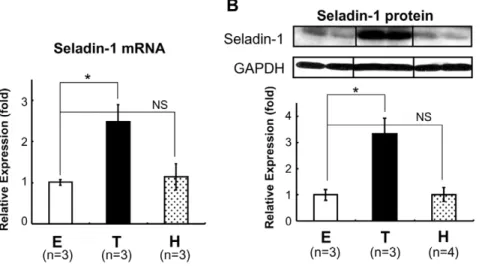
are as follows: 59-GAGUUUGCCUUGCUCAUUG-39; 59 -CGA-CUGAUGUUCCCACGGA-39; 59 -GAACAACUGGGCAU-GAUCG-39; 59-CCUCAAGGAUUUCAGUUAU-39, and the target sequences for LXR-b were as follows: 59 -AACAGCGG-CUCAAGAACUA-39; 59-CUAAGCAAGUGCCUGGUUU-39; 59-GAAGAAGAUUCGGAAACAG-39; 59 -CAACCACGAGA-CAGAGUGU-39. SiRNA for the negative control (ON-TARGET plus Non-targeting pool, D-001810-10-20, Thermo Fisher Scien-tific, Inc., Waltham, MA) was employed. SiRNAs were transfected into HTB185 cells, derived from human medulloblastoma using the lipofection method (Lipofectamine RNAiMAX, Invitrogen). Briefly, in the 6-well format, 50 pmol of siRNAs against each LXR (a or b) or 100 pmol of non-targeting siRNA per well were Figure 1. TH induced Seladin-1 mRNA and protein expression in the mouse forebrain.C57/B6 mice (8- to 12-week-old males) were rendered hypothyroid (H) with MMI and PTU diet or treated with T3(T). E: euthyroid basal status (control). The number of mice involved in each
treatment is indicated in the figures. Forebrain total RNA was isolated and one microgram of total RNA was subjected to reverse transcription (RT). RT real-time PCR analysis for Seladin-1 was performed using mouse forebrain cDNA (A). Relative values (mean6S.E.) normalized by cyclophilin A mRNA levels compared with E are shown as relative expression (fold). Western blot analysis using mouse forebrain whole cell extract was performed (B). Representative Western blots for Seladin-1 are shown in the upper panel. Relative optical densities (O.D.) (mean6S.E.) normalized by GAPDH levels compared with ‘‘E’’ are shown as relative expression (fold). Anasteriskindicates that the difference between the denoted pairs is significant at a confidence level ofp,0.05(*). NS: not significant.
doi:10.1371/journal.pone.0054901.g001
Table 1.Free T4and free T3levels in each thyroid state.
Free T4(ng/dL) Free T3(pg/mL)
E (Euthyroid) 1.2360.03 3.4260.12
T (Thyrotoxic) 0.2860.07** 66.768.3*
H (Hypothyroid) 0.1460.04** 1.4560.13***
Results are expressed as mean6SEM (n = 3).
Anasteriskindicates that the difference compared with ‘‘E’’ is significant at a confidence level ofp,0.05 (*),p,0.01 (**),p,0.001 (***), by Welch’st-testing. doi:10.1371/journal.pone.0054901.t001
transfected into the HTB185 cells. Sixteen hours after transfection, cultures were treated with DMEM containing 10% resin charcoal double-stripped fetal bovine serum for 8 hours in the absence or presence of 1026M TO. After incubation, total RNA was extracted from the cells and real-time quantitative PCR assays were performed as described above. Human Seladin-1, LXR-a
and LXR-b mRNA expression levels were analyzed using TaqmanTM probes (Hs00207388_m1, Hs00172885_m1 and Hs00173195_m1, respectively. Applied Biosystems, Carlsbad, CA).
Chromatin-immunoprecipitation (ChIP) Assay
ChIP assays were performed using a kit (ChIP-ITTM Express) from Active Motif (Carlsbad, CA) in accordance with the manufacturer’s protocol with some modifications. Briefly, in prior to ChIP assays, the mouse Seladin-1 gene promoter (21113/
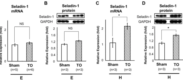
+21 bp) in pGL4 was transfected into the HTB185 cells with Lipofectamine 2000 (Invitrogen, Carlsbad, CA). The transfected HTB185 cells were incubated in medium containing 10% resin-charcoal double-stripped fetal bovine serum with or without 1028M T3. After incubation, formaldehyde (37%) was directly Figure 2. TO induced Seladin-1 mRNA and protein expression in the mouse forebrain in hypothyroid status.TO was administered to mice in euthyroid (E) or hypothyroid (H) status. The number of mice employed is indicated in the figures. Reverse transcription (RT) real-time PCR analysis for Seladin-1 was performed using mouse forebrain cDNA (A, C). Relative values (mean6S.E.) normalized by cyclophilin A mRNA levels compared with Sham are shown as relative expression (fold). Western blot analysis using mouse forebrain whole cell extract was performed (B, D). Representative Western blots for Seladin-1 are shown in the upper panel (B, D). Relative values (mean6S.E.) normalized by GAPDH protein levels compared with Sham are shown as relative expression (fold). Anasteriskindicates that the difference between the denoted pairs is significant at a confidence level ofp,0.05(*). NS: not significant.
doi:10.1371/journal.pone.0054901.g002
Figure 3. TO induced Seladin-1 mRNA and protein expression in the forebrain in TRKI mice.RT real-time PCR analysis for Seladin-1 was performed using mouse forebrain cDNA (A, C). Relative values (mean6S.E.) normalized by cyclophilin A mRNA levels compared with Sham are shown as relative expression (fold). Western blot analysis using mouse forebrain whole cell extract was performed (B, D). Representative Western blots for Seladin-1 are shown in the upper panel (B, D). The number of mice employed is indicated in the figures. Relative values (mean6S.E.) normalized by GAPDH protein levels compared with Sham are shown as relative expression (fold). Anasteriskindicates that the difference between the denoted pairs is significant at a confidence level ofp,0.05(*). NS: not significant.
added to the culture at a final concentration of 1% and the cells were incubated for 15 min at 37uC to crosslink protein to DNA. The cells were pelleted and resuspended in 600ml of lysis buffer
supplemented with 3ml of Protease Inhibitor Cocktail (PIC) and 0.5 mM PMSF and incubated on ice for 10 minutes. The resuspended cells were passed through a 27G syringe 10 times to Figure 4. T3induced the mouse Seladin-1 gene promoter activity via TR-b.A) T3induced the mouse Seladin-1 gene promoter (21936/ +21 bp) coupled to the luciferase reporter construct (pGL4) in CV-1 cells in the presence of TR-b, but not TR-a. Vehicle for T3was distilled water
buffered with 1 mM HEPES (pH 7.5). Relative luciferase activities (mean6S.E., n = 6) compared to the light units with the21936/+21 bp construct in the absence of T3(arbitrary light units divided by cellular protein and byb-gal activity) are shown as relative promoter activity (fold). B) A schematic
representation of the deletion mutants of the mouse Seladin-1 gene promoter. The closed boxes indicate consensus TRE half-sites (site 1 and 2). C) The2136/+21 bp construct was not activated by T3.Relative luciferase activities (mean6S.E.) compared to the light units with the21936/+21 bp
construct in the absence of T3(arbitrary light units divided by cellular protein and byb-gal activity) are shown as relative promoter activity (fold). The
number of samples is indicated in the figure. D) Hatched boxes indicate mutation in the TRE half-sites. E) Mutation in site 2 abolished the activation of the mouse Seladin-1 gene promoter by T3. Relative luciferase activities (mean6S.E.) compared to the light units with the21936/+21 bp construct
and expression vector (pSG5) in the absence of T3(arbitrary light units divided by cellular protein and byb-gal activity) are shown as relative
promoter activity (fold). The number of samples is indicated in the figure. F) The mouse Seladin-1 gene promoter (21936/+21 bp) reporter was
co-transfected into CV-1 cells with several types of TR-b1 mutant. Relative luciferase activities (mean6S.E., n = 3) compared with the light units with wild-type TR-b1 in the absence of T3(arbitrary light units divided by cellular protein and byb-gal activity) are shown as relative promoter activity
(fold). An asterisk indicates that the difference between the denoted pairs is significant at a confidence level ofp,0.05(*), p,0.01(**), and p,0.0001(****). NS: not significant.
doi:10.1371/journal.pone.0054901.g004
aid nucleus release. The nucleus pellet was resuspended in 210ml of Shearing Buffer (supplemented with 1.05ml of PIC) and the samples were placed on ice. The nucleus lysate was sonicated 4 times with 10-sec pulses using a sonicator set at 50% of maximum power to reduce DNA length to between 200 and 1000 bp. Sheared chromatin solution (50ml) was used for each ChIP assay with 2mg of a mouse anti-TR-b monoclonal antibody (PP-H3825A-00, Perseus proteomics, Tokyo, Japan), mouse
anti-LXR-a monoclonal antibody (PP-K8607-00, Perseus proteomics, Tokyo, Japan) and rabbit anti-RXR-a polyclonal antibody (sc-774, Santa Cruz Biotechnology, CA). As a negative control, normal rabbit IgG (#2729, Cell Signaling Technology, Denver, CO) was used. For Ct value-based evaluation of ChIP results, quantitative PCR was performed with power SYBR Green (#4367659, Applied Biosystems) in Applied Biosystems 7500 sequence detector by using 1ml of immunoprecipitate or input according to the manufacturer’s specified parameters. The primers used for the region between2226 bp and+21 bp for quantitative PCR were as follows: forward 59 -CTCCAGAGCGAGAGCCC-TAA-39 and reverse 59-GTAAGTCGGGCCCGCCGCCT-39. The predicted PCR product was 247 bp long. The values were corrected using the input values and presented as %input6SEM. All ChIP assays were repeated at least twice with triplicate determinations.
Statistical Analyses
Statistical analysis was performed using ANOVA and paramet-ric Welch’s t-test or nonparametric Mann-Whitney test [46,47] where appropriate to assess statistical differences between means with Prism 5 (GraphPad Software, La Jolla, CA) and JMP (SAS
Institute Inc., Cary, NC). Values are expressed as the mean6
standard error of the mean (SEM).
Results
T3Up-regulates Seladin-1 Gene Expression in the Mouse
Forebrain
To examine whether TH regulates mouse Seladin-1 gene expression, we performed real-time RT-PCR using mouse forebrain steady-state total RNA. For this purpose, we first rendered the B6 mice in a hypothyroid state (H) with an MMI/ PTU diet, or made them thyrotoxic (T) with T3 administration intraperitoneally (Figure 1A, 1B). TH levels (free T4 and free T3) are shown in Table 1. As shown in Figure 1A, when T3 was administered to 8- to 12-week-old B6 mice intraperitone-ally, Seladin-1 gene expression levels in the forebrain were increased about 2.5-fold. Unexpectedly, mice in a hypothyroid state did not show significant down-regulation of Seladin-1 gene compared to the control. We obtained a similar result for protein expression levels (Figure 1B).
TO Up-regulates Seladin-1 Gene Expression in the Mouse Forebrain Under Hypothyroid Status and in the Forebrain of TRKI Mice
Based on the data that hypothyroid treatment did not reduce Seladin-1 gene expression, from a perspective of TR-LXR crosstalk as previously reported [5,37,48], we speculated that LXR would compensate to maintain the gene expression under hypothyroid status. Unexpectedly, as shown in Figure 2A, TO did not change Seladin-1 gene expression levels in the forebrain of B6 mice in the basal status. However, in a hypothyroid state, TO Figure 6. The up-regulation of Seladin-1 gene expression by TO requires LXRs in HTB185 cells.A) TO failed to induce the Seladin-1 gene expression in LXR-knockdown HTB185 cells. Vehicle for TO was DMSO. Relative values (mean6S.E., n = 5) normalized by cyclophilin A mRNA levels compared with the expression levels of Seladin-1 mRNA with non-targeting siRNA (sicontrol) are shown as relative expression (fold). B) SiRNAs against LXR-aand LXR-bsignificantly reduced each gene expression level in HTB185 cells. Relative values (mean6S.E., n = 6) normalized by cyclophilin A mRNA levels compared with the expression levels of LXR-amRNA with sicontrol are shown as relative expression (fold). C) TO induced the mouse Seladin-1 gene promoter (21936/+21 bp) in CV-1 cells in the presence of LXR-aor LXR-b. Relative luciferase activities (mean6S.E., n = 3) compared to the light units with the21936/+21 bp construct in the absence of TO (arbitrary light units divided by cellular protein and byb-gal activity) are shown as relative promoter activity (fold). Anasteriskindicates that the difference between the denoted pairs is significant at a confidence level of p,0.05(*),p,0.01(**) andp,0.001(***). NS: not significant.
doi:10.1371/journal.pone.0054901.g006
Figure 7. TO induced the mouse Seladin-1 gene promoter activity but LXR-adid not bind to site 2.A) A schematic representation of the deletion mutants and site 2 mutant (mut 2) of the mouse Seladin-1 gene promoter. The closed boxes indicate site 1 and 2. The hatched box indicates mutation in the TRE half-site. TO induced the mouse Seladin-1 gene promoter (21936/+21 bp) coupled to the luciferase reporter construct (pGL4) in
CV-1 cells in the presence of LXR-a. Vehicle for TO was DMSO. Relative luciferase activities (mean6S.E.) compared with the light units of the wild mouse Seladin-1 promoter (21936/+21 bp) reporter with LXR-ain the absence of TO (arbitrary light units divided by cellular protein and byb-gal activity) are shown as relative promoter activity (fold). The number of samples is indicated in the figure. Anasteriskindicates that the difference between the denoted pairs is significant at a confidence level ofp,0.01(**) andp,0.0001(****). NS: not significant. B) LXR-aor RXR-a/LXR-adoes not bind to site 2. Six microliters (indicated as ‘+’) ofin vitro-translated human TR-b1 and/or human RXR-aand/or mouse LXR-aprotein were incubated with32P-radiolabeled DNA probes. For competition experiments, 40-fold molar excess of cold oligonucleotides was included as indicated. NS:
increased Seladin-1 gene expression levels in the forebrain (by 2.1 fold, Figure 2C). We obtained a similar result for protein expression levels (Figure 2B, 2D). As previously reported [41], free T4 levels of TRKI mice (homozygous mice), which demonstrates severe thyroid dysfunction were extremely high (over 7.77 ng/dl) because of RTH (data not shown). However, Seladin-1 mRNA and protein levels in the forebrain of TRKI mice were comparable to those of wild-type (WT) mice (Figure 3A, 3B). On the other hand, as shown in Figure 3C, administration of TO significantly increased Seladin-1 gene expression in the forebrain of TRKI mice compared with that of Sham group about 1.2-fold. We obtained a similar result for protein expression levels (Figure 3D). These lines of evidence supported our hypothesis; LXRs compensate to maintain Seladin-1 gene expression in hypothyroidism and/or in TR-bdysfunction.
T3Activates Mouse Seladin-1 Gene Promoter via Novel
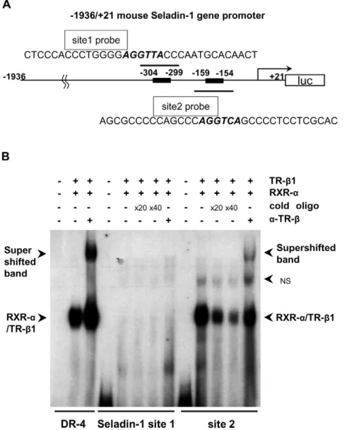
Positive Thyroid Hormone Response Element (TRE), Site 2 We subcloned mouse Seladin-1 gene promoter from21936 to
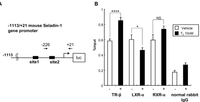
+21 base pair (bp) s and inserted it into luciferase reporter vector, pGL4 (Promega). We employed this reporter construct and performed luciferase assays with CV-1 cells, which are known to be deficient for endogenous TRs [45]. When we co-transfected the gene promoter (21936/+21 bp) with TR-b1 into CV-1 cells, T3 significantly activated the promoter activity, however the up-regulation was not observed with co-transfection of TR-a1 (Figure 4A). These data indicated that TH activates mouse Seladin-1 gene promoter via TR-b. Next, we prepared a series of deletion constructs of the mouse Seladin-1 gene promoter (Figure 4B), which were subjected to transfection into CV-1 cells with these reporters together with TR-b1. As shown in Figure 4C, the2136/+21 construct was not activated by T3. We identified two TRE half-sites, one is located from2304 to2299 bp and the other one is located from2159 to2154 bp in the mouse Seladin-Figure 8. TR-bis recruited to the site 2 region in the mouse Seladin-1 gene promoter in a T3-dependent manner.The location of the
PCR primers is indicated in an image above the data as arrows. The closed boxes indicate site 1 and 2 (A). Real-time PCR was performed to determine the relative value of the enrichment of each nuclear receptor. The value is shown as % of input (mean6S.E., n = 8) (B). TR-b, RXR-aand LXR-aare recruited to the region from2226 bp to+21 bp. The recruitment of TR-band RXR-ais increased upon T3administration. The normal rabbit IgG was
used as a negative control. Anasteriskindicates that the difference between the denoted pairs is significant at a confidence level ofp,0.05(*) and p,0.0001(****). NS: not significant.
doi:10.1371/journal.pone.0054901.g008
Figure 9. Schematic diagram illustrating the hypothetical mechanism of TR and LXR crosstalk on the mouse Seladin-1 gene promoter.
doi:10.1371/journal.pone.0054901.g009
1 promoter and hypothesized that these TRE half site regions could be a novel positive TRE. We referred to the upstream and downstream region as site 1 and 2, respectively. We introduced mutation into the TRE half-site in site 1 and 2 in the context of the 21936/+21 reporter (Figure 4D, upper panel). As shown in Figure 4E, lower panel, the positive regulation by T3deteriorated in the mutt 2 reporter, indicating that the TRE half-site in site 2 is functionally important.
TR-b1 DNA Binding is Essential for Positive Regulation of Mouse Seladin-1 Gene Promoter
We co-transfected several types of mutant TR-b1 and the 21936/+21 reporter into CV-1 cells. As shown in Figure 4F, GS125, a DNA binding mutant [49],D337T, a ligand binding mutant [50,51], and R427Q [51,52], a heterodimerization mutant with RXR-a, did not activate the promoter, whereas E457A, a co-activator binding mutant [53], positively regulated mouse Seladin-1 gene promoter activity as wild-type TR-b1. These data suggest that ligand binding, TR-b1 DNA binding, and heterodimerization with RXR-aare required for the positive regulation.
RXR-a/TR-b1 Heterodimer Binds to Site 2
Since we found that site 2 is functionally important for the positive regulation of the mouse Seladin-1 gene promoter based on the reporter assay data (Figure 4E), we hypothesized that TR-b1 could bind to site 2 in the mouse Seladin-1 gene promoter. To investigate this, we employed oligonucleotides for site 1, encom-passing2318 to2285 bp and for site 2, from2173 to2140 bp in the mouse Seladin-1 gene promoter, including the TRE consensus half-site motif. We performed electrophoretic mobility-shift assays (EMSAs) with the radiolabeled site 1 and 2 probes (Figure 5A). As shown in Figure 5B, it was revealed that RXR-a/ TR-b1 heterodimer bound to site 2 but not to site 1. Anti-TR-b
antibody shifted the heterodimer band, indicating that the binding was TR-b-specific. In addition, either RXR-a/TR-a1 heterodi-mer or TR-a1 homodyne did not bind to site 2 (data not shown).
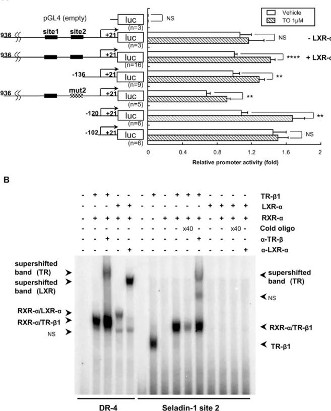
TO Increased Seladin-1 Gene Expression in HTB185 Cells through Intrinsic LXRs
On the basis of the data that TO increased mouse Seladin-1 gene expression in the forebrain in hypothyroid state and in TRKI mice, we examined whether TO potentiate to increase Seladin-1 gene expression in HTB185 cells and requires intrinsic LXRs. As shown in Figure 6A, TO significantly increased human Seladin-1 gene expression about 1.5 fold (left graph). In addition, induction of siRNA against LXR-a/binto HTB185 cells revealed that TO did not increase the gene expression in LXRs knockdown-HTB185 cells (Figure 6A, right graph). These data indicate that TO increases Seladin-1 gene expression in HTB185 cells through intrinsic LXRs. We confirmed LXR-a and -b gene expression levels in the HTB 185 cells, which siRNA against LXR-a/bwas induced (Figure 6B). It revealed that both LXR isoform gene expression levels were significantly reduced in the LXRs knock-down cells. In addition, TO induced the mouse Seladin-1 gene promoter (21936/+21 bp) in CV-1 cells in the presence of LXR-a
or LXR-bindicating that both isoforms potentiate to activate the gene promoter upon TO administration (Figure 6C). However, as shown in Figure 6B, since the gene expression of LXR-a was robust compared to that of LXR-b, we concluded that LXR-ais the dominant isoform in HTB185 cells. Thus, we employed
LXR-afor further analysis of the up-regulation of mouse Seladin-1 gene promoter by TO.
TO Activates Mouse Seladin-1 Gene Promoter, However LXR-aor RXR-a/LXR-aHeterodimer did not Bind to Site 2
As shown in Figure 7A, TO activates mouse Seladin-1 gene promoter (21936 to+21 bp) in CV-1 cells upon co-transfection with LXR-a, which is a dominant isoform of LXRs in HTB185 cells. An analysis of deletion constructs of the gene promoter revealed that the2102/+21 construct was not activated by TO indicating that the region from2120 to2102 bp but not site 2 is responsible for the positive regulation (Figure 7A). Moreover, EMSA demonstrated that LXR-aor RXR-a/LXR-aheterodimer did not bind to site 2 (Figure 7B). We performed EMSA employing several probes with different sequence including the region from 2120 to2102 bp; however, LXR-aor RXR-a/LXR-a hetero-dimer did not bind to any probes (data not shown).
TR-bis Recruited to the Site 2 Region in the Mouse Seladin-1 Gene Promoter in a T3-dependent Manner
To elucidate interaction between TR-band the mouse Seladin-1 gene promoter, we performed chromatin immunoprecipitation (ChIP) assays using a set of primers amplifying the2226 to+21 bp region, which includes site 2, in the mouse Seladin-1 gene promoter followed by quantitative PCR (Figure 8A). We introduced the mouse Seladin-1 gene promoter (21113 to
+21 bp) in HTB185 cells by transient transfection. As shown in Figure 8B, in HTB185 cells, the recruitment of intrinsic TR-bto the 2226 to +21 bp region significantly increased upon T3 administration. Since the set of PCR primer covers the region from2120 to2102 bp, with which LXR-ashould interact,
LXR-arecruitment to the region was observed in the absence of TH, but TH significantly reduced the recruitment.
Discussion
neurotropic and neuroprotective effects in vitro [55,56] and increase Seladin-1 gene expression in fetal neuroepithelial cells [57]. Similar to site 2 for TR, half-palindromic estrogen response element (ERE) in Seladin-1 gene promoter is functionally responsible for the promoter activation by ER [58].
The ChIP assays using HTB185 cells revealed that TR-b is recruited to site 2 flanking region and that the recruitment is augmented upon T3administration. Generally, TR-bas a type 2 nuclear receptor should constitutively bind to DNA in the gene promoter in the presence or absence of T3; however, recently, Liu et al. reported that TR-bwas recruited to the TRE in a T3 -dose-dependent manner in a time-course study [59]. Our data regarding TR-brecruitment is congruent with this report by Liu et al. The study employing several types of TR-bmutant revealed that the positive regulation of the promoter by TR-b required ligand binding, DNA binding, and heterodimerization with
RXR-a, and that co-activator binding does not seem to be necessarily required for the regulation (Figure 4F).
Crosstalk between TR-b and LXR-a has been reported, especially on lipid metabolism-related genes. Recently, several types of crosstalk between TRs and LXRs have been identified and crosstalk has also been observed in other physiological systems such as central nervous system other than lipid metabolism [5,41,48]. Therefore, we hypothesized that LXR-a could compensate to maintain mouse Seladin-1 gene expression in hypothyroid status and in the TRKI mice. Supporting our hypothesis, TO, an artificial agonist of LXRs, induced the gene expression in a hypothyroid state (Figure 2C, 2D) and in TRKI mice (Figure 3C, 3D), but not in euthyroid state (Figure 2A, 2B). Since TO clearly increased Seladin-1 gene expression in HTB185 cells through intrinsic LXRs and induced the mouse Seladin-1 gene promoter activity in CV-1 cells, Seladin-1 is considered as an LXR target gene. Wang et al. reported that Seladin-1 gene expression in the brain of LXR-b knockout mice was not decreased compared to that of wild-type mice [34]. We speculate that it is due to that TR-b dominantly regulates Seladin-1 gene expression in mice. Another speculation is that LXR-bcould not be required for the regulation of mouse Seladin-1 gene in forebrain.
We demonstrated that LXR-a is a dominant isoform in HTB185 cells, in which Seladin-1 gene expression was positively regulated by TO (Figure 6), therefore we employed LXR-a to elucidate a molecular mechanism for the positive regulation of the gene. Based on the data obtained by the analysis of deletion constructs of the gene promoter, LXR-a activated the mouse Seladin-1 gene promoter and the responsible region should be located from2120 to2102 bp. The exact sequence of the region is GAGCATCCCGGTTCCCGC, which does not match either canonical TRE or LXRE. We performed EMSAs employing various probes with different sequence in the region; however, LXR-a or RXR-a/LXR-a heterodimer did not bind to any probes. To examine LXR-a recruitment in vivo, we tried to performin vivoChIP assays using mouse forebrain as we previously reported with liver [37,41,43], however, it did not work probably due to excessed lipid contents in the tissue. Therefore, alternatively we employed HTB185 cells to recapitulate physiological environ-ment utilizing intrinsic nuclear receptors. Since HTB185 cells are
derived from human medulloblastoma, we had to introduce the mouse Seladin-1 gene promoter as described above. The ChIP assays demonstrated that LXR-a was clearly recruited to the region from 2226 to +21 bp. Even though RXR-a was also recruited to the region, it remains unclear whether LXR-a
recruitment requires RXR-ain the current study. Collectively, we speculated that LXR-a is recruited to the region without DNA binding. Since site 2 is located upstream of the region from2120 to2102 bp, TR-b and LXR-a do not share the same response element in the mouse Seladin-1 gene promoter. The ChIP assays also demonstrated that LXR-arecruitment was deteriorated upon TH administration indicating that in thyrotoxic state, LXR-a
could not induce the gene promoter. On the basis of these several lines of evidence, we speculate that in a thyrotoxic state, TR-b
binds to site 2 and regulates Seladin-1 gene expression; on the other hand, LXR-amay functionally compensate for TR-bbeing recruited to the region from2120 to2102 bp under hypothyroid state or in the presence of mutant TR-b to maintain Seladin-1 gene expression (Figure 9). In hypothyroid status, co-repressors such as NCoR and SMRT [60,61] are hard to dissociate from
TR-b because of lack of T3. Co-repressors are tethered to TR-b
D337T mutant, which TRKI mice harbor, because of total deficiency of ligand binding of the mutant receptor. Indeed, NCoR binds to both TR-band LXR-aand is a key regulator of TR and LXR target genes [62]. Therefore, considering the data obtained from analysis of TRKI mice and luciferase assay using TR-b D337T mutant, we hypothesize that in hypothyroid state and in RTH, co-repressors should be tethered to TR-b in a squelching fashion and LXR-a are relatively free from co-repressors, which enables to activate the mouse Seladin-1 gene promoter upon administration of the LXR specific ligand.
Nowadays, the concept of subclinical hypothyroidism is widely accepted [63,64], and it is assumed that AD patients with subclinical hypothyroidism could be increasing in number. On the basis of the data in the current study, for these patients, LXR agonists could be applicable as novel molecular therapeutic agents against AD since LXR compensates Seladin-1 gene expression under hypothyroid status.
In conclusion, mouse Seladin-1 gene expression is positively regulated by TR-b and LXR-a at the transcriptional level and LXR-a compensates the gene expression in hypothyroidism and RTH. Thus, we have, for the first time, demonstrated TR/LXR crosstalk on the Seladin-1 gene promoter in the mouse forebrain. We believe that further analysis of the crosstalk could shed light on the role of TH and LXR agonist against AD.
Acknowledgments
We thank Prof. Fredric E. Wondisford in Johns Hopkins Medical Institute for generous providing TRKI mice.
Author Contributions
Joined the discussion for the data in the current study: SO TS MY MM. Conceived and designed the experiments: KH. Performed the experiments: EI. Analyzed the data: EI KH. Contributed reagents/materials/analysis tools: SO TS MY MM. Wrote the paper: KH.
References
1. Blennow K, de Leon MJ, Zetterberg H (2006) Alzheimer’s disease. Lancet 368: 387–403.
2. Di Paolo G, Kim TW (2011) Linking lipids to Alzheimer’s disease: cholesterol and beyond. Nature reviews Neuroscience 12: 284–296.
3. Nunez J, Celi FS, Ng L, Forrest D (2008) Multigenic control of thyroid hormone functions in the nervous system. Molecular and cellular endocrinology 287: 1– 12.
4. Konig S, Moura Neto V (2002) Thyroid hormone actions on neural cells. Cellular and molecular neurobiology 22: 517–544.
5. Hashimoto K, Mori M (2011) Crosstalk of thyroid hormone receptor and liver X receptor in lipid metabolism and beyond. Endocrine journal 58: 921–930. 6. Flamant F, Samarut J (2003) Thyroid hormone receptors: lessons from knockout
and knock-in mutant mice. Trends in endocrinology and metabolism 14: 85–90. 7. Jones I, Srinivas M, Ng L, Forrest D (2003) The thyroid hormone receptor beta gene: structure and functions in the brain and sensory systems. Thyroid 13: 1057–1068.
8. Forrest D, Sjoberg M, Vennstrom B (1990) Contrasting developmental and tissue-specific expression of alpha and beta thyroid hormone receptor genes. The EMBO journal 9: 1519–1528.
9. Mellstrom B, Naranjo JR, Santos A, Gonzalez AM, Bernal J (1991) Independent expression of the alpha and beta c-erbA genes in developing rat brain. Molecular endocrinology 5: 1339–1350.
10. Bradley DJ, Towle HC, Young WS III (1992) Spatial and temporal expression of alpha- and beta-thyroid hormone receptor mRNAs, including the beta 2-subtype, in the developing mammalian nervous system. The Journal of neuroscience: the official journal of the Society for Neuroscience 12: 2288–2302. 11. Dugbartey AT (1998) Neurocognitive aspects of hypothyroidism. Archives of
internal medicine 158: 1413–1418.
12. Davis JD, Stern RA, Flashman LA (2003) Cognitive and neuropsychiatric aspects of subclinical hypothyroidism: significance in the elderly. Current psychiatry reports 5: 384–390.
13. Hashimoto K, Curty FH, Borges PP, Lee CE, Abel ED, et al. (2001) An unliganded thyroid hormone receptor causes severe neurological dysfunction. Proceedings of the National Academy of Sciences of the United States of America 98: 3998–4003.
14. Fu AL, Zhou CY, Chen X (2010) Thyroid hormone prevents cognitive deficit in a mouse model of Alzheimer’s disease. Neuropharmacology 58: 722–729. 15. Alzoubi KH, Gerges NZ, Aleisa AM, Alkadhi KA (2009) Levothyroxin restores
hypothyroidism-induced impairment of hippocampus-dependent learning and memory: Behavioral, electrophysiological, and molecular studies. Hippocampus 19: 66–78.
16. Rivas M, Naranjo JR (2007) Thyroid hormones, learning and memory. Genes, brain, and behavior 6 Suppl 1: 40–44.
17. Breteler MM, van Duijn CM, Chandra V, Fratiglioni L, Graves AB, et al. (1991) Medical history and the risk of Alzheimer’s disease: a collaborative re-analysis of case-control studies. EURODEM Risk Factors Research Group. International journal of epidemiology 20 Suppl 2: S36–42.
18. Ganguli M, Burmeister LA, Seaberg EC, Belle S, DeKosky ST (1996) Association between dementia and elevated TSH: a community-based study. Biological psychiatry 40: 714–725.
19. Kalmijn S, Mehta KM, Pols HA, Hofman A, Drexhage HA, et al. (2000) Subclinical hyperthyroidism and the risk of dementia. The Rotterdam study. Clinical endocrinology 53: 733–737.
20. de Jong FJ, den Heijer T, Visser TJ, de Rijke YB, Drexhage HA, et al. (2006) Thyroid hormones, dementia, and atrophy of the medial temporal lobe. The Journal of clinical endocrinology and metabolism 91: 2569–2573.
21. Greeve I, Hermans-Borgmeyer I, Brellinger C, Kasper D, Gomez-Isla T, et al. (2000) The human DIMINUTO/DWARF1 homolog seladin-1 confers resistance to Alzheimer’s disease-associated neurodegeneration and oxidative stress. The Journal of neuroscience : the official journal of the Society for Neuroscience 20: 7345–7352.
22. Crameri A, Biondi E, Kuehnle K, Lutjohann D, Thelen KM, et al. (2006) The role of seladin-1/DHCR24 in cholesterol biosynthesis, APP processing and Abeta generation in vivo. The EMBO journal 25: 432–443.
23. Kuehnle K, Crameri A, Kalin RE, Luciani P, Benvenuti S, et al. (2008) Prosurvival effect of DHCR24/Seladin-1 in acute and chronic responses to oxidative stress. Molecular and cellular biology 28: 539–550.
24. Waterham HR, Koster J, Romeijn GJ, Hennekam RC, Vreken P, et al. (2001) Mutations in the 3beta-hydroxysterol Delta24-reductase gene cause desmoster-olosis, an autosomal recessive disorder of cholesterol biosynthesis. American journal of human genetics 69: 685–694.
25. Martin M, Dotti CG, Ledesma MD (2010) Brain cholesterol in normal and pathological aging. Biochimica et biophysica acta 1801: 934–944.
26. Cecchi C, Rosati F, Pensalfini A, Formigli L, Nosi D, et al. (2008) Seladin-1/ DHCR24 protects neuroblastoma cells against Abeta toxicity by increasing membrane cholesterol content. Journal of cellular and molecular medicine 12: 1990–2002.
27. Benvenuti S, Luciani P, Cellai I, Deledda C, Baglioni S, et al. (2008) Thyroid hormones promote cell differentiation and up-regulate the expression of the seladin-1 gene in in vitro models of human neuronal precursors. The Journal of endocrinology 197: 437–446.
28. Apfel R, Benbrook D, Lernhardt E, Ortiz MA, Salbert G, et al. (1994) A novel orphan receptor specific for a subset of thyroid hormone-responsive elements and its interaction with the retinoid/thyroid hormone receptor subfamily. Molecular and cellular biology 14: 7025–7035.
29. Stulnig TM, Steffensen KR, Gao H, Reimers M, Dahlman-Wright K, et al. (2002) Novel roles of liver X receptors exposed by gene expression profiling in liver and adipose tissue. Molecular pharmacology 62: 1299–1305.
30. Whitney KD, Watson MA, Collins JL, Benson WG, Stone TM, et al. (2002) Regulation of cholesterol homeostasis by the liver X receptors in the central nervous system. Molecular endocrinology 16: 1378–1385.
31. Koldamova RP, Lefterov IM, Staufenbiel M, Wolfe D, Huang S, et al. (2005) The liver X receptor ligand T0901317 decreases amyloid beta production in
vitro and in a mouse model of Alzheimer’s disease. The Journal of biological chemistry 280: 4079–4088.
32. Riddell DR, Zhou H, Comery TA, Kouranova E, Lo CF, et al. (2007) The LXR agonist TO901317 selectively lowers hippocampal Abeta42 and improves memory in the Tg2576 mouse model of Alzheimer’s disease. Molecular and cellular neurosciences 34: 621–628.
33. Burns MP, Vardanian L, Pajoohesh-Ganji A, Wang L, Cooper M, et al. (2006) The effects of ABCA1 on cholesterol efflux and Abeta levels in vitro and in vivo. Journal of neurochemistry 98: 792–800.
34. Wang Y, Rogers PM, Stayrook KR, Su C, Varga G, et al. (2008) The selective Alzheimer’s disease indicator-1 gene (Seladin-1/DHCR24) is a liver X receptor target gene. Molecular pharmacology 74: 1716–1721.
35. Enmark E, Gustafsson JA (2001) Comparing nuclear receptors in worms, flies and humans. Trends in pharmacological sciences 22: 611–615.
36. Berkenstam A, Farnegardh M, Gustafsson JA (2004) Convergence of lipid homeostasis through liver X and thyroid hormone receptors. Mechanisms of ageing and development 125: 707–717.
37. Hashimoto K, Yamada M, Matsumoto S, Monden T, Satoh T, et al. (2006) Mouse sterol response element binding protein-1c gene expression is negatively regulated by thyroid hormone. Endocrinology 147: 4292–4302.
38. Hashimoto K, Ishida E, Matsumoto S, Okada S, Yamada M, et al. (2009) Carbohydrate response element binding protein gene expression is positively regulated by thyroid hormone. Endocrinology 150: 3417–3424.
39. Zhang Y, Yin L, Hillgartner FB (2001) Thyroid hormone stimulates acetyl-coA carboxylase-alpha transcription in hepatocytes by modulating the composition of nuclear receptor complexes bound to a thyroid hormone response element. The Journal of biological chemistry 276: 974–983.
40. Gullberg H, Rudling M, Forrest D, Angelin B, Vennstrom B (2000) Thyroid hormone receptor beta-deficient mice show complete loss of the normal cholesterol 7alpha-hydroxylase (CYP7A) response to thyroid hormone but display enhanced resistance to dietary cholesterol. Molecular endocrinology 14: 1739–1749.
41. Hashimoto K, Cohen RN, Yamada M, Markan KR, Monden T, et al. (2006) Cross-talk between thyroid hormone receptor and liver X receptor regulatory pathways is revealed in a thyroid hormone resistance mouse model. The Journal of biological chemistry 281: 295–302.
42. Huuskonen J, Vishnu M, Pullinger CR, Fielding PE, Fielding CJ (2004) Regulation of ATP-binding cassette transporter A1 transcription by thyroid hormone receptor. Biochemistry 43: 1626–1632.
43. Hashimoto K, Matsumoto S, Yamada M, Satoh T, Mori M (2007) Liver X receptor-alpha gene expression is positively regulated by thyroid hormone. Endocrinology 148: 4667–4675.
44. Hashimoto K, Yamada M, Monden T, Satoh T, Wondisford FE, et al. (2005) Thyrotropin-releasing hormone (TRH) specific interaction between amino terminus of P-Lim and CREB binding protein (CBP). Molecular and cellular endocrinology 229: 11–20.
45. Satoh T, Yamada M, Iwasaki T, Mori M (1996) Negative regulation of the gene for the preprothyrotropin-releasing hormone from the mouse by thyroid hormone requires additional factors in conjunction with thyroid hormone receptors. The Journal of biological chemistry 271: 27919–27926.
46. Moser BK, Stevens GR, Watts CL (1989) The 2-Sample T-Test Versus Satterthwaite Approximate F-Test. Communications in Statistics-Theory and Methods 18: 3963–3975.
47. Ruxton GD (2006) The unequal variance t-test is an underused alternative to Student’s t-test and the Mann-Whitney U test. Behavioral Ecology 17: 688–690. 48. Tan XJ, Fan XT, Kim HJ, Butler R, Webb P, et al. (2010) Liver X receptor beta and thyroid hormone receptor alpha in brain cortical layering. Proceedings of the National Academy of Sciences of the United States of America 107: 12305– 12310.
49. Shibusawa N, Hollenberg AN, Wondisford FE (2003) Thyroid hormone receptor DNA binding is required for both positive and negative gene regulation. The Journal of biological chemistry 278: 732–738.
50. Usala SJ, Menke JB, Watson TL, Wondisford FE, Weintraub BD, et al. (1991) A homozygous deletion in the c-erbA beta thyroid hormone receptor gene in a patient with generalized thyroid hormone resistance: isolation and character-ization of the mutant receptor. Molecular endocrinology 5: 327–335. 51. Safer JD, Langlois MF, Cohen R, Monden T, John-Hope D, et al. (1997)
Isoform variable action among thyroid hormone receptor mutants provides insight into pituitary resistance to thyroid hormone. Molecular endocrinology 11: 16–26.
52. Flynn TR, Hollenberg AN, Cohen O, Menke JB, Usala SJ, et al. (1994) A novel C-terminal domain in the thyroid hormone receptor selectively mediates thyroid hormone inhibition. The Journal of biological chemistry 269: 32713–32716. 53. Collingwood TN, Rajanayagam O, Adams M, Wagner R, Cavailles V, et al.
(1997) A natural transactivation mutation in the thyroid hormone beta receptor: impaired interaction with putative transcriptional mediators. Proceedings of the National Academy of Sciences of the United States of America 94: 248–253. 54. Morvan-Dubois G, Demeneix BA, Sachs LM (2008) Xenopus laevis as a model
for studying thyroid hormone signalling: from development to metamorphosis. Molecular and cellular endocrinology 293: 71–79.
56. Maggi A, Ciana P, Belcredito S, Vegeto E (2004) Estrogens in the nervous system: mechanisms and nonreproductive functions. Annual review of physiology 66: 291–313.
57. Benvenuti S, Luciani P, Vannelli GB, Gelmini S, Franceschi E, et al. (2005) Estrogen and selective estrogen receptor modulators exert neuroprotective effects and stimulate the expression of selective Alzheimer’s disease indicator-1, a recently discovered antiapoptotic gene, in human neuroblast long-term cell cultures. The Journal of clinical endocrinology and metabolism 90: 1775–1782. 58. Luciani P, Deledda C, Rosati F, Benvenuti S, Cellai I, et al. (2008) Seladin-1 is a fundamental mediator of the neuroprotective effects of estrogen in human neuroblast long-term cell cultures. Endocrinology 149: 4256–4266.
59. Liu Y, Xia X, Fondell JD, Yen PM (2006) Thyroid hormone-regulated target genes have distinct patterns of coactivator recruitment and histone acetylation. Molecular endocrinology 20: 483–490.
60. Chen JD, Evans RM (1995) A transcriptional co-repressor that interacts with nuclear hormone receptors. Nature 377: 454–457.
61. Horlein AJ, Naar AM, Heinzel T, Torchia J, Gloss B, et al. (1995) Ligand-independent repression by the thyroid hormone receptor mediated by a nuclear receptor co-repressor. Nature 377: 397–404.
62. Astapova I, Lee LJ, Morales C, Tauber S, Bilban M, et al. (2008) The nuclear corepressor, NCoR, regulates thyroid hormone action in vivo. Proceedings of the National Academy of Sciences of the United States of America 105: 19544– 19549.
63. Fatourechi V (2009) Subclinical hypothyroidism: an update for primary care physicians. Mayo Clinic proceedings Mayo Clinic 84: 65–71.
64. Surks MI, Ortiz E, Daniels GH, Sawin CT, Col NF, et al. (2004) Subclinical thyroid disease: scientific review and guidelines for diagnosis and management. JAMA : the journal of the American Medical Association 291: 228–238.