Cop
yright
© ABE&M t
odos os dir
eit
os r
eser
vados
.
Short-term effects of triiodothyronine
on thyroid hormone receptor alpha
by PI3K pathway in adipocytes, 3T3-L1
Efeitos rápidos da triiodotironina no receptor de hormônio tireoidiano alfa pela via PI3K em adipócitos, 3T3-L1
Miriane de Oliveira1, Regiane Marques Castro Olimpio1, Maria Teresa De Sibio1,
Fernanda Cristina Fontes Moretto1, Renata de Azevedo Mello Luvizotto2,
Célia Regina Nogueira1
ABSTRACT
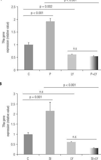
Objective: The present study aimed to examine the effects of thyroid hormone (TH), more precisely triiodothyronine (T3), on the modulation of TH receptor alpha (TRα) mRNA expression and the involvement of the phosphatidyl inositol 3 kinase (PI3K) signaling pathway in adipo-cytes, 3T3-L1, cell culture. Materials and methods: It was examined the involvement of PI3K pathway in mediating T3 effects by treating 3T3-L1 adipocytes with physiological (P = 10nM) or supraphysiological (SI = 100 nM) T3 doses during one hour (short time), in the absence or the presence of PI3K inhibitor (LY294002). The absence of any treatment was considered the control group (C). RT-qPCR was used for mRNA expression analyzes. For data analyzes ANOVA comple-mented with Tukey’s test was used at 5% signiicance level. Results: T3 increased TRα mRNA expression in P (1.91 ± 0.13, p < 0.001), SI (2.14 ± 0.44, p < 0.001) compared to C group (1 ± 0.08). This increase was completely abrogated by LY294002 in P (0.53 ± 0.03, p < 0.001) and SI (0.31 ± 0.03, p < 0.001). To examine whether TRα is directly induced by T3, we used the translation inhibitor cycloheximide (CHX). The presence of CHX completely abrogated levels TRα mRNA in P (1.15 ± 0.05, p > 0.001) and SI (0.99 ± 0.15, p > 0.001), induced by T3. Conclusion: These results demonstrate that the activation of the PI3K signaling pathway has a role in T3-mediated indirect TRα gene expression in 3T3-L1 adipocytes. Arq Bras Endocrinol Metab. 2014;58(8):833-7
Keywords
Triiodothyronine; adipocytes; TRα; PI3K
RESUMO
Objetivo: O objetivo do presente estudo foi analisar os efeitos do hormônio tireoidiano (HT), triiodotironina (T3), na modulação da expressão de mRNA do receptor alfa (TRα) de HT e o en-volvimento da via de sinalização da via fosfatidilinositol 3-quinase (PI3K) em adipócitos, 3T3-L1.
Materiais e métodos: Foi examinado o envolvimento da via PI3K nos efeitos do T3 nos trata-mentos de adipócitos, 3T3-L1, nas doses isiológica (P = 10nM) ou supraisiológica (SI = 100 nM) durante uma hora (tempo curto), na ausência ou na presença do inibidor da PI3K (LY294002). A ausência de qualquer tratamento foi considerada o grupo controle (C). RT-qPCR foi utilizado para analisar a expressão do mRNA. Para as análises dos dados, utilizou-se ANOVA comple-mentada com o teste de Tukey a 5% de signiicância. Resultados: O T3 aumentou a expressão de mRNA de TRα em P (1,91 ± 0,13, p < 0,001) e SI (2,14 ± 0,44, p < 0,001) em comparação com o grupo C (1 ± 0,08). Esse aumento foi completamente abolido por LY294002 em P (0,53 ± 0,03, p < 0,001) e SI (0,31 ± 0,03, p < 0,001). Para examinar se a expressão de TRα foi diretamente induzida pelo T3, utilizou-se o inibidor de tradução, ciclohexamida (CHX). A presença de CHX reduziu os níveis de mRNA de TRα em P (1,15 ± 0,05, p > 0,001) e SI (0,99 ± 0,15, p > 0,001), induzidos pelo T3. Conclusão: Esses resultados demonstram que a ativação da via de sinalização de PI3K tem um papel importante na expressão do gene TRα mediada indiretamente pelo T3, em adipócitos 3T3-L1. Arq Bras Endocrinol Metab. 2014;58(8):833-7
Descritores
Triiodotironina; adipócitos; TRα; PI3K
1 Department of Internal Medicine,
Botucatu School of Medicine, University of São Paulo State (Unesp), Botucatu, SP, Brazil
2 Institute of Health Sciences,
Universidade Federal do Mato Grosso (UFMT), Sinop, MT, Brazil
Correspondence to:
Miriane de Oliveira Botucatu Medical School, Universidade Estadual Paulista Distrito de Rubião Jr s/n 18618-000 – Botucatu, SP, Brazil [email protected]
Received on Feb/20/2014 Accepted on Aug/18/2014
Cop
yright
© ABE&M t
odos os dir
eit
os r
eser
vados
.
INTRODUCTION
T
hyroid hormone (TH) inluences the metabo-lism and development of adipose tissue (AT) and modulates the proliferation and differentiation of adipocytes (1), mainly 3,5,3’-triiodothyronine (T3) (2). TH also stimulates lipolysis and lipogenesis (3) and is involved in regulation of lipid and carbo-hydrate metabolism in liver, skeletal muscle and heart tissues (4).TH receptors (THR) are proteins that belong to the nuclear hormone receptor superfamily and origi-nate from TH receptor alpha (TRα) and TH receptor beta (TRβ) genes (5,6) located on chromosomes 17 and 3 in humans, respectively. These nuclear receptors act on certain genes according to nucleotide sequences called TH responsive elements (TRE) located in the promoter sites of target genes (7). Although THR are primarily nuclear receptors, approximately 10% are located in the cytoplasm (8). All isoforms of THR, TRα1, TRα2, and TRβ1, are expressed in white and brown adipose tissue, being TRα1 the predominant THR isoform (9,10). T3 and other hormones regulate the different TR isoforms.
TH may function through other mechanisms than THR/TRE (11). Alternative mechanisms may be re-ferred as a non-classical or non-genomic because initia-tion sites are located in the plasma membrane, such as activation of αvβ3 integrin, or the cytoplasm where TH may activate mitogen-activated protein (MAPK) or the phosphatidyl inositol 3-kinase (PI3K) pathway. Initia-tion sites are proteins that are characterized as iodothy-ronine receptors (12,13).
As lipid metabolism is closely associated with a number of health problems, the regulation of adipo-cytes represents an area of emerging interest. So far, TR has been implicated as a major factor in the regulation of the development and function of adipose tissue (14-16). In this study it was assessed the effects of different levels of T3 (physiological and supraphysiological) on TRα gene expression in adipocyte cell culture, 3T3-L1, by real-time PCR (RT-qPCR), during one-hour expo-sure. The inluence of protein synthesis on regulation of TRα transcription by T3 and possible activation of a non-classical pathway (PI3K), by T3, to modulate this gene was also evaluated. Our observations suggest that mRNA TRα levels modulated by T3 depends on acti-vation of the PI3K pathway.
MATERIALS AND METHODS
Chemicals
Isobutylmethylxanthine (IBMX), dexamethasone, in-sulin, cycloheximide (CHX), triiodothyronine (T3), LY294002 (LY), dimethyl sulfoxide (DMSO), sodium hydroxide (NaOH) and Charcoal Stripped fetal bovine serum (FBS) were purchased from Sigma (St Louis, MO, USA). Dulbecco’s modiied Eagle’s medium (DMEM), fetal bovine serum and antibiotic-antimy-cotic 100X solution were purchased from Gibco BRL (Grand Island, NY, USA).
Cell culture and differentiation
The experimental protocol was approved by the Ethi cs Committee on Animal Experiments of the Botucatu School of Medicine-Unesp (protocol nº 752).
Mouse 3T3-L1 pre-adipocytes were obtained from the Cell Bank of Rio de Janeiro (Rio de Janeiro, RJ, Brazil) and grow in polystyrene 6-well plates at 37ºC in DMEM supplemented with 10% FBS and 1% antibi-otic-antimycotic 100× solution. Upon cell confluence (designated as day 0), differentiation was initiated with 1 µg/mL insulin, 1 µM DEX, and 0.5 mM IBMX in DMEM containing 10% FBS. After a 4-day incubation, culture media was replaced by DMEM supplemented with 10% FBS and 1 µg/mL insulin, and the cells were fed every two days with DMEM containing 10% FBS. 3T3-L1 cells were fully differentiated by day 8. Af-ter differentiation, cells were incubated for 24 hours in DMEM supplemented with 10% charcoal stripped FBS (to deplete T3) and 1 µg/ml insulin. After incuba-tion, cells were treated with physiological T3 (10 nM; P group) or supraphysiological T3 (100 nM; SI) (17-19) for one hour. A non-treated group, that received 0.1% NaOH (diluent T3), served as the control (C). P and SI groups were also treated with CHX (10 μg/mL) (13) and LY (50 μM) (13) for one hour.
LY was used to determine if the non-classical path-way (PI3K) was involved in T3 action on TRα mRNA levels. CHX was used to determine if T3 directly or in-directly modulates TRα during the one-hour exposure.
Oil red O staining
formalde-Cop
yright
© ABE&M t
odos os dir
eit
os r
eser
vados
.
hyde for 30 minutes at room temperature, and washed twice again with PBS. After fixation, cells were stained for two hours at room temperature with a filtered oil red O solution (0.5 g oil red O (Sigma) in 100 mL isopropanol), washed twice with distilled water, and vi-sualized to conirm differentiation.
Gene expression
Whole RNA was extracted from 3T3-L1 cells using Trizol (Invitrogen) according to the manufacturer’s instructions. A high capacity cDNA reverse transcrip-tion kit for RT-PCR® (Invitrogen, Sao Paulo, Brazil) was used to synthesize 20 μL of complementary DNA (cDNA) from 1,000 ng of whole RNA.
TRα (assay Mm00617505_m1 – Applied Bio-systems) mRNA levels were determined by real-time polymerase chain reaction (RT-qPCR). Quantitative measurements were determined with the Applied Bio-systems StepOne Plus detection system using a Taq-Man qPCR commercial kit (Invitrogen) according to the manufacturer’s instructions. Cycling condi-tions were as follows: enzyme activation at 50°C for 2 min, denaturation at 95°C for 10 min, cDNA pro-duct amplification for 40 cycles of denaturation at 95°C for 15 s, and annealing/extension at 60°C for 1 min. Gene expression was quantiied relative to C group val-ues after normalization by an internal cyclophilin (assay Mm00434759_m1- Applied Biosystems) control using the 2 −ΔΔCt methodas previously described (20).
Statistical analysis
Gene expression was analyzed using analysis of variance (ANOVA) followed by Tukey’s test. Data are expressed as mean ± standard deviation. The signiicance level was set at 5%.
RESULTS
Different T3 concentrations up-regulation TRα mRNA by PI3K pathway
Figure 1 shows the up-regulation of TRα levels in 3T3-L1 adipocytes, and to verify PI3K pathway involvement in mediating the action of T3 on TRα mRNA expres-sion the P and SI groups were treated with the PI3K pathway inhibitor LY. We showed that LY associated with T3 led to decreased TRα mRNA levels (Figure 1A and B).
Effect of inhibition of protein synthesis on T3-induced modulation of TRα mRNA levels
3T3-L1 adipocytes were cultured with T3 at doses that inluenced TRα mRNA levels to determine the need for protein synthesis during one hour exposure. CHX was added to the P and SI groups. Complete abroga-tion of the T3-induced mRNA increase by CHX indi-cates that the TRα gene is indirectly induced by TH and requires prior protein synthesis (Figure 2A and B). However, CHX alone had signiicant inluence on TRα mRNA levels.
DISCUSSION
AT is a target of TH and expresses THR that are impor-tant factors in regulating tissue development and
func-Figure 1. Effects of T3 and LY294002 on modulation of TRα mRNA levels in one hour. P = 10 nM T3, SI = 100 nM T3, C = no T3, and LY = 50 µM LY294002. (A) T3 inluence (10 nM) on TRα gene expression in the presence/absence of LY. (B) T3 inluence (100 nM) on TRα gene expression in the presence/absence of LY. ANOVA with Tukey’s test. n.s. = non-signiicant; n = 3 per treatment.
n.s p < 0.001
p = 0.002
p < 0.001
TR
α
gene
expression (relative value)
2.5
2
1.5
1
0.5
0
C P LY P+LY
TR
α
gene
expression (relative value)
p < 0.001
n.s
p = 0.001 3
2.5
2
1.5
1
0.5
0
C SI LY SI+LY
n.s
A
Cop
yright
© ABE&M t
odos os dir
eit
os r
eser
vados
.
tion (16,21). In this paper we evaluated AT responses to different T3 levels based on TRα mRNA expression without interference from systemic factors that occur in vivo. As an experimental model, we used 3T3-L1 cells
(embryonic Mus musculus cells) that were
differenti-ated in vitro into adipocytes. TRα mRNA levels were
quantiied using RT-qPCR.
Elements (TRE) (11). These mechanisms can be called non-classical or non-genomic because their initiation sites may be in the plasma membrane, like the activa-tion of integrin αvβ3 pathway, or in the cytoplasm, where the TH activates the mitogen-activated protein kinase (MAPK) or PI3K pathway. The initiation sites are proteins that are characterized as iodothyronine re-ceptors (13,21).
PI3K participates in a wide variety of cellular pro-cess, including intracellular traficking, organization of the cytoskeleton, cell growth and transformation, and prevention of apoptosis (24,25). PI3K has a role in differentiation of several cell lines (26,27), in-cluding adipocytes. PI3K pathway activation by TH originates in the cytoplasm and involves TRα or TRβ, resulting in speciic gene transcription, including hy-poxia inducing factor (HIF-1α), glucose transporter 1 (GLUT1), calcineurin inhibitor (ZAKI4α) and leptin (11,13,21,22). In this study, we used the LY294002 inhibitor to evaluate the need for PI3K pathway ac-tivation in modulating TRα mRNA by T3 during a one hour time period. Within the one hour period, the increased on TRα mRNA levels in the P and SI groups were suppressed by LY (Figure 1A and B), proving that pathway activation is necessary for T3 to increase TRα levels. However, the inhibition of PI3K decreased basal TRα mRNA expression demonstrat-ing a need this pathway to TRα mRNA expression in normal cell condition.
In addition, inhibition of protein synthesis with CHX completely blocked T3-induced increase in TRα mRNA levels (Figure 2A and B), showing that it is a gene indirectly up-regulated by T3, depending on the synthesis of a yet unknown protein. Moreover, CHX group (without T3) compared with control group de-creases the basal levels of TRα mRNA, indicating the existence of certain short-lived proteins essential for the expression of this gene in normal conditions. Monden and cols. (28) demonstrated that CHX blocks the re-duction in TRα levels caused by T3 in HTB-185 cells, suggesting that down-regulation of TRα by T3 re-quires synthesis of a new protein.
In summary, during one hour of treatment, in-creased TRα levels in the P and SI groups were indi-rectly modulated by T3 and depended on activation of the PI3K pathway. This is the irst study to demonstrate TRα modulation by T3 in a very short time period (one hour) and assess PI3K pathway activation by TH on this gene in adipocytes, 3T3-L1.
Figure 2. Effects of T3 and CHX on modulation of TRα mRNA levels in one hour. P = 10 nM T3, SI = 100 nM T3, C = no T3, and CHX = 10 μg/mL. (A) T3 inluence (10 nM) on TRα gene expression in the presence/absence of CHX. (B) T3 inluence (100 nM) on TRα gene expression in the presence/absence of CHX. ANOVA with Tukey’s test. n.s. = non-signiicant; n = 3 per treatment.
p < 0.001
p < 0.001 2.5
2
1.5
1
0.5
0
C P CHX P+CHX
p < 0.001
p < 0.001
TR
α
gene
expression (relative value)
p < 0.001
2.5 3
2
p = 0.01 p = 0.01
p < 0.001
1.5
1
0.5
0
C SI CHX SI+CHX
TR
α
gene
expression (relative value)
3T3-L1 pre-adipocytes represent a well-established model for adipogenesis (22). TRα expression is higher than TRβ in 3T3-L1 adipocytes (18), which is in accor-dance with the indings of Jiang and cols. (2004) (23). Studies from our group showed that a T3 physiological and supraphysiological dose, at different times, increase TRα in adipocytes, 3T3-L1 (18).
It is known that TH may act by mechanisms other than the classical TR/Thyroid Hormone Responsive
A
Cop
yright
© ABE&M t
odos os dir
eit
os r
eser
vados
.
Acknowledgments: we are grateful to Sueli A. Clara, José C. Geor-gete, Mário B. Bruno, Camila R. C. Camacho and Keize Nagamati Junior for their technical support. We also would like to thank Dr. Luis C. O. Magalhães for the translation of the manuscript into English. This manuscript has been proofread by native English spe-akers with related background in BioMed Proofreading. We thank CAPES and Fapesp (# 2010/16911-4) for inancial support. The authors declare no conlicts of interest in the present study.
Funding: this work was supported by São Paulo Research Foun-dation (Fapesp), process number 2010/16911-4, and Coorde-nação de Aperfeiçoamento de Pessoal de Nível Superior (Capes).
Disclosure: no potential conlict of interest relevant to this article was reported.
REFERENCES
1. Darimont C, Gaillard D, Ailhaud G, Negrel R. Terminal differentia-tion of mouse preadipocyte cells: adipogenic and antimitogenic role of triiodothyronine. Mol Cell Endocrinol. 1993;98:67-73. 2. Ailhaud G, Grimaldi P, Negrel R. Cellular and molecular aspects
of adipose tissue development. Annu Rev Nutr. 1992;12:207-33. 3. Oppenheimer JH, Schwartz HL, Lane JT, Thompson MP.
Function-al relationship of thyroid hormone-induced lipogenesis, lipolysis, and thermogenesis in the rat. J Clin Invest. 1991;87:125-32. 4. Santini F, Marsili A, Mammoli C, Valeriano R, Scartabelli G, Pelosini
C, et al. Serum concentrations of adiponectin and leptin in patients with thyroid dysfunctions. J Endocrinol Invest. 2004;27:RC5-7. 5. Williams GR. Cloning and characterization of two novel thyroid
hormone receptor beta isoforms. Mol Cell Biol. 2000;20:8329-42. 6. Macchia PE, Takeuchi Y, Kawai T, Cua K, Gauthier K, Chassande
O, et al. Increased sensitivity to thyroid hormone in mice with complete deiciency of thyroid hormone receptor alpha. Proc Natl Acad Sci U S A. 2001;98:349-54.
7. Glass CK. Differential recognition of target genes by nuclear receptor monomers, dimers, and heterodimers. Endocr Rev. 1994;15:391-407. 8. Baumann CT, Maruvada P, Hager GL, Yen PM. Nuclear cytoplas-mic shuttling by thyroid hormone receptors. Multiple protein interactions are required for nuclear retention. J Biol Chem. 2001;276:11237-45.
9. Hernandez A, Obregon MJ. Presence and mRNA expression of T3 receptors in differentiating rat brown adipocytes. Mol Cell Endo-crinol. 1996;121:37-46.
10. Bianco AC, Silva JE. Cold exposure rapidly induces virtual satura-tion of brown adipose tissue nuclear T3 receptors. Am J Physiol. 1988;255:E496-503.
11. Moeller LC, Broecker-Preuss M. Transcriptional regulation by non-classical action of thyroid hormone. Thyroid Res. 2011;4 Suppl 1:S6.
12. Cheng SY, Leonard JL, Davis PJ. Molecular aspects of thyroid hormone actions. Endocr Rev. 2010;31:139-70.
13. Moeller LC, Dumitrescu AM, Refetoff S. Cytosolic action of thy-roid hormone leads to induction of hypoxia-inducible factor-1al-pha and glycolytic genes. Mol Endocrinol. 2005;19:2955-63. 14. Flores-Delgado G, Marsch-Moreno M, Kuri-Harcuch W. Thyroid
hormone stimulates adipocyte differentiation of 3T3 cells. Mol Cell Biochem. 1987;76:35-43.
15. Chawla A, Lazar MA. Induction of Rev-ErbA alpha, an orphan receptor encoded on the opposite strand of the alpha-thyroid hormone receptor gene, during adipocyte differentiation. J Biol Chem. 1993;268:16265-9.
16. Yen PM. Physiological and molecular basis of thyroid hormone action. Physiol Rev. 2001;81:1097-142.
17. Yoshida T, Monkawa T, Hayashi M, Saruta T. Regulation of ex-pression of leptin mRNA and secretion of leptin by thyroid hor-mone in 3T3-L1 adipocytes. Biochem Biophys Res Commun. 1997;232:822-6.
18. de Oliveira M, Luvizotto R de A, Olimpio RM, de Sibio MT, Silva CB, Conde SJ, et al. Modulation of thyroid hormone receptors, TRα and TRβ, by using different doses of triiodothyronine (T3) at different times. Arq Bras Endocrinol Metabol. 2013;57:368-74. 19. de Oliveira M, Luvizotto Rde A, Olimpio RM, De Sibio MT, Conde
SJ, Biz Rodrigues Silva C, et al. Triiodothyronine increases mRNA and protein leptin levels in short time in 3T3-L1 adipocytes by PI3K pathway activation. PLoS One. 2013;18;8(9):e74856. 20. Livak KJ, Schmittgen TD. Analysis of relative gene expression
data using real-time quantitative PCR and the 2(-Delta Delta C(T)) method. Methods. 2001;25:402-8.
21. Cheng SY, Leonard JL, Davis PJ. Molecular aspects of thyroid hormone actions. Endocr Rev. 2010;31:139-70.
22. Green H, Meuth M. An established pre-adipose cell line and its differentiation in culture. Cell. 1974;3:127-33.
23. Jiang W, Miyamoto T, Kakizawa T, Sakuma T, Nishio S, Takeda T, et al. Expression of thyroid hormone receptor alpha in 3T3-L1 adipocytes; triiodothyronine increases the expression of lipogenic enzyme and triglyceride accumulation. J Endocrinol. 2004;182:295-302.
24. Toker A, Cantley LC. Signalling through the lipid products of phosphoinositide-3-OH kinase. Nature. 1997;387:673-6.
25. Vanhaesebroeck B, Leevers SJ, Panayotou G, Waterield MD. Phosphoinositide 3-kinases: a conserved family of signal trans-ducers. Trends Biochem Sci. 1997;22:267-72.
26. Aubin D, Gagnon A, Sorisky A. Phosphoinositide 3-kinase is re-quired for human adipocyte differentiation in culture. Int J Obes. 2005;29:1006-9.
27. Fang J, Ding M, Yang L, Liu LZ, Jiang BH. PI3K/PTEN/AKT sig-naling regulates prostate tumor angiogenesis. Cell Signal. 2007;19:2487-97.