D
EPARTAMENTO DEB
IOLOGIAV
EGETALT
HE EFFECTS OF LOW
DOSE IONIZING RADIATION
ON ANGIOGENESIS
I
NÊSS
OFIAB
ATISTAV
ALAS
ILVA DEO
LIVEIRAD
OUTORAMENTO EMB
IOLOGIA(B
IOLOGIAC
ELULAR)
2011
D
EPARTAMENTO DEB
IOLOGIAV
EGETALT
HE EFFECTS OF LOW
DOSE IONIZING
RADIATION ON ANGIOGENESIS
INÊS SOFIA BATISTA VALA SILVA DE OLIVEIRA
Dissertation submitted to obtain a PhD Degree in Biology, speciality of Cellular Biology by the Universidade de Lisboa 2011 Supervisor Susana Constantino Rosa Santos, PhD. Principal Investigator of Instituto de Medicina Molecular and Auxiliary Professor at Faculdade de Medicina, Universidade de Lisboa. CoSupervisor Rita Maria Pulido Garcia Zilhão, PhD. Auxiliary Professor at Faculdade de Ciências, Universidade de Lisboa.
Ao Cláudio e ao Artur
i PREFACE The present thesis embraces the data obtained during my PhD research project.
The experimental work was developed under the supervision of Prof. Dr. Susana Constantino Rosa Santos at the Angiogenesis Unit, Instituto de Medicina Molecular, Lisboa, Portugal.
This PhD was also supervised by Prof. Dr. Rita Maria Pulido Garcia Zilhão from the Departamento de Biologia Vegetal, Faculdade de Ciências de Lisboa, Universidade de Lisboa, Lisboa, Portugal. The financial support was provided by the Fundação para a Ciência e Tecnologia, through a PhD fellowship grant SFRH/BD/27541/2006. This dissertation is organized in five chapters, which are preceded by a summary, both in Portuguese and in English. Chapter I consist of a general introduction to blood vessels, with particular emphasis on the angiogenic process, approached from the early embryonic development to adulthood, in physiology and pathology. A brief overview on radiotherapy and some cellular and molecular effects of ionizing radiation is also presented.
Chapter II specifically indicates the main objectives of the research proposal that led to the work presented in this thesis.
Chapter III and IV include the experimental work developed through the research project. Chapter II, low doses of ionizing radiation promote tumor angiogenesis and metastasis by enhancing angiogenesis, includes some already published work (presented in the publication format) and some complementary data to the article. Chapter III, combined
effect of vasoprost® and low‐dose ionizing radiation on angiogenesis, presents some results from ongoing work that is currently being developed in our lab and has not yet been published. Each one of these chapters includes a specific introduction, the results obtained in the work developed, and a focused discussion, as well as the methods, acknowledgements and references.
Chapter IV comprises the concluding remarks and future perspectives.
iii ACKNOWLEDGEMENTS Quase sinto o barulho do tempo a passar por mim...
Mais do que um desafio académico, estes últimos anos têm sido uma jornada pessoal, cheia de surpresas, momentos de alegria e entusiasmo, mas também com algumas lágrimas, dias de tristeza e sentimentos de dúvida. Esta tese, eu devo‐a não só ao meu esforço pessoal mas, sem dúvida, também à amizade e confiança de muitas outras pessoas que me acompanharam e tornaram este desafio um pouco mais fácil de ser vivido.
Gostaria de começar por agradecer à minha orientadora, Susana Constantino, pelo seu suporte e encorajamento ao longo destes anos. Obrigada por me teres recebido na Unidade de Angiogénese, onde aprendi quase tudo o que sei sobre ciência, por me teres tentado animar quando as coisas corriam menos bem, por teres acreditado nas minhas capacidades, e pela orientação crítica ao longo de todo o doutoramento e escrita desta tese. Obrigada Professora Rita Zilhão, por ter aceitado ser minha orientadora pela Faculdade de Ciências, e por todo o apoio e encorajamento ao longo destes anos.
Obrigada à Fundação para a Ciência e Tecnologia, pela minha bolsa de doutoramento (SFRH / BD / 27541 / 2006), financiada por fundos nacionais do Ministério da Ciência, Tecnologia e Ensino Superior.
Agradeço também ao Departamento de Radioterapia, do Hospital de Santa Maria, sem o qual não teria sido possível executar este trabalho. Gostaria de agradecer particularmente a ajuda da Professora Isabel Monteiro Grillo que possibilitou a
realização deste trabalho, e da Isabel Diegues, da Céu, da Ana Monserrate e da Antonieta, que me aturaram ao longo de todos estes anos.
Obrigada também ao João Barata, parte do meu comitê de tese, pelas discussões e sugestões críticas no desenvolvimento do trabalho.
Obrigada Inês, minha fiel companheira de bancada e, sobretudo, minha amiga, que de tão perto acompanhaste estes últimos anos e todas as coisas boas e “menos boas” que com eles vieram. Obrigada pelo teu apoio constante, pelo teu carinho e ajuda. Obrigada pela nossa amizade que prevaleceu ás ínfimas horas que passámos fechadas num laboratório.
Obrigada Raquel. A tua chegada ao laboratório serviu como “uma lufada de ar fresco”, e a tua boa disposição, ânimo e companheirismo foram imprescindíveis ao longo dos últimos anos que passei no laboratório.
Obrigada Heleninha pela paciência e muitos ensinamentos. Obrigada pelo carinho e pelas inúmeras conversas de “pé de orelha”.
Obrigada Catarina, pela paciência, preocupação e pela ajuda que me deste no laboratório.
Lara, obrigada pela tua ajuda imprescindível com o “mundo aquático”, e pelo teu sempre optimismo! Quase me conseguiste fazer gostar de peixes!
Obrigada Leila, pela tua preciosa contribuição neste trabalho, e também a ti, Dolores, pela ajuda com os ratinhos e pela simpatia e companheirismo com que sempre me recebeste.
v Não poderia esquecer o Ricardo Henriques que me iniciou no maravilhoso mundo da microscopia, e o Rino que sempre se mostrou disponível para me ajudar.
Obrigada Moisés pelos teus preciosos conselhos e críticas durante a escrita desta tese.
Tânia, tu sim, és inspiradora...! Não há palavras para agradecer o teu apoio, a disponibilidade e encorajamento, os teus preciosos conselhos no trabalho e na vida e, sobretudo, a amizade que te fez aparecer nas horas certas, mesmo quando te disse que não queria companhia... Obrigada também por teres lido a minha tese e pelas tuas sugestões.
Paulo, meu amigo de todas as horas! Dificilmente conseguiria mencionar tudo o que te devo e pelo qual te agradeço... Obrigada pelo ombro amigo sempre disponível, pela força e pela disponibilidade. Obrigada pela força nos momentos mais difíceis e por teres partilhado também comigo os dias de maior felicidade. És único!
Obrigada Filipe, Sofia e Sara, pela vossa amizade, por todos os momentos de descontração, e por saber que estão sempre presentes.
Obrigada Mãe, por me teres transmitido os valores por que me guio, por teres estado sempre presente quando necessitei e me teres dado algum espaço quando precisei de aprender a conduzir o meu próprio caminho.
Obrigada Raquel, e minha linda sobrinha Lu, pelas pequenas coisas que tornam os nossos dias um pouco melhor. Obrigada por estares presente e por seres um exemplo de coragem.
Obrigada avó, tio Carlos, Sílvia e Rafinha, pela porta sempre aberta, e por me ajudarem a perceber que as melhores coisas da vida dependem das pessoas com quem as partilhamos.
Céu, Zé Manel e Luís, por me terem acolhido como parte da vossa família, por todo o vosso apoio e carinho, muito obrigado!
A vocês, Cláudio e Artur, eu dedico esta tese, e a minha felicidade! A ti Cláudio, pela tua paciência, disponibilidade, compreensão, encorajamento, ajuda e força. Pelo teu ombro, pelo teu abraço e, sobretudo, por tudo o que me fazes sentir com apenas um sorriso. Pela tua presença, mesmo quando estás a quilómetros de distância, e por me teres ensinado que “se estás a atravessar um inferno... continua a andar!”. És um exemplo de coragem e perseverança. Obrigada por me fazeres feliz. A ti Artur, minha estrelinha cintilante, que tornas os dias soturnos cheios de luz e alegria e colocas um sorriso de felicidade em mim sempre que olho para ti.
vii RESUMO
A angiogénese é o processo de formação de novos vasos sanguíneos a partir de vasos pré‐existentes. Em situações fisiológicas a angiogénese ocorre durante o desenvolvimento embrionário, crescimento de órgãos e, no adulto, em processos de cicatrização de feridas e ciclo reprodutivo. Nestas condições, o processo angiogénico é fortemente controlado por um equilíbrio complexo entre factores estimuladores (pró‐ angiogénicos) e inibidores (anti‐angiogénicos).
A angiogénese pode, no entanto, ocorrer em situações patológicas onde há uma perda do equilíbrio entre factores pró‐ e anti‐angiogénicos, resultando numa vascularização excessiva ou deficiente. O cancro é uma das patologias que se caracterizam por um excesso de angiogénese.
A radioterapia é frequentemente aplicada ao tratamento do cancro. Porém, tem vindo a ser observado que doentes submetidos a esta terapia têm um risco aumentado de desenvolver metástases. Esta situação constitui um desafio para a clínica e os mecanismos celulares e moleculares que estão na origem deste problema têm vindo a ser investigados.
É geralmente assumido que a metastização e recidiva tumoral após a terapia se devem ao aparecimento de células tumorais resistentes à radiação ionizante. No entanto, há evidências de que doses terapêuticas de radiação ionizante promovem alterações ao nível do microambiente tumoral, podendo também contribuir para o processo de radioresistência.
A vasculatura providencia oxigénio e nutrientes ao tumor, sendo essencial para o seu desenvolvimento. Contudo, favorece também a metastização, na medida em que as células tumorais entram em circulação através de vasos sanguíneos. A contribuição da vasculatura irradiada na invasão e metastização após radioterapia é, portanto, de extrema importância. Por este motivo, ao longo dos últimos anos, têm surgido diversos estudos com o objectivo de perceber através de que mecanismos, doses de radiação
ionizante induzem a angiogénese na área tumoral e qual poderá ser a sua contribuição no processo de invasão e metastização.
Estes estudos têm‐se focado em doses de radiação ionizante que são administradas diariamente, em pequenas fracções, até que a dose potencialmente curativa seja acumulada no interior da área a tratar, com o objectivo de minimizar o dano provocado nos tecidos saudáveis. Para além disso, a administração em baixas doses e a convergência de diversos feixes que garantem a distribuição homogénea das curvas de isodose em radioterapia externa, contribuem para a existência de uma menor dose de radiação ionizante fora da área a tratar. Os efeitos biológicos e moleculares destas baixas doses de radiação ionizante nos tecidos que rodeiam a área a tratar são ainda desconhecidos.
O nosso trabalho centrou‐se, de forma inovadora, na vasculatura que rodeia o tumor e que recebe doses relativamente baixas de radiação ionizante. O principal objectivo, foi investigar o efeito destas baixas doses de radiação ionizante na angiogénese, e compreender a sua contribuição para a recidiva tumoral, invasão e metastização.
Investigámos assim o efeito das baixas doses de radiação ionizante in vitro, em células endoteliais humanas de microvasculatura de pulmão (HMVEC‐L, lung human microvascular endothelial cells) e células endoteliais de veia umbilical (HUVEC, human umbilical vein endothelial cells). Constatámos que doses iguais ou inferiores a 0.8 Gy promovem a migração de células endoteliais, sem afectar a sobrevivência e o ciclo celular, activam o receptor‐2 do factor de crescimento endotelial vascular (VEGF, vascular endothelial growth factor) e, em condições de hipóxia, promovem o aumento da expressão do próprio VEGF.
A utilização do peixe‐zebra como modelo de estudo permitiu‐nos confirmar in vivo a indução da angiogénese em resposta a baixas doses de radiação ionizante. Observámos que doses de 0.5 Gy aceleram o processo angiogénico durante o desenvolvimento embrionário e promovem um aumento do número de vasos durante a regeneração da barbatana caudal dos adultos.
ix Para estudarmos a contribuição das baixas doses de radiação ionizante no crescimento tumoral e metastização, utilizámos dois modelos experimentais de ratinho: um modelo de leucemia, e um modelo metastático de cancro de mama. Verificámos que baixas doses de radiação ionizante promovem o crescimento tumoral e metastização através de um mecanismo dependente do receptor do VEGF.
O efeito das baixas doses de radiação no desenvolvimento tumoral também foi estudado utilizando um modelo de melanoma em peixe‐zebra. Neste modelo, os peixes‐zebra mutantes em p53 (protein 53) e BRAF (raf murine sarcoma viral oncogene homolog B1) são expostos a baixas doses de radiação ionizante antes do melanoma ser detectado. De acordo com os nossos resultados, não publicados, é desenvolvido um maior número de melanomas em peixes zebra irradiados. Verificámos igualmente, que os melanomas nestes peixes irradiados apresentam um tamanho superior em relação aos melanomas desenvolvidos em peixes‐zebra não irradiados. Estudos adicionais estão a ser efectuados com o objectivo de caracterizar os melanomas que surgem em ambos os grupos experimentais.
Finalmente, e com o objectivo de identificar os mecanismos através dos quais as baixas doses de radiação ionizante induzem uma resposta pró‐angiogénica, investigámos o perfil de expressão génica de HMVEC‐L irradiadas versus não irradiadas. Os nossos resultados indicam a modulação da expressão génica de mediadores moleculares envolvidos na resposta angiogénica.
No seu conjunto, o nosso trabalho permite compreender o efeito das doses de radiação ionizante que estão presentes nos tecidos que rodeiam a área tumoral e sua importância na angiogénese, e consequentemente na progressão tumoral e metastização, pelo que poderá ser um contributo importante na optimização dos actuais protocolos de radioterapia.
Assim, de acordo com os nossos resultados as baixas doses de radiação ionizante induzem angiogénese in vivo; não existe, contudo, prova de que induzam angiogénese terapêutica em doentes com doença isquémica, sendo este um dos objectivos de investigação do nosso laboratório.
A isquémia crítica dos membros inferiores é uma das manifestações clínicas da doença arterial periférica em que se descreve doentes com dor em repouso ou com lesões tróficas cutâneas, sejam elas úlceras ou gangrena. A Isquemia crítica dos membros inferiores envolve uma perturbação grave tanto ao nível da microcirculação como da macrocirculação.
O vasoprost® é frequentemente utilizado no tratamento da doença arterial periférica. O princípio activo do vasoprost® é a prostaglandina E1 (alprostadil), cujas propriedades hemodinâmicas e acção anti‐agregante plaquetária justificam a sua indicação no tratamento da doença vascular periférica grave.
No entanto, a literatura não é unânime quanto à sua função como indutor angiogénico. Por este motivo, e através da realização de um conjunto de ensaios in vitro, em HUVEC, começámos por clarificar este assunto. Os nossos resultados sugerem que o vasoprost® funciona como um agente pró‐angiogénico, induzindo a migração, proliferação e sobrevivência endotelial.
O uso de vasoprost® na clínica apresenta, contudo, limitações terapêuticas. Assim, a amputação surge como última alternativa terapêutica, apesar das taxas de morbilidade e mortalidade associadas. O objectivo de preservar o membro tem estimulado a investigação de tratamentos alternativos, incluindo a angiogénese terapêutica.
Propusemo‐nos então a averiguar se baixas doses de radiação ionizante poderiam potenciar os resultados obtidos pelo tratamento com o vasoprost®. Avaliámos a acção combinada destes dois agentes e verificámos in vitro que doses de radiação ionizante inferiores a 0.8 Gy potenciam o efeito pró‐angiogénico do vasoprost®.
Os nossos resultados sugerem deste modo, que a combinação da radiação ionizante e vasoprost® devem ser considerados em estudos futuros, de forma a avaliar o seu potencial terapêutico na doença arterial periférica.
xi Palavras‐chave:
Angiogénese; Células endoteliais, Metástases, Radiação ionizante; Radioterapia; Vasoprost.
xiii ABSTRACT Angiogenesis is the formation of new blood vessels from pre‐existing ones. This process is regulated by a balance between pro‐ and anti‐angiogenic molecules and is derailed in various diseases, such as cancer. Radiotherapy is a commonly‐used treatment for cancer. However, recent studies suggest that ionizing radiation (IR) doses delivered inside the tumor target volume during fractionated radiotherapy can stimulate invasion and metastasis through effects on cancer cells but also on other elements of the microenvironment. Furthermore, radiotherapy results also in the delivery of doses lower that the therapeutic ones to the tissues surrounding the tumor area, and the biological effects of these low IR doses remain largely undetermined.
Our overall goal was to investigate the effects of these low IR doses on angiogenesis, and consequently in tumor progression and metastasis.
We showed that low‐dose IR induces an angiogenic response both in vitro and in vivo. Doses equal or lower than 0.8 Gy promote endothelial cell migration without causing cell cycle arrest or apoptosis, activate vascular growth factor (VEGF) receptor‐2 and up‐ regulate the expression of VEGF. In zebrafish, low‐dose IR accelerates sprouting angiogenesis during development and enhances angiogenesis during regeneration. In mice, we showed that low‐dose IR promotes angiogenesis resulting in accelerated tumor growth and metastasis formation in a VEGFR‐dependent manner. Additionally, we demonstrated that low‐dose IR modulates the gene expression of molecular mediators involved in the angiogenic response.
Our observations provide novel insights into the biological effects of low‐dose IR relevant to tumor biology, which may serve as basis for the prevention of possible tumor‐ promoting effects of current radiotherapy protocols.
Therefore, according to our findings low‐dose IR induces angiogenesis in vivo but, there is no evidence that it produces therapeutic angiogenesis in ischemic disease patients. In the
second part of this work we showed that low‐dose IR potentiates the pro‐angiogenic effect of vasoprost®, commonly used in the treatment of peripheral arterial disease treatment (PAD).
Our results suggest that the combinatory use of both vasoprost® and low‐dose IR should be considered for future studies concerning its clinical therapeutic potential in pathologies such as PAD. Keywords Angiogenesis; Endothelial cells; Ionizing radiation; Metastasis; Radiotherapy; Vasoprost.
xv TABLE OF CONTENTS Preface i Acknowledgements / Agradecimentos iii Resumo vii Abstract xiii Table of contents xv Abbreviations xix List of Figures xxiii List of Tables xxvii I. GENERAL INTRODUCTION 1 1. Circulatory system and structural properties of blood vessels: an overview 3 2. Vasculogenesis and angiogenesis during embryonic development 6 2.1. Early blood vascular development 6 2.2. Maturation of blood vessels 7 2.2.1. Arterial and venous systems 9 2.2.2. Homotypic and heterotypic junctions 11 2.2.3. Local specialization of endothelial cells 12 2.2.4. Vessel regression 13 3. Postnatal Neovascularization 14 3.1. Angiogenic Regulators 14 3.1.1. Angiopoietins 19 3.1.2. FGFs and FGFRs 20 3.1.3. TGFβ 24 3.1.4. VEGF and VEGFRs 26 3.1.5. CYR61 29 3.2. Physiological angiogenesis 31 3.3. Pathological angiogenesis 33 3.3.1. Tumor angiogenesis 35 3.3.1.1. Role of the endothelium in tumor cell metastasis 38
3.3.1.2. Role of hypoxia in tumor angiogenesis and metastasis 40 3.3.2. Peripheral arterial disease 42 4. Angiogenesis as a therapeutic target 44 4.1. Pro‐angiogenic therapy 45 4.2. Anti‐angiogenic therapy 46 5. Radiotherapy 50 5.1. Molecular basis of ionizing radiation response 51 5.2. Role of hypoxia in radiation therapy 54 5.3. Unexpected effects of radiotherapy in blood vessels and metastasis 56 5.4. Ionizing radiation combined therapy 58 References 61 II. OBJECTIVES 79 III. RESEARCH WORK: LOW DOSES OF IR PROMOTE TUMOR GROWTH AND METASTASIS BY ENHANCING ANGIOGENESIS 83 Introduction of the chapter 85 Article 89 Complementary results 105 Discussion of the chapter 128 Complementary Material and Methods 143 Complementary references 146 IV. RESEARCH WORK: COMBINED EFFECT OF VASOPROST® AND LOW‐DOSE IONIZING RADIATION ON ANGIOGENESIS 155 Introduction of the chapter 157 Results 160 Discussion of the chapter 165 Material and Methods 168 Acknowledgements 170
xvii References 171 V. CONCLUDING REMARKS AND FUTURE PERSPECTIVES 175 References 182
xix ABBREVIATIONS 5‐FU 5‐fluorouracil Akt protein kinase B (also known as PKB) ALK activin receptor‐like kinase ANGPT angiopoietin ANOVA analysis of variance BM basal membrane BRAF raf murine sarcoma viral oncogene homolog B1 CAM chicken chorioallantoic membrane cDNA complementary DNA CHK Csk‐homologous kinase CLI critical limb ischemia CLTC clathrin COUP‐TFII chicken ovalbumin upstream promoter transcription factor II CSFs colony‐stimulating factors CT computed tomography CTV clinical target volume
CYR61 cysteine‐rich protein 61 (also known as CNN1) DAPI 4', 6‐Diamidino‐2‐phenylindole Dll4 delta‐like‐4 DNA deoxyribonucleic acid dpf days post‐fertilization dpmd days post‐melanoma detection DSB double strand breaks e.g. exempli gratia (for example) ECM endothelial cell matrix ECs endothelial cells EDG‐1 endothelial differentiation G‐protein coupled receptor‐1 EGF epidermal growth factor EGFP enhanced green fluorescent protein
ELISA enzyme linked immunosorbent assay eNOS endothelial nitric oxide synthase EPCs endothelial precursor cells EPO Erythropoietin ERK extracellular signal‐related kinase (also known as MAPK) FACS fluorescence activated cell sorting FAK focal adhesion kinases FBS fetal bovine serum FCT fundação para a ciência e tecnologia FGF fibroblast growth factor FGFR fibroblast growth factor receptor FITC fluorescein isothiocyanate foxc forkhead C GFP green fluorescent protein GTV gross tumour volume HEY2 hairy and enhancer of split‐related protein 2 HGF hepatocyte growth factor HIF hypoxia inducible factor HMVEC‐L microvascular endothelial cells HR homologous recombination HSPG heparin sulfate proteoglycan HuR hypoxic‐induced stability factor HUVEC human umbilical vein endothelial cells i.e. id est (that is) IGF insulin‐like growth factor IL interleukin IMM instituto de medicina molecular IPA ingenuity pathway analysis IR ionizing radiation IVIS in vivo imaging system MAPK mitogen‐activated protein kinase (also known as ERK) MEK mitogen‐activated protein kinase kinase (also referred to as MAPKK)
xxi MMP matrix metalloproteinase mRNA messenger RNA NFkB nuclear factor‐kB NHEJ non‐homologous end‐joining NO nitric oxide NP neurpilin PAD peripheral arterial disease PAK p21‐activated kinase PCA principal component analysis PDGF platelet‐derived growth factor PDGFR platelet‐derived growth factor receptor PECAM platelet endothelial cell adhesion molecule PEDF pigment epithelium‐derived factor PF platelet factor PGE1 Prostaglandin E1 PGs prostaglandins PI propidium iodide PI3K phosphatidylinositol‐3‐kinase PKC protein kinase C PLC phospholipase C PlGF placenta growth factor PTEN phosphatase and tensin homolog PTK/ZK PTK787/ZK222584 PTV planning target volume qPCR quantitative real time polymerase chain reaction RNA ribonucleic acid ROS reactive oxygen species RT‐PCR real time polymerase chain reaction S1P1 Sphingosine‐1‐phosphate‐1 SIV sub‐intestinal vessels SMCs smooth muscle cells SSB single strand breaks
TGFβ transforming growth factor‐β TIE tyrosine kinase with immunoglobulin and EGF homology domains TIMP tissue inhibitor of metalloproteinase TKI tyrosine kinase inhibitor TKR tyrosine kinase receptors TNFβ tumor necrosis factor‐α TSP thrombospondin TUBB β‐tubullin TV treatment volume TβR transforming growth factor‐β receptor VCAM vascular cell adhesion molecule VE‐Cadherin vascular endothelial cadherin VEGF vascular endothelial growth factor VEGFR1 vascular endothelial growth factor receptor‐1 (also known as Flt1) VEGFR2 vascular endothelial growth factor receptor‐2 (also known as KDR or Flk1) VHL von Hippel Lindau WB western blot
xxiii LIST OF FIGURES I. GENERAL INTRODUCTION Figure 1| Blood vessels 5 Figure 2| Vasculogenesis and angiogenesis during embryonic development 9 Figure 3| Arterial‐venous differentiation 11 Figure 4| Capillary wall morphology 12 Figure 5| The angiogenic sprouting occurs as a coordinated multistep process 15 Figure 6| Angiopoietin signaling in angiogenesis 21 Figure 7| FGF signaling pathway overview 23 Figure 8| TGFβ/ALK1 and TGFβ/ALK5 signaling pathways in ECs 25 Figure 9| VEGF pathway overview 28 Figure 10| CYR61 regulates angiogenesis both by direct and indirect mechanisms 31 Figure 11| Intussusceptive angiogenesis 33 Figure 12| Contrast between normal and tumor vasculature 35 Figure 13| A few of the molecular and cellular players in the tumor/microvascular microenvironment 37 Figure 14| Tumor metastasis formation: interactions with blood vessels 39 Figure 15| HIF1α regulation in normoxia and hypoxia 41 Figure 16| Proposed role of vessel normalization in the response of tumors to anti‐angiogenic therapy 47 Figure 17| Isodose curves on a pelvic axial slice 51 Figure 18| Time‐scale of the effects of radiation exposure 53
Figure 19| Mechanisms for HIF1 up‐regulation and consequences after radiation therapy 55 Figure 20| Schematic representation of some major pro‐angiogenic signaling from irradiated cancer cells to ECs 58 III. RESEARCH WORK: LOW DOSES OF IR PROMOTE TUMOR GROWTH AND METASTASIS BY ENHANCING ANGIOGENESIS Figure 1| Low‐dose IR promotes endothelial cellmigration without causing cell cycle arrest or apoptosis 91 Figure 2| Low‐dose IR activates PI3K/Akt and MEK/ERK pathways and prevents apoptosis induced by their inhibition 93 Figure 3| Low‐dose IR protects microvasculature from bevacizumab‐induced cell death by inducing VEGFR‐2 activation 94 Figure 4| Low dose IR enhances hypoxia‐induced VEGF expression 95 Figure 5| Low‐dose IR accelerates angiogenic sprouting during zebrafish embryonic development and enhances angiogenesis during fin regeneration 96 Figure 6| Low‐dose IR enhances angiogenesis in matrigel plug assay 97 Figure 7| Low‐dose IR promotes acceleration of tumor growth and metastasis in a VEGF receptor‐dependent manner 98 Figure S1| Low doses of IR induce phosphorylation of H2AX 102 Figure S2| Low doses of IR do not protect the microvasculature from 5‐FU‐, gemcitabine‐ or paclitaxel‐induced cell death 103 Figure S3| Low‐dose IR promotes endothelial cell migration by activating VEGFR‐2 104
xxv Figure C1| Quantification of H2AX phosphorylation induced by IR in HMVEC‐L 106 Figure C2| Low‐dose IR activates PI3K/Akt and MEK/ERK pathways in HMVEC‐L 107 Figure C3| Treatment with either Ly294002 or U0126 decreases the migration capacity of ECs 108 Figure C4| Low‐dose IR induce the migration of ECs in the presence of Ly294002 and U0126 109 Figure C5| Low doses of IR protect endothelial cells from serum withdraw‐induced cell death 110 Figure C6| Low‐dose IR induces the phosphorylation or the expression of adhesion molecules in HMVEC‐L 111 Figure C7| Low‐dose IR promotes HUVEC migration without causing cell cycle arrest or apoptosis 112 Figure C8| Low‐dose IR modulates tyrosine phosphorylation levels and activates PI3K/Akt and MEK/ERK pathways in HUVEC 113 Figure C9| Low‐dose IR protects HUVEC from bevacizumab‐induced cell death 114 Figure C10| Low doses of IR protect HUVEC from gemcitabine‐ and paclitaxel‐induced cell death 60 h post‐treatment 115 Figure C11| Low doses of IR are not able to prevent the HUVEC arrest induced by 5‐FU, gemcitabine or paclitaxel 116 Figure C12| Low doses of IR are not able to prevent the HMVEC‐L arrest induced by 5‐FU, gemcitabine or paclitaxel 117 Figure C13| Principal component analysis (PCA) of irradiated and unirradiated HMVEC‐L 119 Figure C14| Ingenuity pathway analysis showing canonical pathways significantly modulated by low‐dose IR in HMVEC‐L 120 Figure C15| Ingenuity pathway analysis showing the cellular biological functions significantly modulated by low‐dose IR in HMVEC‐L 121
Figure C16| Low doses of IR modulate the expression of several pro‐angiogenic targets and cytoskeleton‐related proteins in HMVEC‐L 122 Figure C17| Low doses of IR modulate the expression of VEGFR2, VEGFR1 and CYR61 In HUVEC 124 Figure C18| Low‐Dose IR accelerates zebrafish development 125
Figure C19| p53 ‐/‐ BRAFV600E zebrafish develop spontaneous melanomas 126 Figure C20| p53 ‐/‐ BRAFV600E zebrafish exposed to low‐dose IR seem to present
Bigger tumors with an accelerated growth 126 IV. RESEARCH WORK: COMBINED EFFECT OF VASOPROST® AND LOW‐DOSE IONIZING RADIATION ON ANGIOGENESIS Figure 1| Vasoprost® promotes endothelial cell migration 161 Figure 2| The combination of vasoprost® and low‐dose IR improves the migratory response of ECs 162 Figure3| The combination of vasoprost® and low‐dose IR improves the proliferation response of ECs 163 Figure 4| The combination of vasoprost® and low‐dose IR maximizes the EC protection from serum withdrawal‐induced cell death 164
xxvii LIST OF TABLES I. GENERAL INTRODUCTION Table 1| Major stimulators of angiogenesis and their role in the formation of blood vessels 16 Table 2| Major endogenous inhibitors of angiogenesis and their role in the formation of blood vessels 18 Table 3| Selected list of diseases characterized or caused by abnormal/excessive or insufficient angiogenesis 34 III. RESEARCH WORK: LOW DOSES OF IR PROMOTE TUMOR GROWTH AND METASTASIS BY ENHANCING ANGIOGENESIS Table C1| Primers used for quantitative RT‐PCR 145
Ineˆ s Sofia Vala1, Leila R. Martins2, Natsuko Imaizumi3, Raquel J. Nunes1, Jose´ Rino4, Franc¸ois Kuonen3,Lara M. Carvalho5, Curzio Ru¨ egg3, Isabel Monteiro Grillo6, Joa˜o Taborda Barata2, Marc Mareel7, SusanaConstantino Rosa Santos [Digite o nome da empresa]
I. GENERAL
INTRODUCTION
This chapter contains a general introduction to the subjects approached during the experimental work presented in this thesis
3 1 1.. CCIIRRCCUULLAATTOORRYY SSYYSSTTEEMM AANNDD SSTTRRUUCCTTUURRAALL PPRROOPPEERRTTIIEESS OOFF BBLLOOOODD VVEESSSSEELLSS:: AANN O OVVEERRVVIIEEWW
All vertebrates require an efficient circulatory system, able to distribute oxygen and nutrients to tissues and, simultaneously remove carbon dioxide and other metabolic waste products. This task is carried out by two main networks: the blood vessels and the lymphatic vessels, both formed by endothelial cells (ECs) (Adams and Alitalo, 2007). Additionally to the gases, liquids and nutrients transport, the vascular system is also important in the regulation of body temperature and systemic pH (Carmeliet, 2005). In the cardiovascular system, oxygenated blood is pumped from the heart through the arteries and capillaries to the tissues where exchanges occur. The blood is then returned to the heart via the venous system (Eichmann et al., 2005).
As a result of the high arterial pressure, blood plasma leaks from the capillaries into the extracellular space, becoming interstitial fluid. The majority of the extravasated fluid is reabsorbed by post‐capillary venules driven by osmotic forces, but the remaining is drained by the lymphatic system, returning it into the venous circulation. Unlike the blood vascular system, the lymphatic system does not feature a central pump. Instead, interstitial fluid (lymph) is moved forward by skeletal muscle action and respiratory movement (Cueni and Detmar, 2008). The lymphatic system is also essential for the immune defense (Eichmann et al., 2005).
Both networks are essential for homeostasis of a healthy organism, and their malformation or dysfunction contributes to many diseases (Eichmann et al., 2005). Blood vessels are divided into three main groups: arteries, veins, and capillaries. Arteries and veins are further divided, according to caliber, into large, medium, and small blood vessels. The vascular system is subjected to varying degrees of hydrostatic pressure, and the structure of vessels varies in an adaptive fashion. Blood vessels are thickest and their walls more complex in the immediate vicinity of the heart, where hydrostatic pressure is greatest (Nussenbaum and Herman, 2010). As blood vessels decrease in caliber, their wall becomes thinner and less complex.
Heterogeneity in vessel wall composition is evident between vessels of different sizes and between arterial and venous vessels (Figure 1). Large vessel vascular walls are composed of the tunica intima, which consists of the endothelium, basal membrane and an internal elastic layer; the tunica media, a thick layer of smooth muscle with reticular fibers, elastin and proteogycans; and the tunica adventitia, which consists of connective tissue with both elastic and collagenous fibers (Cleaver and Melton, 2003). Since veins conduct blood back to the heart, the pressure exerted by the heartbeat on them is much less than in the arteries. The middle muscular wall of a vein is therefore much thinner than that of an artery (Bergers and Song, 2005). Veins differ from arteries also in that they have semi‐ lunar valves, which prevent the blood from flowing backwards (Cleaver and Melton, 2003).
Small blood vessels are composed of ECs surrounded by a basal lamina covered by pericytes. Pericytes exhibit long cytoplasmic processes that not only can contact numerous endothelial cells and thus integrate signals along the length of the vessel, but can also extend to more than one capillary in the vasculature (Bergers and Song, 2005). Pericytes are functionally significant; when vessels lose pericytes, they become hemorrhagic and hyperdilated, leading to conditions such as edema, diabetic retinopathy, and even embryonic lethality (Hellstrom et al., 2001). Recently, pericytes have gained new attention as functional and critical contributors to tumor angiogenesis and therefore as potential new targets for anti‐angiogenic therapies.
5 Figure 1| Blood vessels. Blood vessels are divided into three main groups: arteries, veins, and
capillaries. They all present particular cellular differences, which are highlighted above. Tunica intima (ECs, BM) Tunica media (SMCs and ECM) Tunica adventia (fibrous conective tissue) Elastic tissue lumen
Artery Capillary Vein
ECs, BM pericytes
2
2.. VVAASSCCUULLOOGGEENNEESSIISS AANNDD AANNGGIIOOGGEENNEESSIISS DDUURRIINNGG EEMMBBRRYYOONNIICC DDEEVVEELLOOPPMMEENNTT
Two mechanisms account for the formation of blood vessels, vasculogenesis and angiogenesis. Unfortunately, the terms vasculogenesis and angiogenesis literally have the same general meaning, i.e., the genesis of blood vessels. Despite the nomenclature, the two processes are clearly distinct. Vasculogenesis is the process of de novo blood vessel formation driven by the recruitment and differentiated of mesodermal cells into the endothelial lineage and the de novo assembly of such cells into blood vessels. Angiogenesis is the generation of new blood vessels from pre‐existing ones, a process driven by EC proliferation. 2.1. EARLY BLOOD VASCULAR DEVELOPMENT Given the importance of the vascular system, in any organ and tissue, its establishment has to occur early in embryogenesis (Eichmann et al., 2005).
During development, the vasculature is established both by vasculogenesis and angiogenesis.
The first evidence of blood vessel development first appears on the extra‐embryonic yolk sac, and intraembryonic tissue, where groups of splanchnic mesoderm cells, specified to become hemangioblasts, aggregate and condense, forming what is known as blood islands. Here, hemangioblasts, the precursors of blood cells and ECs, start to differentiate. Cells at the perimeter of the blood islands become angioblasts, the precursors of the ECs. Those at the center constitute the hematopoietic precursors of all the blood cells (Conway et al., 2001).
As the yolk sac begins to form, angioblasts migrate to distant sites, multiply and differentiate forming a primitive network of simple endothelial tubes, called primary vascular plexus. This process where blood vessels are created de novo from endothelial precursor cells (EPCs), in response to local signals such as growth factors, is known as vasculogenesis. During this process, ECs undergo specification, proliferation, migration,
7 differentiation and finally fuse to form the inside layer of nascent vessels (Conway et al., 2001; Jain, 2003; Risau, 1997).
The subsequence growth, expansion and remodeling of the primitive vessels into a mature vascular network, that develops into arteries, veins, and capillaries, is referred to as angiogenesis (Conway et al., 2001; Jain, 2003; Lin et al., 2007).
Vascular endothelial growth factor (VEGF), and its receptor‐2 (VEGFR2) are the most critical drivers of embryonic vessel formation, since knock‐out mice for either of these molecules result in embryonic lethality between 8.5 and 9.5 days post‐coitum. Embryos deficient for the receptor tyrosine kinase VEGFR2 or VEGF fail to develop blood islands, and therefore ECs (Carmeliet et al., 1996; Shalaby et al., 1995). In contrast, in mice missing vascular endothelial growth factor receptor‐1 (VEGFR1), vessels do form but exhibit excessive levels of ECs, which obstruct the lumen of abnormal vascular channels (Fong et al., 1995). Thus, although both VEGFR1 and VEGFR2 are expressed on hematopoietic stem cells and ECs, only VEGFR2 is absolutely critical for the earliest stages of vasculogenesis. VEGFR1 becomes involved later on, acting as a negative regulator of VEGF activity during early angiogenesis (Fong et al., 1995; Risau, 1997).
2.2. MATURATION OF BLOOD VESSELS
After the establishment of a primitive vascular plexus by vasculogenesis, sprouting angiogenesis starts, and new vessels form from the sides and ends of pre‐existing ones. At this point, sprouting angiogenesis is facilitated by hypoxia, which up‐regulates a number of genes involved in vessel formation, patterning and maturation, such as endothelial nitric oxide synthase (eNOS), VEGF and angiopoetin‐2 (ANGPT2). Existing vessels dilate in response to nitric oxide (NO) (a product of eNOS), and become leaky in response to VEGF, which indirectly controls the redistribution of intracellular adhesion molecules (e.g. platelet endothelial cell adhesion molecule‐1 (PECAM1) and vascular endothelial cadherin (VE‐Cadherin). As the endothelial cell matrix (ECM) dissolves in response to activation of matrix metalloproteinases (MMPs) (e.g. MMP2, MMP3, MMP9)
and suppression of tissue inhibitor of metalloproteinases (TIMPs) (e.g. TIMP2), plasma proteins leaked from these nascent vessels serve as a provisional matrix. Since the physical barriers are dissolved, ECs are free to migrate establishing interactions between their integrins and matrix proteins, simultaneously proliferating in response to VEGF and other endothelial mitogens, as platelet‐derived growth factors (PDGFs), fibroblast growth factors (FGFs), angiopoetin‐1 (ANGPT1) and ANGPT2 (in the presence of VEGF) (Conway et al., 2001; Jain, 2003). The selection of ECs for sprouting is a highly regulated process, where Notch signaling also plays an important role (Adams and Alitalo, 2007). The maturation of new vessels, involves the recruitment of mural cells and the expansion of the surrounding matrix. The regulation of this process involves 4 main molecular pathways: (1) PDGFB/PDGFRβ (PDGF receptor‐β), (2) ANGPT1/TIE2 (tyrosine kinase with immunoglobulin and epidermal growth factor homology domains), (3) S1P1 (sphingosine‐ 1‐phosphate‐1)/EDG‐1 (endothelial differentiation G‐protein coupled receptor‐1), and (4) TGFβ (transforming growth factor‐β) signaling. PDGFB has an important role in the recruitment of mural cells. In response to VEGF, PDGFβ is secreted by ECs recruiting PDGFRβ positive mural cells around vessel sprouts, which will stabilize the new vessels by inhibiting EC proliferation and migration (Bergers and Song, 2005; Hellstrom et al., 2001). ANGPT1 is secreted by pericytes and interacts with TIE2, which is specifically expressed in ECs, mediating an appropriate interaction between ECs and pericytes (Bergers and Song, 2005; Loughna and Sato, 2001). S1P1/EDG1 and TGFβ have been shown to be involved in the recruitment and differentiation of pericytes (Jain, 2003; Pepper, 1997; Risau, 1997). The last step in the maturation process is the organ‐specific specialization, where ECs acquire highly specialized characteristics to provide the functional needs within specific tissues. This process includes arterial‐venous determination, formation of homotypic and heterotypic junctions and EC differentiation to form organ‐specific capillary structures (Conway et al., 2001).
Figure 2 represents a schematic summary of the major steps involved in the vasculogenesis and angiogenesis during embryonic development.
9 Figure 2| Vasculogenesis and angiogenesis during embryonic development. During
vasculogenesis, hemangioblasts aggregate and condense, differentiating into angioblasts, which migrate, proliferate and differentiate into ECs, forming a primitive network of simple endothelium tubes (primary vascular plexus). Due to an orchestrated chain of growth factors and matrix remodeling molecules, ECs proliferate and migrate, giving rise to new sprouting vessels (angiogenesis). Maturation of blood vessels, involves the recruitment of mural cells (pericytes and SMCs), the development of the surrounding matrix, and specialization of ECs. 2.2.1. ARTERIAL AND VENOUS SYSTEMS After endothelial progenitors differentiate into ECs to further form the primate vascular plexus, remodeling into a more complex network requires the discrimination of artery and vein boundaries. Although the process of artery and vein specification is not well understood, it is known that most of the molecules that seem to be involved, are also expressed in the nervous SMCs SMCs Pericytes Imature vessels sprouting, remodeling and regression Mature vessels Hemangioblasts Angioblasts ECs
(proliferation, migration and differentiation) Primitive vascular plexus Naked EC Vessel stabilization and maturation ECM Mature vessels Growth factors and matrix derived components (e.g. VEGF, ANGPT2, MMPs) Ang1, PDGFB, S1P1, TGFβ FGF2, VEGF VEGF Vasculogenesis Angiogenesis
system, regulating cell fate decisions and axonal migration guidance (Eichmann et al., 2005).
It is known that the Notch pathway, with its ligands (Delta‐like‐4, Jagged‐1, and Jagged‐2) and receptors (Notch‐1, Notch‐3 and Notch‐4), promotes arterial fate of ECs by repressing venous differentiation (Figure 3). Upstream of Notch, we find VEGF and Foxc1 and Foxc2 (forkhead C1 and C2), which activate Notch pathway, while HEY2 (hairy and enhancer of split‐related protein 2) and EphrinB2 act downstream to determine arterial fate (Adams and Alitalo, 2007; Carmeliet, 2003; Lin et al., 2007).
Moreover, COUP‐TFII (chicken ovalbumin upstream promoter transcription factor II), specifically expressed in venous endothelium, and phosphatidylinositol‐3‐kinase (PI3K) / protein kinase B (Akt) signaling have also been implicated in the specification of vein identity. The first by suppressing the Notch pathway, and the second, by blocking ERK (extracellular signal‐related kinase) signaling, which is preferentially detected in angioblasts that are fated to become arteries (Lin et al., 2007).
EphrinB2 ligand and its receptor, EphB4, also contribute for the formation of arterial‐ venous anastomoses, since EphrinB2 marks only arteries and EphB4 is vein specific (Adams and Alitalo, 2007; Carmeliet, 2003).
11 Figure 3| Arterial‐venous differentiation. VEGF binds to VEGFR2/neurpilin‐1 (NP1) complex,
activating both ERK and Notch pathways, which results in the expression of Ephrin‐B2 and thus, arterial fate. Foxc1 and Foxc2 also activate the Notch pathway through the expression of Delta‐ like‐4 (Dll4). However, COUP‐TFII and PI3K/Akt signaling are able to inhibit arterial fate through the inhibition of both Notch and ERK signaling pathways, respectively, resulting in the expression of the venous marker EphB4. Question markers indicate unconfirmed interactions. Adapted (Lin et al., 2007). 2.2.2. HOMOTYPIC AND HETEROTYPIC JUNCTIONS
Tight‐junctions (formed by occludins, claudins and zona occludens) and adherens junctions (formed by cadherins, with special attention to VE‐cadherin), promote EC‐EC communication (Dejana et al., 1995) and provide mechanical strength and tightness, thereby establishing a permeability barrier to plasma solutes and leukocytes (Carmeliet, 2003). In addition, gap junctions (clusters of connexins) provide both EC‐EC and EC‐ perivascular cells communication, allowing the direct exchange of ions and small molecules between neighboring cells (Dejana et al., 1995; Jain, 2003). Other adhesion
Arterial‐fated angioblast Venous‐fated angioblast VEGF VEGFR2 PLC‐γ ↓ PKC ↓ Raf ↓ MEK1/2 ↓ ERK1/2 Notch, Dll4 HEY2 Ephrin‐B2 Eph‐B4 Foxc1/2 NP1 ? ? VEGF VEGFR2 PLC‐γ ↓ PKC ↓ Raf ↓ MEK1/2 ↓ ERK1/2 Notch, Dll4 HEY2 Ephrin‐B2 Eph‐B4 Foxc1/2 NP1 ? ? COUP‐TFII PI3K ↓ Akt ?
molecules such as PECAM1 and integrins play also an important role by promoting both homotypic and heterotypic adhesion (Dejana, 2004). When ECs migrate during vessel sprouting, these contacts are transiently dissolved but later re‐established.
2.2.3. LOCAL SPECIALIZATION OF ENDOTHELIAL CELLS
Capillaries in different tissues exhibit different cellular morphology (Figure 4), associated with distinct levels of permeability: (a) continuous capillaries that occur for instance in muscle and nervous tissue where ECs form a continuous internal lining without openings in their walls; (b) fenestrated capillaries that occur in endocrine glands and gastrointestinal mucosa where although the continuous BM, ECs are pierced by pores (fenestrations) allowing the rapid passage of macromolecules; (c) discontinuous capillaries found in spleen and liver with large openings and a discontinuous or absent BM (Cleaver and Melton, 2003).
Figure 4| Capillary wall morphology. Capillaries in different tissues exhibit different cellular
morphology. (A) Continuous capillaries have no openings in their walls and are lined continuously with the endothelial cell body. (B) Fenestrated capillaries have small openings, called fenestrae, of about 80−100 nm in diameter. Fenestrae are covered by a small, non‐membranous, permeable diaphragm, and allow the rapid passage of macromolecules. The BM of ECs is continuous over the Lumen EC nucleus Basement membrane Intercellular gap Fenestrae Intercellular gap
Continuous Fenestrated Discontinuous
13 fenestrae. (C) Discontinuous capillaries have a large lumen, many fenestrations with no diaphragm and a discontinuous or absent BM. Adapted (Cleaver and Melton, 2003). 2.2.4. VESSEL REGRESSION Vessel regression is a physiological mechanism, and occurs when the nascent vasculature consists of too many vessels. Removal of angiogenic stimuli is, in many cases, enough to promote vessel regression, especially if they are still immature (Carmeliet, 2003). Insufficient perfusion of blood, absence of pericytes or presence of anti‐angiogenic factors are other stimulus that can contribute to this process (Risau, 1997).
3
3.. PPOOSSTTNNAATTAALL NNEEOOVVAASSCCUULLAARRIIZZAATTIIOONN
Until 1997, it was generally accepted that vasculogenesis could only occur during embryogenesis. More recently, the existence of a postnatal vasculogenesis has been supported by the evidence that EPCs circulate postnatally in the peripheral blood (Reed et al., 2007), and that may be recruited from the bone marrow and incorporated into sites of active neovascularization (Asahara et al., 1999; Shi et al., 1998). The recruitment and integration of EPCs involves chemoattraction, active arrest, migration to the interstitial space, incorporation into the vasculature, and differentiation into mature ECs (Hillen and Griffioen, 2007). Although this multistep process includes the active role of many molecules, once more, VEGF seems to be the most important factor in the control of this process in adulthood (Grunewald et al., 2006; Ribatti et al., 2001).
Although vasculogenesis may occur during adulthood, often associated with pathological conditions, new vessels in the adult arise mainly through angiogenesis (Conway et al., 2001; Jain, 2003; Lin et al., 2007).
3.1. ANGIOGENIC REGULATORS
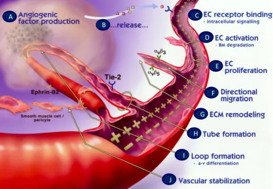
As it was previously referred, angiogenesis requires a precise coordination of multiple steps (Figure 5), which are regulated by a delicate balance between pro‐ and anti‐ angiogenic factors. A selective list of some of the most important angiogenic stimulators and inhibitors is shown in Table 1 and Table 2, respectively.
15
Figure 5| The angiogenic sprouting occurs as a coordinated multistep process. (A) diseased or
injured tissues produce and release angiogenic growth factors (e.g. VEGF) that diffuse into the nearby tissues; (B) the angiogenic growth factors bind to specific receptors (e.g. VEGFR2) located on the ECs of nearby preexisting blood vessels; (C) once growth factors bind to their EC receptors, a cascade of intracellular signaling is activated; (D) pericytes detach and blood vessels dilate before the basement membrane and extracellular matrix is degraded by ECs produced enzymes (MMPs); (E) ECs proliferate and (F) migrate towards the growth factor producer tissue through the action of integrins (e.g. αvβ3, αvβ5) that serve as grappling hooks to help pull the sprouting new blood vessel sprout forward; (G) additional enzymes (MMPs) are produced to dissolve and remodel the ECM around the vessel; (H) sprouting ECs roll up to form tubes which (I) differentiate in arterial‐venous systems and connect to form blood vessel loops that can circulate blood; (J) Newly formed blood vessel tubes are stabilized by SMCs and pericytes that provide structural support. Adapted (Angiogenesis Foundation website (http://www.angio.org/understanding/ process.php). c d e f a b g h i j A B C D E F G H I J
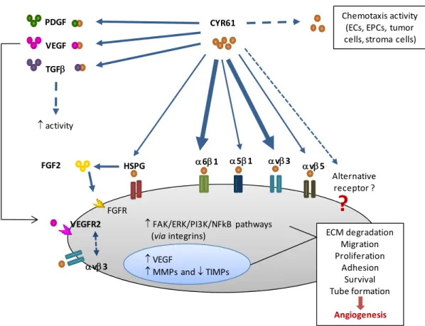
Table 1| Major stimulators of angiogenesis and their role in the formation of blood vessels
CLASS FACTOR BIOLOGICAL FUNCTIONS REFERENCE Growth factors, Cytokines and Chemokines Angiotropin ↑EC migration Angiogenesis in vivo (Hockel et al., 1988) Angiopoietin‐1 (ANGPT1) ↓EC apoptosis EC sprouting Vessel stabilization (Loughna and Sato, 2001; Nussenbaum and Herman, 2010; Tait and Jones, 2004; Yancopoulos et al., 2000) Angiopoietin‐2 (ANGPT2)* ↑EC proliferation ↑EC migration EC sprouting only in the presence of VEGF (Loughna and Sato, 2001; Nussenbaum and Herman, 2010; Tait and Jones, 2004; Yancopoulos et al., 2000) Epidermal growth factor (EGF) ↑EC proliferation ↑VEGF Angiogenesis in vivo (van Cruijsen et al., 2006) Erythropoietin (EPO) ↑EC proliferation Angiogenesis in vivo (Yasuda et al., 2002) Fibroblast growth factors (FGFs) family ↑Plasminogen activators ↑EC proliferation ↑EC migration ↑αvβ3 integrin ↓EC apoptosis Angiogenesis in vivo (Beenken and Mohammadi, 2009; Cross and Claesson‐ Welsh, 2001; Distler et al., 2003; Presta et al., 2005; Turner and Grose, 2010) Hepatocyte growth factor (HGF) ↑EC proliferation ↑EC migration Angiogenesis in vivo (Taniyama et al., 2001) Insulin‐like growth factor‐1 (IGF1) ↑EC proliferation ↓EC apoptosis ↑VEGF ↑Plasminogen activators (Delafontaine et al., 2004) Interleukin‐8 (IL8) ↑EC proliferation ↑EC migration ↓EC apoptosis (Li et al., 2005) Platelet‐derived growth factor (PDGF) ↑SMCs and pericyte proliferation ↑VEGF Vessel stabilization (Distler et al., 2003; Hellberg et al., 2010; Nussenbaum and Herman, 2010) Transforming growth factor‐ ↑EC proliferation ↑EC migration (Bertolino et al., 2005; Distler et al., 2003;
17 β (TGFβ)* ↓EC apoptosis ↑PDGF and eNOS Tube formation Vessel stabilization Angiogenesis in vivo Kaminska et al., 2005; Laverty et al., 2009; Pepper, 1997) Vascular endothelial growth factor (VEGF) family ↑Permeability; ↑Plasminogen activators ↑EC proliferation ↑EC migration ↓EC apoptosis Angiogenesis in vivo (Ferrara, 2001; Ferrara, 2002; Kuwano et al., 2001; Nussenbaum and Herman, 2010; Takahashi and Shibuya, 2005; Yancopoulos et al., 2000) Matrix proteins and adhesion molecules Cysteine‐rich protein 61 (CYR61) ↑EC proliferation ↑EC migration ↓EC apoptosis Tube formation (Brigstock, 2002; Chen and Lau, 2009; Chen and Du, 2007; Leask and Abraham, 2006) Integrins EC attachment ↑EC migration ↓EC apoptosis FGF induced angiogenesis (Avraamides et al., 2008; Nussenbaum and Herman, 2010) Platelet endothelial cell adhesion molecule‐1 (PECAM1) EC aggregation ↑EC migration Tube formation Vessel stabilization FGF induced angiogenesis (Distler et al., 2003) Vascular endothelial‐ cadherin (VE‐cadherin) ↓EC apoptosis Vessel stabilization Angiogenesis in vivo (Dejana et al., 1999) Proteases Matrix metalloproteinases (MMPs)* ECM degradation (van Hinsbergh and Koolwijk, 2008)
Others Angiogenin ↑EC proliferation (Wiedlocha, 1999)
Ephrin ↑EC proliferation ↑EC migration Vessel stabilization (Mosch et al., 2010) Nitric oxide (NO) ↑Permeability ↑EC proliferation ↑EC migration ↑FGF and VEGF (Ziche and Morbidelli, 2000) * Can show opposite effects depending upon doses and environmental conditions
Table 2 ‐ Major endogenous inhibitors of angiogenesis and their role in the formation of blood vessels
CLASS FACTOR BIOLOGICAL FUNCTIONS REF Growth factors, Cytokines and Chemokines Angiopoietin‐2 (ANGPT2)* ↑EC apoptosis Vessel destabilization (Loughna and Sato, 2001; Nussenbaum and Herman, 2010; Tait and Jones, 2004; Yancopoulos et al., 2000) Interleukin‐12 (IL12) ↓FGF mediated angiogenesis (Kerbel and Hawley, 1995) Interferon−α, −β, −γ ↓MMPs ↓FGF ↓IL8 mediated angiogenesis ↓Plasminogen activators (Nussenbaum and Herman, 2010; Nyberg et al., 2005) Platelet factor‐4 (PF4) ↓FGF mediated angiogenesis (Nussenbaum and Herman, 2010; Nyberg et al., 2005) Transforming growth factor‐β (TGFβ)* ↓EC proliferation ↓EC migration ↑EC apoptosis ↑TIMPs ↓Plasminogen activators (Bertolino et al., 2005; Distler et al., 2003; Kaminska et al., 2005; Laverty et al., 2009; Pepper, 1997) Matrix proteins and adhesion molecules Arresten ↓EC proliferation ↓EC migration ↓Tube formation (Nyberg et al., 2005) Endostatin ↓EC proliferation ↓MMPs (Nussenbaum and Herman, 2010; Nyberg et al., 2005) Thrombospondin‐1 and ‐2 (TSP1, TSP2) ↓EC migration ↑EC apoptosis ( Distler et al., 2003) Proteases Matrix metalloproteinases (MMPs)* Generate angiostatin (van Hinsbergh and Koolwijk, 2008) tissue inhibitors of metalloproteinases (TIMPs) ↓MMPs (Van Hinsbergh and Koolwijk, 2008; Nussenbaum and Herman, 2010)
Others Angiostatin ↓EC proliferation
↓EC migration ↑EC apoptosis ↓Tube formation (Distler et al., 2003; Nussenbaum and Herman, 2010; Nyberg et al., 2005) * Can show opposite effects depending upon doses and environmental conditions