i
Outubro de 2018
Maria Beatriz Tomaz Ferreira
[Nome completo do autor]
[Nome completo do autor]
[Nome completo do autor]
[Nome completo do autor]
[Nome completo do autor]
[Nome completo do autor]
[Nome completo do autor]
Licenciada em Química Aplicada
[Habilitações Académicas]
[Habilitações Académicas]
[Habilitações Académicas]
[Habilitações Académicas]
[Habilitações Académicas]
[Habilitações Académicas]
[Habilitações Académicas]
A rational approach towards novel modulators of
necroptosis
[Título da Tese]
Dissertação para obtenção do Grau de Mestre em
Química Bioorgânica
Dissertação para obtenção do Grau de Mestre em
[Engenharia Informática]
Orientador: Carlos Alberto Mateus Afonso, Professor Catedrático, Faculdade de
Farmácia da Universidade de Lisboa
Co-orientador:
João Manuel Janeiro Martins Ravasco, Investigador, Faculdade de
Farmácia da Universidade de Lisboa
Júri:
Presidente: Professora Doutora Ana Maria Ferreira da Costa Lourenço
Arguente: Professora Doutora Paula Cristina de Sério Branco Vogal: Professor Doutor Carlos Alberto Mateus Afonso
iii
A rational approach towards novel modulators of necroptosis
Copyright © Maria Beatriz Tomaz Ferreira, Faculdade de Ciências e Tecnologia,
Universidade Nova de Lisboa.
A Faculdade de Ciências e Tecnologia e a Universidade Nova de Lisboa têm o direito,
perpétuo e sem limites geográficos, de arquivar e publicar esta dissertação através de
exemplares impressos reproduzidos em papel ou de forma digital, ou por qualquer
outro meio conhecido ou que venha a ser inventado, e de a divulgar através de
repositórios científicos e de admitir a sua cópia e distribuição com objetivos
educacionais ou de investigação, não comerciais, desde que seja dado crédito ao
autor e editor.
v
Agradecimentos
A todas as pessoas que tornaram possível a realização desta dissertação e a minha evolução enquanto química, gostava de deixar aqui o meu mais profundo agradecimento.
Em primeiro lugar, quero agradecer ao Professor Carlos Afonso, por me ter recebido tão bem no seu laboratório e pela sua orientação científica e preocupação durante este ano.
Ao João Ravasco por toda a paciência que teve para mim durante este ano e o tempo perdido para me ajudar e ensinar tanto no laboratório como na elaboração desta dissertação
Ao grupo de investigação da Professora Cecília Rodrigues, em especial à Sara Oliveira, pela realização dos ensaios biológicos.
À Professora Maria de Jesus Perry pela disponibilidade e ajuda na realização dos ensaios de estabilidade.
Quero agradecer à Késsia, ao João e à Milene, os meus companheiros ao longo deste ano, obrigado por estarem sempre presente nas boas e nas más colunas.
A todos os colegas do laboratório, que me acompanharam durante este ano, um obrigado gigante pela ajuda, pela disponibilidade e pela partilha de conhecimento que me fizeram crescer muito como química. Quero agradecer especialmente ao Rafael, por todos os conselhos e sugestões sobre as reações que tanto me ajudaram, à Dra. Filipa Siopa, por todo o apoio e motivação, que tão importante foi durante este ano e ainda ao Dr. Carlos Monteiro que apesar de chato, ajudou-me sempre que precisei.
A todos os meus amigos, que estiveram sempre presentes e me apoiaram, em especial, à minha xuxua Gabriela, alem de uma colega, tornou-se uma amiga, umas das melhores coisas que a FCT me proporcionou e apesar de distância física durante este ano, estiveste sempre presente, obrigado por tornares estes 5 anos muito mais fáceis. Ao meu carequinha favorito, Edgar Castanheira, obrigado por estares presente desde do primeiro dia de faculdade, por me tirares duvidas quando na percebia nada da matéria, por seres o amigo que és. À Joana e à Filipa, as minhas companheiras de jantares, obrigado por me ajudarem a desanuviar e a largar o estudo.
Ao Dinis, por me acompanhar e apoiar nesta e em tantas outras etapas, por não se chatear com os meus n atrasos e por me ouvir mesmo quando não percebe nada do que estou a falar.
À minha família, em especial aos meus pais, pela confiança que depositam em mim, pela ajuda e pelo apoio em todas as minhas decisões e acima de tudo por estarem sempre presentes nos bons e maus momentos. Um obrigado do tamanho do mundo.
vi
Quero agradecer ao iMED.ULisboa por ceder as suas instalações para a realização desta dissertação e à Fundação para a Ciência e Tecnologia (UID/DTP/04138/2013, COMPETE Programme (SAICTPAC/0019/2015) pelo suporte financeiro.
vii
Resumo
A necroptose é uma forma regulada de necrose que ocorre em resposta a um dano celular ou uma infeção. Esta é independente de caspases, ocorrendo em condições nas quais a via clássica de morte celular por apoptose está inibida. A execução deste tipo de morte celular envolve o recrutamento de cinases como a RIPK1 e a RIPK3, que dão origem a um complexo denominado necrossoma e levam ao recrutamento da MLKL e consequente fosforilação, desencadeando o processo de necroptose. A RIPK1 e a RIPK3 encontram-se envolvidas numa grande variedade de patologias para as quais atualmente ainda carecem de tratamentos eficazes, como é o caso do acidente vascular cerebral, do enfarte do miocárdio ou da pancreatite aguda.
A Nec-1 foi o primeiro inibidor conhecido para a necroptose, atuando através do bloqueio da atividade da RIPK1. No entanto, esta apresenta alguns problemas tais como inespecificidade para o alvo. Como tal, novos inibidores da RIPK1 têm sido desenvolvidos, procurando apresentar melhores propriedades farmacocinéticas e farmacodinâmicas, com foco na sua estabilidade e atividade in vivo.
Devido à grande relevância destes novos alvos no tratamento de patologias nas quais está envolvida a necroptose, e com base num estudo preliminar de 21 derivados de Oxazol-5-(4H)-onas em linhagens de células microglia BV2 e L292, foi encontrado um composto líder, a OXA 12, que apresenta uma atividade inibitória semelhante à Nec-1. A partir da OXA 12, e utilizando a reação de Erlenmeyer-Plöch foram sintetizadas novas OXAs com a introdução de diversos grupos. Além destas OXAs ainda foram sintetizadas outras, alterando o ácido hipúrico por outros grupos como foi o caso das hidrazonas. Estes novos compostos seguiram posteriormente para avaliação biológica.
ix
Abstract
Necroptosis is a regulated form of necrosis that occurs in response to a cellular damage or infection. It is independent of caspases, occurring under conditions in which the classical pathway of cell death by apoptosis is inhibited. The execution of this type of cell death involves the recruitment of kinases as the RIPK1 and the RIPK3, which give rise to a complex necrosome and lead to the recruitment of the MLKL and consequent phosphorylation, triggering a necroptosis process. RIPK1 and RIPK3 are involved in a wide variety of pathologies for which they currently lack effective treatments, such as stroke, myocardial infarction or acute pancreatitis.
Nec-1 was the first known inhibitor for necroptosis, acting through the blockade of RIPK1 activity. However, this presents some problems such as non-specificity for the target. As such, new inhibitors of RIPK1 have been developed, seeking to present better pharmacokinetic and pharmacodynamic properties, focusing on their stability and activity in vivo.
Due to the great relevance of these new targets in the treatment of pathologies in which necroptosis is involved and based on a preliminary study of 21 Oxazol-5-(4H)-one derivatives in BV2 and L292 microglial cell lines, a lead compound, OXA 12, was found has an inhibitory activity similar to Nec-1. From OXA 12 and using the Erlenmeyer-Plöch reaction new OXAs were synthesized with the introduction of several groups. Besides these OXAs were further synthesized, altering the hippuric acid by other groups such as the hydrazones. These new compounds were subsequently used for biological evaluation.
xi
Subject Index
1. Introduction ... 1 1.1. Cell cycle ... 3 1.1.1. Interphase ... 3 1.1.2. Mitotic phase ... 4 1.2. Cell death ... 6 1.2.1. Apoptosis ... 6 1.2.2. Necrosis... 81.3. Necroptosis as therapeutic target ... 11
1.3.1. Ischemia-reperfusion injury ... 11 1.3.3. Inflammatory diseases ... 13 1.4. Necroptosis inhibitors ... 15 1.4.1. RIPK1 ... 15 1.4.2. RIPK3 ... 16 1.4.3. MLKL ... 17 1.5. Oxazol-5(4H)-ones ... 19 1.5.1. Erlenmeyer-Plochl reaction... 19 1.5.2. Uses of oxazolones ... 21
1.5.3. Oxazolones as necroptosis inhibitors ... 23
2. Objectives ... 25
3. Results and Discussion ... 29
3.1. Synthesis of 4 or 5-substituted-2-methyl-benzimidazoles ... 31
3.2. Synthesis of 1,2-dimethyl-1H-benzo[d]imidazole ... 32
3.3. Synthesis of 4-(2-(4 or 5-substituted-benzimidazol-2- yl)vinyl)benzaldehyde ... 33
3.4. Synthesis of (E)-4-(2-(2H-imidazol-2-yl)vinyl)benzaldehyde ... 37
3.5. Synthesis of Oxazol-5(4H)-ones ... 37
3.6. Synthesis of (E)-(4-(2-(1H-benzo[d]imidazol-2-yl)vinyl)phenyl)methanol ... 40
3.7. Synthesis of hydrazones or oxime ... 41
3.8. Attempts hydrogenation with Pd/C ... 43
3.9. Synthesis of hippuric acid derivatives ... 45
3.9.1. Synthesis of N-acetylglycine ... 45
3.9.2. Synthesis of (pyrazine-2-carbonyl)glycine and nicotinoylglycine s ... 45
3.10. Stability assays ... 52
3.10.1. UV spectra ... 52
3.10.2. Development of a method for the detection of OXA 12 ... 53
3.10.3. Metabolic stability assays using human plasma and PBS with 20% acetonitrile ………54
3.11. Biological assays ... 57
a) Substituents at C-4 position of oxazolone moiety (A) ... 58
b) Benzimidazole ring moiety (B) ... 59
c) Hippuric acid moiety (C) ... 61
xii
5. Materials and methods ... 69
5.1. General remarks ... 71
5.2. Synthetic proceduresa ... 73
5.2.1. General procedure for the preparation of 4 or 5-substituted-2-methyl-benzimidazoles ... 73
5.2.2. Procedure for the preparation of 1,2-dimethyl-1H-benzo[d]imidazole ... 74
5.2.3. General procedure for the preparation of 4-(2-(4 or 5-substituted-benzimidazol-2- yl)vinyl)benzaldehyde ... 75
5.2.4. Attempt procedure for the preparation of (E)-4-(2-(1H-imidazol-2-yl)vinyl)benzaldehyde ... 78
5.2.5. General procedure for the preparation of Oxazol-5(4H)-ones ... 79
5.2.6. Procedure for the preparation of (E)-(4-(2-(1H-benzo[d]imidazol-2-yl)vinyl)phenyl)methanol ... 81
5.2.7. General procedure for the preparation of hydrazones and oximes... 82
5.2.8. General procedure for catalytic hydrogenation attempts ... 84
5.2.9. Preparation of hippuric acid derivates ... 85
5.2.9.1. N-acetylglycine50 ... 85
5.3. Metabolic Stability Assays ... 90
5.3.1. UV spectra ... 90
5.3.2. Method for detecting OXA 12 ... 90
5.3.3. Metabolic Stability Assays ... 90
5.3.3.1. PBS and 20% Acetonitrile ... 90 5.3.3.2. Human plasma ... 91 5.4. Biological assays ... 92 6. Bibliography ... 97 7. Attachments ... 101 7.1. Appendix 1 - 5-fluoro-2-methyl-1H-benzo[d]imidazole (1) ... 101 7.2. Appendix 2 – 5-(tert-butyl)-2-methyl-1H-benzo[d]imidazole (2) ... 103 7.3. Appendix 3 - 2,4-dimethyl-1H-benzo[d]imidazole (3) ... 105 7.4. Appendix 4 - 1,2-dimethyl-1H-benzo[d]imidazole (4) ... 107 7.5. Appendix 5 - (E)-4-(2-(1H-benzo[d]imidazol-2-yl)vinyl)benzaldehyde (5) ... 109 7.6. Appendix 6 - (E)-4-(2-(5-fluoro-1H-benzo[d]imidazol-2-yl)vinyl)benzaldehyde (6) ………..111 7.7. Appendix 7 - (E)-4-(2-(5-(tert-butyl)-1H-benzo[d]imidazol-2-yl)vinyl)benzaldehyde (7) ... 113 7.8. Appendix 8 - (E)-4-(2-(4-methyl-1H-benzo[d]imidazol-2-yl)vinyl)benzaldehyde (8) ………..115 7.9. Appendix 9 - (E)-4-(2-(1-methyl-1H-benzo[d]imidazol-2-yl)vinyl)benzaldehyde (9) ……….117
7.10. Appendix 10 -Attempt synthesis of (E)-4-(2-(1H-imidazol-2-yl)vinyl)benzaldehyde (10) ... 119
7.11. Appendix 11 - 4-((Z)-4-((E)-2-(1H-benzo[d]imidazol-2-yl)vinyl)benzylidene)-2-phenyloxazol-5(4H)-one (11) ... 120
7.12. Appendix 12 - 4-((E)-4-((E)-2-(4-methyl-1H-benzo[d]imidazol-2-yl)vinyl)benzylidene)-2-phenyloxazol-5(4H)-one (12) ... 121
xiii
7.13. Appendix 13 - 4-((Z)-4-((E)-2-(1-methyl-1H-benzo[d]imidazol-2-yl)vinyl)benzylidene)-2-phenyloxazol-5(4H)-one (13) ... 122 7.14. Appendix 14 - 4-((E)-4-((E)-2-(1H-benzo[d]imidazol-2-yl)vinyl)benzylidene)-2-methyloxazol-5(4H)-one (14) ... 123 7.15. Appendix 15 - (E)-(4-(2-(1H-benzo[d]imidazol-2-yl)vinyl)phenyl)metanol (15) 125 7.16. Appendix 16 - 2-((E)-4-((E)-(2-benzylhydrazineylidene)methyl)styryl)-1H-benzo[d]imidazole (16) ... 127 7.17. Appendix 17 - 2-((E)-4-((E)-(2-phenylhydrazineylidene)methyl)styryl)-1H benzo[d]imidazole (17) ... 129 7.18. Appendix 18 - (E)-4-((E)-2-(1H-benzo[d]imidazol-2-yl)vinyl)benzaldehyde O-benzyl oxime (18) ... 1317.19. Appendix 19 – Attempt to synthetized (E)-4-(4-(2-(1H-benzo[d]imidazol-2-yl)ethyl)benzylidene)-2-phenyloxazol-5(4H)-one ... 133
7.20. Appendix 20 - 4-(2-(1H-benzo[d]imidazol-2-yl)ethyl)benzaldehyde ... 134
7.21. Appendix 21 - N-acetylglycine (21) ... 135
7.22. Appendix 22 - Methyl (pyrazine-2-carbonyl)glycinate (22) ... 137
7.23. Appendix 23 - (Pyrazine-2-carbonyl)glycine (23) ... 139
xv
Figure Index
Figure 1.1. Phases of cell cycle. [Adapted from Ref. 3]3 ... 3
Figure 1.2. Extrinsic (a) and intrinsic (b) apoptotic pathway... 7
Figure 1.3. Signalling pathway to lead cell survival (a), apoptosis (b) or necroptosis (c) ... 9
Figure 1.4. Necroptosis involved in injury to different organs ... 11
Figure 1.5. Chemical structure of identified necroptosis inhibitor targeting RIPK1 ... 16
Figure 1.6. Chemical structure of identified necroptosis inhibitor targeting RIPK3 ... 17
Figure 1.7. Chemical structure of identified necroptosis inhibitor targeting MLKL ... 18
Figure 1.8. General structure of oxazolones, with their reactive sites ... 19
Figure 1.9. Chemical structure of the 21 oxazolone derivatives developed in this team by Catarina Rodrigues and tested in cell lines. ... 23
Figure 1.10. Chemical structure of compounds developed on the basis of OXA 12 structure .... 24
Figure 2.1. Structure of OXA 12 ... 27
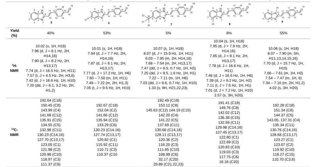
Figure 3.1. 1H-NMR spectrum (a) and 13C-NMR spectrum (b) of compound 5 ... 34
Figure 3.2. a) 1H-NMR of the compound 11, b) 1H-NMR of the reaction of hydrogenation of compound 11 ... 44
Figure 3.3. a) 1H-NMR of the compound 5, b) 1H-NMR of the reaction of hydrogenation of compound 5 ... 44
Figure 3.4. a) 1H-NMR spectrum of urea resulting from an intramolecular attack of the intermediate formed with DCC, b) 1H-NMR spectrum of compound 22, synthesized from the reaction between glycine methyl ester hydrochloride and pyrazine-2-carboxylic acid via a coupling using DCC and DMAP, containing DCU. ... 47
Figure 3.5. 1H-NMR spectrum of 22 (b), synthesized from the reaction between glycine methyl ester hydrochloride and pyrazine-2-carboxylic acid via a coupling using DCC and DMAP, with formation of DCU (a) as by-product. Compound 23 (c), resulted from the hydrolysis of compound 23 using KOH. ... 48
Figure 3.6. UV spectra of OXA 12 in acetonitrile ... 52
Figure 3.7. Run in isocratic mode using as mobile phase H2O/MeOH (0.5:9.5) and the retention time of OXA 12 was 1.82 min. ... 53
Figure 3.8. Run in isocratic mode using as mobile phase H2O/MeOH (1:9) and the retention time of OXA 12 was 2.42 min. ... 54
Figure 3.9. Graph showing the results corresponding to human plasma assay with OXA 12, containing the area of the peak of each injection as a function of the time of the assay. ... 55
Figure 3.10. Graph showing the results corresponding in PBS assay, containing the area of the peak of each injection as a function of the time of the assay... 55
Figure 3.11. Structure of OXA 12 containing five moieties. In grey, substituents at the C-4 position of the oxazolone ring (A), benzimidazole ring moiety in blue (B) and green hippuric acid moiety (C). In this moiety there is the ring of oxazolone in purple (D) and orange, the phenyl ring terminal (E). ... 58
xvi
Figure 7.1. 1H-NMR spectra of compound 1, in CDCl3 ... 101
Figure 7.2. 13C-NMR of compound 1, in CDCl3 ... 101
Figure 7.3. IR spectrum of compound 1, in ATR ... 102
Figure 7.4. 1H-NMR spectra of compound 2, in CDCl3 ... 103
Figure 7.5. 13C-NMR spectra of compound 2, in CDCl3 ... 103
Figure 7.6. IR spectrum of compound 2, in ATR ... 104
Figure 7.7. 1H-NMR spectra of compound 3, in CDCl3 ... 105
Figure 7.8. 13C-NMR spectra of compound 3, in CDCl3 ... 105
Figure 7.9. IR spectrum of compound 3, in ATR ... 106
Figure 7.10. 1H-NMR spectra of compound 4, in DMSO ... 107
Figure 7.11. 13C-NMR spectra of compound 4, in CDCl3 ... 107
Figure 7.12. IR spectrum of compound 4, in ATR... 108
Figure 7.13. 1H-NMR spectra of compound 5, in DMSO ... 109
Figure 7.14. 13C-NMR spectra of compound 5, in DMSO ... 109
Figure 7.15. IR spectrum of compound 5, in ATR... 110
Figure 7.16. 1H-NMR spectra of compound 6, in DMSO ... 111
Figure 7.17. 13C-NMR spectra of compound 6, in DMSO ... 111
Figure 7.18. IR spectrum of compound 6, in ATR... 112
Figure 7.19. 1H-NMR spectra of compound 7, in DMSO ... 113
Figure 7.20.13C-NMR spectra of compound 7, in DMSO ... 113
Figure 7.21. IR spectrum of compound 7, in ATR ... 114
Figure 7.22. 1H-NMR spectra of compound 8, in DMSO ... 115
Figure 7.23. 13C-NMR spectra of compound 8, in DMSO ... 115
Figure 7.24. IR spectrum of compound 8, in ATR... 116
Figure 7.25. 1H-NMR spectra of compound 9, in Acetone ... 117
Figure 7.26. 13C-NMR spectra of compound 9, in Acetone ... 117
Figure 7.27. IR spectrum of compound 9, in ATR ... 118
Figure 7.28. 1H-NMR spectra of the attempt to synthetize compound 10, in DMSO ... 119
Figure 7.29. 1H-NMR spectra of the attempt to synthetize compound 10, in DMSO ... 119
Figure 7.30. 1 H-NMR spectra of compound 11, in DMSO ... 120
Figure 7.31. 13C-NMR spectra of compound 11, in DMSO... 120
xvii
Figure 7.33. IR spectrum of compound 12, in ATR ... 121
Figure 7.34. -1H-NMR spectra of compound 13, in Acetone ... 122
Figure 7.35. IR spectrum of compound 13, in ATR... 122
Figure 7.36. 1H-NMR spectra of compound 14, in DMSO ... 123
Figure 7.37. 13C-NMR spectra of compound 14, in DMSO ... 123
Figure 7.38. HSQC spectra of compound 14, in DMSO ... 124
Figure 7.39. IR spectrum of compound 14, in ATR... 124
Figure 7.40. 1H-NMR spectra of compound 15, in DMSO ... 125
Figure 7.41. 13 C-NMR spectra of compound 15, in DMSO ... 125
Figure 7.42. IR spectrum of compound 15, in ATR... 126
Figure 7.43.1H-NMR spectra of compound 16, in DMSO ... 127
Figure 7.44. 13C-NMR spectra of compound 16, in DMSO... 127
Figure 7.45. IR spectrum of compound 16, in ATR ... 128
Figure 7.46. 1HNMR spectra of compound 17, in DMSO ... 129
Figure 7.47. 1H-NMR spectra of compound 17, in DMSO ... 129
Figure 7.48. IR spectrum of compound 17, in ATR ... 130
Figure 7.49. 1H-NMR spectra of compound 18, in DMSO ... 131
Figure 7.50.1H-NMR spectra of compound 18, in DMSO ... 131
Figure 7.51. IR spectrum of compound 18, in ATR ... 132
Figure 7.52.1H-NMR spectra of the attempt to synthetize compound 19, in DMSO ... 133
Figure 7.53. 1H-NMR spectra of the attempt to synthetize compound 20, in DMSO ... 134
Figure 7.54. 1H- NMR spectra of compound 21, in D2O ... 135
Figure 7.55. 13 C-NMR spectra of compound 21, in D2O ... 135
Figure 7.56. IR spectrum of compound 21, in ATR ... 136
Figure 7.57. 1H-NMR spectra of compound 22, in CDCl3 ... 137
Figure 7.58. 13C-NMR spectra of compound 22, in CDCl3... 137
Figure 7.59. IR spectrum of compound 22, in ATR ... 138
Figure 7.60. 1H-NMR spectra of compound 23, in D2O ... 139
Figure 7.61. 13C-NMR spectra of compound 23, in D2O ... 139
Figure 7.62. IR spectrum of compound 23, in ATR ... 140
Figure 7.63. 1H-NMR spectra of compound 26, in D2O ... 141
xix
Scheme Index
Scheme 1.1. Conditions of the Erlenmeyer-Plöchl reaction ... 20
Scheme 1.2. Influence of EDGs on hippuric acid ... 20
Scheme 1.3. Influence of EWGs on aldehyde ... 20
Scheme 1.4. Reactive sites of the oxazolone skeleton [Adapted from Ref 35]35 ... 22
Scheme 1.5. Diversity of compounds produced from oxazolone [Adapted from Ref. 36]36 ... 22
Scheme 3.1. Mechanism of formation of 4 or 5-substituted-2-methylbenzimidazole 1-3... 31
Scheme 3.2. Alkylation mechanism for the formation of 1,2-dimethylbenzimidazole 4 by reaction of 2-metilbenzimidazole with iodomethane. ... 32
Scheme 3.3. Mechanism for the formation of compounds 5-9 by reaction of 4 or 5-substituted-2-methyl-benzimidazoles (1-4) with the terephthalaldehyde. ... 33
Scheme 3.4. Attempt to synthesize the compound (E)-4-(2-(2H-imidazol-2-yl)vinyl)benzaldehyde ... 37
Scheme 3.5. A reaction mechanism for the formation of the oxazolone ring from hippuric acid or its derivatives ... 38
Scheme 3.6. A mechanism for forming OXAs (11-14) by reacting the oxazolone ring formed above with compound 5. ... 38
Scheme 3.7. Mechanism of reduction the aldehyde of compound 5 to alcohol 15 using LiAlH4.40 Scheme 3.8. Mechanism of reduction the aldehyde of compound 5 to alcohol 15 using NaBH4 ... 40
Scheme 3.9. Proposed mechanism for the reaction between the aldehyde 5 and a hydrazine or hydroxylamine with formation of a hydrazone (16-17) or oxime (18). ... 41
Scheme 3.10. Stability of the compound obtained by the displacement of electrons from the electronegative atom (N or O) to the carbon making it low susceptible to nucleophilic attacks. 42 Scheme 3.11. Attempts of the hydrogenation reaction using palladium on charcoal as catalyst. ... 43
Scheme 3.12. Mechanism for the formation of N-acetyl glycine 21 through the reaction of glycine with acetic anhydride. ... 45
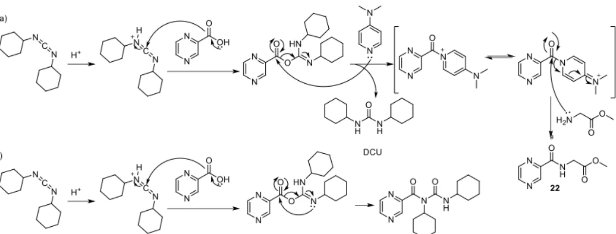
Scheme 3.13. a) Formation of compound 22 from the carboxylic acid and through a coupling using DCC and DMAP as the catalyst, b) Possible mechanism of formation of a urea as a by-product resulting from an intramolecular reaction of the intermediate formed in the reaction with the DCC ... 47
Scheme 3.14. Hydrolysis of compound 22 using KOH as the base, for the formation of compound 23 ... 49
Scheme 3.15. Formation of the acid chloride 24-25 from the carboxylic acid using oxalyl chloride and dimethylformamide (DMF) as catalyst, a) formation of Vilsmeier-Haack reagent through reaction between oxalyl chloride and DMF; b) reaction of Vilsmeier-Haack reagent with carboxylic acid with formation of acid chloride. ... 50
xx
Scheme 3.17. Mechanism of formation of compound 22 and 26 through reaction between acid chloride 24 or 25 and glycine or derivative, through of SN2 reaction com expulsion of chloride.. ... 51
xxi
Table
Index
Table 1.1. Inhibitors reported for each target molecule ... 18
Table 3.1. Characterizations of compounds 1-3, synthetized through of condensation between 4 or 5-subtitutedbenzeno-1,2-diamine and triethyl orthoacetate.a ... 32 Table 3.2. Characterization of compound 4, synthetized through alkylation of
2-methylbenzimidazolea ... 33 Table 3.3. Characterizations of compounds 5-9, synthetized by reaction between
2-methylbenzimidazole and its derivatives 1-4 with terephthalaldehyde.a ... 36 Table 3.4. Characterization of compounds 11-14, synthetized through of reaction of compound 5-9 with hippuric acid or N-acetyl glycine (21) a ... 39 Table 3.5. Characterization of compound 15, synthesized by reduction of aldehyde 5a ... 41 Table 3.6. Characterization of compounds 16-18 from reaction between aldehyde 5 and
hydrazine or hydroxylamine a ... 42 Table 3.7. Characterization of compound 21, synthesized through an reaction between glycine and acetic anhydride.a ... 45 Table 3.8. Results obtained for OXA 12 using CNS MPO ... 46
Table 3.9. Characterization of compound 22, synthesized from the reaction between glycine methyl ester hydrochloride and pyrazine-2-carboxylic acid via a coupling using DCC and DMAP.a ... 48 Table 3.10. Characterization of compound 23, synthetized through hydrolysis of compound 22 using KOHa ... 49 Table 3.11. Characterization of compound 26, synthetized through reaction between acid chloride and glycinea ... 51 Table 3.12. Results of biological assays to OXA derivatives in BV2 murine microglia cells ... 57
Table 3.13. Percent protection against necroptosis for each structure synthesized with substituents at C-4 position of oxazolone moiety, using BV2 murine microglia cells for the performance of the assay ... 58 Table 3.14. Percent protection against necroptosis for each structure synthesized with
introduction of substituents in 4 or 5-position of benzimidazole, using BV2 murine microglia cells for the performance of the assay... 60 Table 3.15. Percent protection against necroptosis for each structure synthesized with alteration in the benzimidazole heterocycle, using BV2 murine microglia cells for the performance of the assay. ... 61 Table 3.16. Percent protection against necroptosis for each structure synthesized with
modifications in hippuric acid, using BV2 murine microglia cells for the performance of the assay. ... 61 Table 3.17. Percent protection against necroptosis for each structure synthesized with
alterations in hippuric acid, using BV2 murine microglia cells for the performance of the assay. ... 62
xxiii
Abbreviations
13C-NMR Nuclear Magnetic Resonance of Carbon 1H-NMR Proton Nuclear Magnetic Resonance of Proton ACN Acetonitrile
AcOEt Ethyl acetate
Apaf1 Apoptotic protease activating factor 1
Bz Benzene
CARD Caspase recruitment domain CNS Central nervous system CYLD Cylindromatosis
d Doublet
DCC N,N'-Dicyclohexylcarbodiimide DCM Dichloromethane
DCU Cyclohexylurea
DISC Death-inducing signalling complex DMAP 4-Dimethylaminopyridine
DMF Dimethylformamide DMSO Dimethyl sulfoxide DNA Deoxyribonucleic acid EDG Electron donating group
EtOH Ethanol
EWG Electron withdrawing group
FADD Fas-associated protein with death domain FasL Fas ligand
Hex Hexane
HMBC Heteronuclear multiple bond correlation HMQC Heteronuclear single quantum coherence HPLC High-performance liquid chromatography IR Infra-red spectroscopy
J Coupling constant
xxiv
M.p. Melting pointMeOH Methanol
MLKL Mixed Lineage Kinase Domain Like Pseudokinase Nec-1 Necrostatin-1
NEMO NF-kappa-B essential modulator
NF-kB Nuclear factor kappa-light-chain-enhancer of activated B cells NMR Nuclear magnetic resonance
NSA Necrosulfonamide
Oxa Oxazolone
Ph Phenyl
q Quartet
Rf Retention factor
RIPK1 Receptor-interacting serine/threonine-protein kinase 1 RIPK3 Receptor-interacting serine/threonine-protein kinase 3
RT Retention time
s Singlet
t Triplet
THF Tetrahydrofuran
TLC Thin Layer Chromatography TNF Tumor necrosis factor TNF-α Tumor necrosis factor alpha TPA Two photons absorption
TRADD Tumour necrosis factor receptor type 1-associated death domain protein
TRAIL TNF-related apoptosis-inducing ligand UV/Vis Ultraviolet- visible spectroscopy δ Chemical shift
xxv
Units
µL Microliter Equiv. Equivalents g Gram h Hour Hz Hertz M Molar m/z Mass-to-charge ratio mg Milligrams MHz Megahertz min Minute mL Millilitre mmol 10-3 mole mol Mole ºC Degree Celsiusppm Parts per million
1
3
1.1.
Cell cycle
An organism is formed through growth and cell division. At this stage, the duplication of the cellular content occurs and later the division into two daughter cells. The cycle that leads to the production of these daughter cells through division and duplication of cellular content is called the cell cycle, and can vary depending on the organism or its stage of life.1,2
For this cycle to be complete, it will be necessary to make the minimum number of processes for the genetic information to pass to the next generation, thus producing the two daughter cells identical to each other and to the parent cell.1
This cycle is composed of two main phases: the interphase, where DNA duplication occurs and the mitotic phase, longer phase of the cell cycle, where cell division occurs.1,2
Figure 1.1. Phases of cell cycle. [Adapted from Ref. 3]3
1.1.1. Interphase
a) G1 phaseThe interphase starts with the G1 phase, which occurs after the end of the previous mitotic phase and until the beginning of the S phase (DNA synthesis). This is the first checkpoint, allows to find and repair DNA damage before the cells enters to S phase. At this stage there is still a continuous cell growth without replication.1,2
4
b) S phaseThis duplication is a complex process in which it is necessary to ensure that chromosomal characteristics are inherited by the daughter cells by the creation of two exactly identical semi-conserved chromosomes
.
1DNA replication is the main stage of this phase, beginning at the origins of replication that are scattered at various locations on the chromosome.1
c) G2 phase
The G2 phase, the last of the interphase, occurs between S phase and mitosis, and just as G1 phase is a checkpoint, preventing the start of mitosis if aberrant genetic modifications occured.1,2
1.1.2. Mitotic phase
1.1.2.1. Mitosis
Mitosis is a very complex and regulated phase that occurs after the G2 phase and where the sister chromatids are separated and distributed into a pair of identical daughter nuclei each containing a copy of the original genome. It can be divided into five different stages: prophase, prometaphase, metaphase, anaphase and telophase, each one corresponding to the completion of one specific set of activities.1,2
After the conclusion of mitosis, the cytokinesis begins, through which the cytoplasm divides and occurs the formation of two daughter cells each containing a nucleus.1,2
a) Prophase
In the prophase occurs the condensation of the replicated chromosomes and these are long, thin and thread-like. Each chromosome has two chromatids. The two chromatids are joined at the centromere. There is the formation of the mitotic spindle outside the nucleus and between the two centrosomes.1,2
b) Prometaphase
After the conclusion of the prophase there is a transition period until the beginning of the metaphase which is called the prometaphase. This phase begins with the disintegration of the nuclear envelope and cellular microtubules attach to chromosomal kinetochores in the centromere.1,2
5
c) MetaphaseAfter prometaphase, the chromosomes are drawn to the opposite ends of the cell by the centrosomes and the metaphase is reached when the alignment of the chromosomes occurs in the equatorial plane, in the middle of the poles.1,2
d) Anaphase
The transition from metaphase to anaphase occurs by breaking the connection between the sister chromatids and the formation of the daughter chromosomes. These newly formed daughter chromosomes are separated and pulled to opposite ends of the cell by shortening the kinetochore microtubules.1,2
e) Telophase
In the last phase of mitosis, the daughter chromosomes reach the poles and decondense. There is also the formation of a new nuclear envelope thus completing the formation of two new nuclei.1,2
1.1.2.2. Cytokinesis
At the end of mitosis, cytokinesis begins. This process occurs by dividing the cytoplasm into two, leading to the formation of two daughter cells each containing its own nucleus.1,2
6
1.2.
Cell death
Cell death plays a very important role in the development of the human, animals and plants, staying throughout life. In a tissue, cell division and cell death are regulated in order to achieve an equilibrium.1
Cell death can occurs in different ways, such as apoptosis, when a cell activates an intracellular death program which occurs in a programmed manner or by necrosis, in the case of an inflammatory response that occurs in a disorganized way.1,4
1.2.1. Apoptosis
Apoptosis is a type of programmed cell death in which cells activate an intracellular death, in a controlled manner. However, this is not the only form of programmed cell death.
In this type of cell death, characteristic morphological changes occur in cells. Apoptotic cells shrink and condense, the cytoskeleton collapses, the nuclear envelope disassembles and the nuclear chromatin condenses and breaks up into fragments.1
These fragments, the surface of the cell and apoptotic bodies, are chemically altered and thus easier to identify by macrophages or neighboring cells. Such features enhance these apoptotic cells to be phagocytosed by the macrophages before their contents are released. Through this process, the cell dies and is rapidly eliminated without causing a harmful inflammatory response, contrary to necrosis.1
As apoptosis is a highly regulated and controlled process that confers advantages during an organism's lifecycle, this programmed cell death is highly important in the development and homeostatic maintenance animals and plants, through the elimination of abnormal, misplaced, non-functional or potentially dangerous cells.1
At this stage, there is a change in the plasma membrane of the apoptotic cells through the displacement of the phospholipid phosphatidylserine to the outer layer of the lipid bilayer of the plasma membrane, serving as a marker for phagocytosis by neighboring cells or macrophages.1
Apoptosis is performed by a similar intracellular machinery in all animals. Importantly, by definition it is dependent on caspases, a family of proteases containing cysteine residues, capable of cleaving proteins specific in the residue of aspartic acid.1
Caspases are synthesized in the cell and remain as inactive precursors or procaspases. The cascade is initiated by the initiator procaspases, which contain a caspase recruitment domain (CARD) within a long pro-domain, which allows binding to adapter proteins in activation complexes, when a signal for the cell to enter into apoptosis occurs.1
7
In these complexes, the initiator procaspases cleave and activate to each other irreversibly. And once activated, these caspases cleave and activate the executioner procaspases and, thus, the caspase proteolytic cascade is initiated.1
The caspase required for apoptosis depends on the type of cell and the stimulus.1
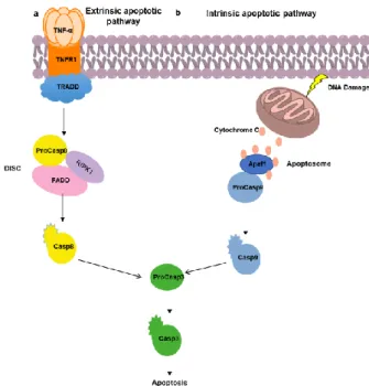
There are two known pathways for activation of the caspase cascade leading to apoptosis, the extrinsic pathway and the intrinsic pathway (Figure 1.2). Each of these pathways uses its own initiator procaspase and its activation complex.1,5
Figure 1.2. Extrinsic (a) and intrinsic (b) apoptotic pathway
1.2.1.1. Extrinsic pathway of apoptosis
The binding of extracellular signalling proteins to death receptors, belonging to the tumor necrosis factor (TNF) family, leads to the activation of the extrinsic apoptosis pathway (Figure 1.2a).1,5
Activation of this pathway may occur by either binding of the Fas ligand (FasL) to the Fas receptor resulting in the binding of the Fas-associated protein with death domain (FADD), or via TNF receptor activation, resulting in the binding of the adapter protein TRADD, and consequent recruitment of FADD and receptor-interacting serine/threonine-protein kinase 1 (RIPK1).1,5
FADD, recruits initiator procaspase (procaspase-8 or procaspase-10) forming a death-inducing signalling complex (DISC). Once activated in the DISC, the initiator procaspases activate executioner procaspases, thereby inducing apoptosis.1,5
8
The extrinsic pathway of apoptosis may be inhibited by the production of decoy receptors by the cells, these having a ligand-binding domain, but not a death domain, inhibiting apoptosis by inhibiting the death receptor. However, they can also produce blocking proteins, such as FLIP, that compete with the initiating procaspases for binding to the DISC complex, inhibiting them. These inhibitory mechanisms serve to prevent unnecessary pathway activation.1,5
1.2.1.2. Intrinsic pathway of apoptosis
Apoptosis can also be activated within the cell in response to certain stimuli. This pathway (Figure 1.2b) depends on the release into the cytosol of mitochondrial proteins that can activate the caspase proteolytic cascade in the cytoplasm.1
One of the crucial proteins of this pathway is cytochrome c, which binds to the procaspase-activating adapter protein, apoptotic protease procaspase-activating factor-1 (Apaf1), leading to apoptosome formation (equivalent to DISC). Apaf1 proteins recruit the initiator procaspases proteins (procaspase-9) in the apoptosome. Once activated, they activate the executioner procaspases resulting in apoptosis.1,5
The regulation of this pathway by the Bcl2 proteins occurs by controlling the release of cytochrome c and other proteins into the cytosol. Pro-apoptotic Bcl2 proteins induce apoptosis by increasing the release of proteins, in the case of the anti-apoptotic Bcl2 proteins, they inhibit apoptosis by blocking the release of these proteins.1,5
Besides the Bcl2 proteins, there are inhibitor of apoptosis (IAP) proteins that are also intracellular regulators of apoptosis. These binds and inhibit the active caspases.1
1.2.2. Necrosis
In contrast to apoptosis, necrosis is an unregulated cell death type that occurs through autolysis caused by external factors such as infection or trauma.6 As it follows a different transduction pathway than apoptosis this type of cell death originates a swelling of the organelles, an early increased permeability of the plasma membrane7,8 and consequently to a loss of intracellular content to the extracellular space, thus causing an inflammatory response.7
Until a few years ago, necrosis was considered an unregulated process unlike apoptosis.9 However, recent studies reveal that there is a type of necrosis that can be regulated but exhibits similar characteristics as necrotic cell death, the necroptosis or necrosis programmed.6
1.2.2.1. Necroptosis
The necroptosis is a term used a few years and refers to a type of programmed necrosis.9 This is caspase-independent and occurs when apoptosis is inhibited.7,8
9
This type of cell death occurs through a variety of intracellular and extracellular stimuli that activate ligands of death receptor family10,11, being involved in a variety of pathologies such as myocardial infraction12, stroke13, atherosclerosis14 or Crohn’s disease15.
1.2.2.1.1. Necroptosis signalling pathway
Figure 1.3. Signalling pathway to lead cell survival (a), apoptosis (b) or necroptosis (c)
Activation of this signaling pathway occurs by the binding of the ligands of the death receptor family (such as TNF-α, FasL and TRAIL) to their receptors on the plasma membrane, triggering the formation of complex I. This complex depending on modifications such as ubiquitination and phosphorylation can determine the life or death of the cell.7,16
Upon ubiquitination RIPK1, recruits the TNF mediators, NEMO and TAK1, activating downstream pathways of NF-κB and MAP (Figure 1.3a).7
However, the inhibition of the NF-kB pathway by the protein synthesis inhibitor, cyclohexamine, and the deubiquitination of RIPK1 by CYLD leads to cell death, inducing the formation of complex IIa (Figure 1.3b). 7,16
Complex IIa is composed of RIPK1 and TRADD, which recruits FADD, and this in turn recruits procaspase-8. This initiates the apoptotic pathway by inhibition of necroptosis through the cleavage of RIPK1 and receptor-interacting serine/threonine-protein kinase 3 (RIPK3).
If apoptosis is inhibited, RIPK1 recruits RIPK3 by binding to RIP-homotypic interacting motif (RHIM) domains to form an amyloid complex, necrossome.4,7,8,16 In this complex, the
10
autophosphorylation of RIPK3, leads to recruitment of its substrate, mixed lineage kinase domain like pseudoquinase (MLKL) (Figure 1.3c).7,8
The MLKL acts as a necroptosis executioner and is found in the cytosol as a monomer when inactive.8 Upon RIPK3 activation, its kinase domain binds to the C-terminal domain of MLKL leading to phosphorylation of MLKL.7,8
Consequently, oligomerization of the N-terminal domain of MLKL occurs through destabilization of the MLKL structure followed by translocation to the plasma membrane, where permeabilization of the membrane.7,8
11
1.3.
Necroptosis as therapeutic target
Necroptosis, a regulated form of necrosis, is associated with many pathologies such as myocardial infarction12 or atherosclerosis14.
1.3.1. Ischemia-reperfusion injury
Ischemia-reperfusion injury is a common type of cell damage. Ischemia occurs when there is a lack of blood supply through the obstruction of a tissue, resulting in the lack of oxygen and nutrients that in the case of being prolonged leads to cell death. Reperfusion occurs following ischemia, through the reintroduction of blood flow to tissues where it has been missing for some time and can lead to inflammation.7,10
This type of injury can be reversible, when it is possible to restore the bloodstream or irreversible, when the deprivation is persistent and leads to cell death.7,10 During restoration of blood flow resulting in the reintroduction of oxygen, the formation of reactive oxygen species (ROS) and pore opening occur, leading to inflammation and death by necrosis.17
In vivo studies administering Necrostatin-1 (Nec-1) before reperfusion in animal models, revealed the inhibition of RIPK1-dependent necrosis, leading to the reduction of infarct size.12
1.3.1.1. Stroke
Stroke occurs due to obstruction of the blood vessels in the brain by thrombosis or embolism or through an intracerebral hemorrhage.10
Liver
- Acute hepatitis54
- Alcoholic and Non-alcoholic liver disease27,52
Kidney - Acute kidney injury54
Spinal cord
- Spinal cord injury54 Brain
- Stroke13
- Traumatic head injury19 Skin - Skin inflammatory diseases28 Bowel - Inflammatory bowel disease17 Pancreas - Acute pancreatitis25
12
Blood vessels obstruction can be prolonged, leading to death of brains cells due to lack of oxygen. In the case of hemorrhage, this occurs through the rupture and leakage of a blood vessel leading to damage in the cells.13
Degterev co-workers demonstrated that necroptosis may play a role in stroke, since that with using Nec-1 has occurred a delay of ischemic neuronal injury.9 Another study, performed by Xingshun Xu co-workers in mices using two protective agents, HMG (anti-apoptotic) and Nec-1 (anti-necrotic), also demonstrated a decrease in infarct size, and combined treatment may be a clinically useful.13
1.3.1.2. Traumatic brain injury
Traumatic brain injury occurs when an external mechanical force is applied to the brain, causing secondary injuries such as ischemia-reperfusion, hemorrhage or edema, leading to difficulties in the functioning or even death of neuronal cells.10
Data obtained in one study revealed that necroptosis may play a role in traumatic brain injury, since the administration of Nec-1 in a mouse model after controlled cortical impact revealed a decrease in tissue injury in the brain and inflammation, increasing motor function and memory space.18
1.3.2. Neurodegenerative diseases
1.3.2.1. Huntington's Disease
Huntington's disease is an autosomal dominant progressive neurodegenerative disease occurring in a single gene called Huntingtin, through of the formation of the mutant protein, result of triple repeats of CAG in the gene encoding Huntingtin protein. This disease is often the result of genetic inheritance and it is characterized by motor, cognitive and psychiatric deficits and may occur at any time in life, however it is more frequent that the symptoms begin in middle age.7,10,19
Assays using Nec-1 in immortalized striatal neuronal line expressing mutant HTT reduces cell death and using in mutant HTT-expressing R6/2 transgenic mouse model delays the onset and progression of disease.20
1.3.2.2. Alzheimer's disease
Alzheimer's disease is a multifactorial chronic neurodegenerative disorder characterized by progressive loss of memory and motor functions and the leading cause of dementia. Although its etiology is still not fully understood, it characterized by the formation of intracellular neurofibrillary tangles and extracellular β-amyloid protein deposits, leading to neuronal cell death in the cerebral cortex and other regions.10
13
In vivo model, administration of Nec-1, in previously treated aluminum mouse resulted in a decrease in neuronal cell death, improved learning and memory, and still decrement expression of Aβ and Tau protein levels.21
1.3.3. Inflammatory diseases
1.3.3.1. Acute pancreatitis
Pancreatitis is an inflammation in the pancreas, which may be acute or chronic.22
Studies in experimental models reported that in acute pancreatitis, acinar cells can die both by apoptosis and by necrosis, varying the severity of pancreatitis proportionally with necrosis and inversely proportional to apoptosis.22
RIPK3 plays an important role in necroptosis. Studies where pancreatitis is induced by cerulein have shown an increase in RIPK3 expression only in pancreatic tissues, or a few hours later there were several areas with acylar cell loss and necrosis. This effect was prevented by using deficient RIPK3 lines and a decrease in serum amylase was still observed in comparison with normal cells. Through this observation and taking into account of pancreatitis induced by cerulein can confirm the involvement of necroptosis in this pathology.23
However, it is recently been reported that administration of Nec-1 has no protective effect on cerulein induced pancreatitis. This even has a contrary effect, accelerating the death of cells. 24
1.3.3.2. Chronic liver diseases
Chronic liver disease can occur through several factors, such as alcohol consumption or fat accumulation. Over time, the disease progresses through several stages between tissue damage and liver dysfunction.
1.3.3.2.1. Alcoholic liver disease
Alcoholic liver disease occurs after a chronic ingestion of alcohol.10
Evidence shows that apoptosis is linked to alcoholic liver disease, however, treatment with a pan-caspase inhibitor is not sufficient to prevent liver damage. Other studies show that RIPK3 expression increases with or without pan-caspase inhibitor after chronic administration of ethanol, the expression of this kinase being evidence of necropsosis.10,25 It has also been reported that mice with RIPK3-deficient show protection against liver damage induced by ethanol. However, when administered Nec-1, it did not modify the expression of RIPK1, not attenuating the liver injury. This may occur if the pathology is independent of RIPK1 or due to pharmacokinetic problems with Nec-1.10,26
14
1.3.3.3. Skin inflammatory diseasesEpidermal keratinocytes are the first line of defense against potentially pathogenic microorganisms since they provide a structural and immunological barrier. When exposed to lesions, they send signals triggering immune responses. Since of responses sent by epidermal keratinocytes is implicated in inflammatory skin disease.10,27
One study found that specific epidermal ablation of FADD leads to spontaneous necrosis of keratinocytes and to the development of severe skin injury. This necrosis was prevented by RIP3 deficiency, thus showing that RIP3-dependent necrosis of FADD-deficient keratinocytes triggers severe inflammation of the skin of mice.10,27 Inflammations can also be triggered in mice by deficient caspase-8 in studies.10,28
It can then be concluded that both FADD and caspase-8 play an important role in preventing skin inflammation.
15
1.4.
Necroptosis inhibitors
Necroptosis is a very complex process, and in order for it to be viably “druggable”, a careful evaluation of the pathway and its intricate interconnexions with other biochemical paths have to be considered. Nowadays, to pharmacologically interfere with RIPK1, RIPK3 and MLKL have been targeted, as they form the necrosome and from there occurs transduction of many other downstream signaling (e.g the translocation of the phosphorylated MLKL to the plasma membrane where it can lead to the swelling of organelles and to membrane rupture).29
Based on these targets, studies have been initiated to discover necroptosis inhibitors. However, so far, no drug has reached the marked, is still in clinical trials.
1.4.1. RIPK1
Necroptosis can be induced by ligands of the death receptor, with RIPK1 being the first site where this pathway can be inhibited.
Necrostatins, small molecules inhibitors of necroptosis, were reported for the first time in 2005 by Degterev et al.9 These compounds were identified through of a screening using TNF-α as pathway inducer and the caspase inhibitor zVAD-fmk to inhibitor apoptosis, leading to necroptosis.
Necrostatins (Figure 1.5a) are selective for RIPK1 receptor, inhibiting their catalytic activity and thus inhibiting necroptosis.14,30 Nec-1, the first known inhibitor of necroptosis, it has been the most studied although there are other necrostatins with inhibitory activity such as 3 and Nec-4.30
Necrostatins were optimized in order to increase activity and metabolic stability. From the optimization of Nec-1, 7-Cl-O-Nec-1 (Nec-1s) was synthesized by addition of a Cl at the 7-position of the indole ring which provided an increase in activity and substitution of thio-hydantoin by hydantoin which increased stability and eliminated toxicity. Furthermore, this molecule presented no off-target activity against indoleamine-2,3-dioxygenase, as happened with the Nec-1 14,30
In order to further improve necrostatin activity, PN10 (Figure 1.5c), a hybrid compound (a combination of Nec-1s and a fragment of Ponatibib), was developed. This compound proved to be highly potent and selective for RIPK1, taking advantage of the best properties of each of the other two inhibitors and increased interactions with the active site.31
Harris et. al. described three novel classes of compounds with inhibitory activity against necroptosis (aminoisoquinolines, pyrrolo[2,3-b]pyridines e furo[2,3-d]pyrimidines) following the screening of GSK kinase inhibitor libraries.30,32 From the crystalline structure of a compound of the first series with RIPK1 it was possible to identify the zones of the interaction of the compound with RIPK1. The X-ray structure showed that 1-aminoisoquinoline forms hydrogen bonds with the
16
Met95 hinge residue of RIPK1. Central urea forms hydrogen bonds with DL15 ASP156 and the Glu63 side chain.32
Of these three series, Cpd 27 (Figure 1.5b), belonging to the last series, was selected for in vivo assays where necroptosis is induced by TNF-α.32
Pazopanib (Figure 1.5d), a selective tyrosine kinase receptor inhibitor, has been more recently identified to decrease necroptosis. This selectively inhibits RIPK1, without decreasing the in vitro activity of RIPK3.33
Figure 1.5. Chemical structure of identified necroptosis inhibitor targeting RIPK1
1.4.2. RIPK3
RIPK3, the second protein required for necroptosis execution, was discovered after RIPK1, its binding partner through the RHIM, that leads to the formation of the necrosome.30
Once the catalytic activity of this kinase or RIPK1 is inhibited, the activation of necroptosis will also be inhibited or canceled, since this protein plays two distinct roles, on one hand it activates the necrotic activity and on the other recruits the MLKL. Phosphorylation of this by RIPK3 is the key step in the execution of necroptosis.30
Once the importance of this protein in the activation of necroptosis has been developed, small inhibitory molecules of this kinase (Figure 1.6).
17
Figure 1.6. Chemical structure of identified necroptosis inhibitor targeting RIPK3
The first three inhibitors to be identified were GSK'840, GSK'843 and GSK'872 (Figure 1.6a) by Kaiser co-workers.34
Of the three compounds identified, GSK'840 was the one that presented a better profile, however this is active in human cells but not in mouse cells, unlike the other two that have inhibitory activity in mouse cells. These results demonstrate that the binding ability of RIPK3 to the compound depends on the cell type and also show the contribution of the activity of this kinase to necroptosis.34
GW'39B (Figure 1.6b), was identified as an inhibitor of RIPK3, exhibiting a structure similar to GSK'872, another inhibitor of RIPK3. This compound blocks the phosphorylation of MLKL and translocation to the plasma membrane by inhibiting RIPK3 in both murine and human cells.34
Dabrafenib (Figure 1.6c), a BRAF inhibitor was also shown to inhibit of RIPK3
,
by disrupting binding of RIPK3 to MLKL. This is the only inhibitor of RIPK3 that has been tested in vivo.Another indirect approach for the inhibition of RIPK3 is found in the complex of Hsp90 with co-chaperone Cdc37 that is associated with RIPK3, so its inhibitors prevent the activation of RIPK3 in the necrosome.30
1.4.3. MLKL
The third hypothesis for the inhibition of necroptosis lies in the necroptosis executioner, the pseudokinase MLKL.14
The discovery of necrosulfonamide (NSA) (Figure 1.7a) as an inhibitor of necroptosis was important for the identification of MLKL since present activity only after necrosome formation. This molecule acts as a Michael acceptor, by coupling α, β-unsaturated enone with the Cys86 residue of MLKL.14 For this reason, it is not possible to use this compound in non-murine cells lacking the cysteine residue.14,30
18
Figure 1.7. Chemical structure of identified necroptosis inhibitor targeting MLKL
More recently, "Compound 1" (Figure 1.7b) has also been reported to target MLKL, preventing the pseudokinase from altering its conformation being
phosphorylated
by RIPK3. However, this compound is toxic to cells and is not selective for MLKL,having its application
limited
.14Table 1.1. Inhibitors reported for each target molecule
Target molecule Necroptosis inhibitors
RIPK1
Nec-1, Nec-3, Nec-4, Nec-5, Nec-1s Cpd27 PN10 Pazopanib GSK’481, GSK2982772 Compound 56 (HIPA-56) Tozasertib GSK2606414, GSK2656157 RIPK3 GSK’840, GSK’843, GSK’872 GW’39B Dabrafenib MLKL NSA Compound 1
19
1.5.
Oxazol-5(4H)-ones
Oxazol-5(4H)-one (hereinafter only referred to as Oxazolones) are small and simple molecules, very valuable and versatile. They have applications in several areas of research, such as organocatalysis or photochemistry, due to the presence of several reactive sites, represented in Figure 1.8 as N-3, C-4 and C-5, where a varied set of modifications can occurs.35,36
Figure 1.8. General structure of oxazolones, with their reactive sites
Plochl et al, synthesized the first oxazolone in 1883, through by the condensation of a benzaldehyde with hippuric acid in the presence of acetic anhydride. It was only in 1900, that Erlenmeyer established the first correct structure of oxazolone (Figure 1.8), containing a 5-membered ring with O and N as the heteroatom, which he called "azolactone".36–38
From this oxazolone, it is possible to easily generate an oxazole enolate through abstraction of the acidic proton at C-4, the formation of a reactive ketene or 1,3-dipolar through the use of a Lewis acid. The ring may also be opened by a nucleophile to the carbonyl.36
1.5.1.
Erlenmeyer-Plochl reaction
The Erlenmeyer-Plöchl reaction was first described in 1883 by Plöchl as the condensation of benzaldehyde with hippuric acid in the presence of acetic anhydride and sodium acetate (Scheme 1.1),38 occurring in the absence of any solvent, it can be said that this reaction follows the principles of green chemistry because it minimizes the use and production of hazardous substances.35
The reaction takes place in two steps, beginning with a cyclization of hippuric acid and its derivatives, followed by a Perkin condensation, thus producing Erlenmeyer azolactone.38
In this reaction competition occurs for the product resulting from the transacylation and condensation of the hippuric acid.39
20
Scheme 1.1.Conditions of the Erlenmeyer-Plöchl reaction
This type of reaction is influenced by the electronic characteristics of the compounds, which are sensitive to the nature of the substituents, with a decrease in the reaction time in the presence of electron donating groups (EDG) in the hippuric acid that increase the reactivity due to high nucleophilicity of the oxygen atom on the amide carbonyl group (Scheme 1.2).39,40
Scheme 1.2.Influence of EDGs on hippuric acid
However, electron withdrawing groups (EWG) in the aldehyde also decrease the reaction time, by increasing the electrophilicity of the carbonyl group (Scheme 1.3).39,40
Scheme 1.3.Influence of EWGs on aldehyde
To better understand the influence of the various substituents in this reaction the Hammett equation can be applied.
21
1.5.1.1. The Hammett equationThe Hammett equation (1) described in 1937 relates the structure and reactivity of benzoic acid and its derivatives with substituents in -meta and -para positions with the rate of reaction.39,41
This equation can be expressed as follows:
𝜌𝜎
𝑥= log
𝑘
𝑥𝑘
𝐻(1)
where ρ is the reaction constant, 𝜎𝑥 is the constant of the substituent and quantifies the
electronic contribution of that substituent, 𝑘𝑥 e 𝑘𝐻 are respectively the velocity constants for the
reaction with the ring substituted X and for the unsubstituted ring.39,41
The 𝜎𝑥 quantifies the electronic contribution of this substituent and the values for each
substituent in the -meta and -para position of the aromatic ring have been tabulated since 1937 by Hammett. The EWGs present positive 𝜎𝑥 values, stabilizing the increase of the negative charge
in the reaction center, while EDGs present negative values of 𝜎𝑥, stabilizing the positive charge
in the reaction center.39,41
In relation to ρ, this one informs on the speed of the step determinant of the reaction and presents positive values of ρ when the reaction speed given by the presence of EWGs, while the negative values of ρ means speed of the reaction by the EDGs.39,41
When the value of ρ is much greater than +1.00 and much less than -1.00 then the reaction under study is sensitive to variations of the substituent, whereas a value of ρ close to 0 means that the reaction is insensitive to variation of the substituent on the aromatic ring.39,41
1.5.2. Uses of oxazolones
Oxazolones are compounds which are of considerable interest, since their structure has several reactive sites and from them it is possible to produce a wide variety of compounds that can be applied in both chemistry and biology.36
22
Scheme 1.4.Reactive sites of the oxazolone skeleton [Adapted from Ref 35]35
The oxazolones contain a proton of acidic nature at C-4, adjacent to the carbonyl, which can easily form an enolate A in a basic medium and it has the possibility to react with several electrophiles. Another possible reaction may occur by the reaction of a Lewis acid with oxazolone and formation of the reactive ketene C or 1,3-dipole B, with the possibility of both reacting by cycloaddition with the formation of several heterocyclic compounds. Finally, the oxazolone further contains a site of electrophilic nature, C-5, where attack by a variety of nucleophiles on the carbonyl can occur and formation of a variety of protected amino acids after ring opening (Scheme 1.4).35,36
Due to reactivity of its structure, oxazolone was considered a good substrate for the production of other heterocycles (Scheme 1.5).36
Scheme 1.5. Diversity of compounds produced from oxazolone [Adapted from Ref. 36]36
23
Due to its reactivity, the oxazolone ring can be used as a substrate for the synthesis of enantiomerically pure amino acids using organocatalysts, or others moiety of biological interest may be formed from this ring (Scheme 1.5).36
The oxazolone ring may also contain an exocyclic double bond at C-4, which induces a new reactive site and allows the synthesis of new structures. With the appearance of this double bond a highly conjugated system was formed allowing higher light absorption and thus the possibility of applications in the field of photochemistry.35,42–44
Oxazolone derivatives have been described as pH45, Fe3+ 46 or glucose47 sensors in solid matrices. More recent studies also point out the possibility of these oxazolones being used as photoswitches42. In this research group studies were realized using oxazolone derivatives, concluding that they can be used in the construction of new TPA (Two Photon Absorption) sensors.39,43,44
1.5.3. Oxazolones as necroptosis inhibitors
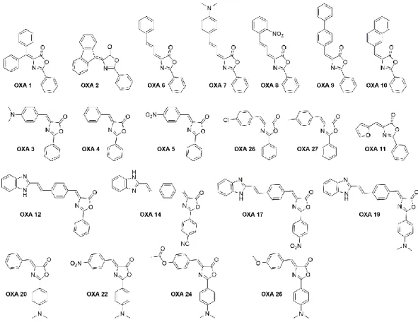
A preliminary screening using a small family of 21 oxazolones (Figure 1.9), developed by Dr. Catarina Rodrigues in the Bioorganic Chemistry Group at iMed.ULisboa, was tested in cell lines (BV2 murine microglia cells and L929 cells).
Figure 1.9. Chemical structure of the 21 oxazolone derivatives developed in this team by Catarina
24
From this screening it was possible to identify a compound, OXA 12, which has inhibitory activity similar to the known necroptosis inhibitor, Nec-1, which as described before presents some disadvantages.
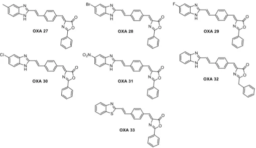
The result of these assays was the starting point for the development of new possible inhibitors of necroptosis based on OXA 12 and to increase its inhibitory activity. From this, were developed in our laboratory by Dr. Mónica Estevão more compounds based on the structure of OXA 12 (Figure 1.10), not yet being possible to obtain any compound with a superior activity. However, the results obtained will be useful for the study structure-activity relationship, giving information about the influence of each group.
Figure 1.10. Chemical structure of compounds developed on the basis of OXA 12 structure
In the first screening only OXA 12 and compounds derived from it, OXA 14, OXA 17 and OXA 19, exhibited inhibitory activity so it can be understood that benzimidazole will be an important group for inhibition, from this fact, the compounds of Figure 1.10. were synthesized
.
25
27
Considering the previous studies performed by this Team, the main purpose of this project Master Thesis is the development of new potential modulators of necroptosis based on the OXA 12 moiety. Through the synthesis of different compounds, containing different substitution patterns based on OXA 12,
The synthesized compounds were further evaluated using BV2 murine microglia cells and the methodology used for the above compounds. In order to establish a structure-activity relationship (SAR) and to find or propose new or optimized modifications or structures, which have an optimized activity, are more potent and/or have better pharmacokinetic/pharmacodynamic properties.
Figure 2.1. Structure of OXA 12
Benzimidazole ring
Oxazolone ring
Hippuric acid moiety