Electrochemical biosensor for phenols and
catecholamines based on tyrosinase immobilized on
gold nanoelectrode ensembles
Ana Isabel Ribeiro de Pinho
Dissertação de Mestrado em Controlo de Qualidade,
área de especialização em Águas e Alimentos
Trabalho realizado sob a orientação da Prof. Doutora Cristina Delerue- Matos,
Co-orientação da Prof.ª Doutora Maria Beatriz Oliveira e do Doutor Viswanathan
Subramanian
ii
É autorizada a reprodução integral desta dissertação apenas para efeitos de
investigação, mediante declaração escrita do interessado, que tal se compromete.
iii
Agradecimentos
À Prof.ª Beatriz por ter sido a mentora deste projecto e por me ter encaminhado para o Isep.
À Prof.ª Cristina por me ter acolhido no seu laboratório, pela disponibilidade dispensada e pelo apoio prestado.
To Dr. Viswanathan, for the long hours that spend with me and for the thoughts that transfer during this project. THANKS VERY MUCH! Without you nothing of this was possible. To meet you was a big pleasure!
Às minhas colegas de laboratório, pela preocupação, carinho e apoio que demonstraram.
À minha amiga Patrícia pelo apoio, preocupação e “divulgação” deste projecto
À minha colega de trabalho, Patrícia pelo apoio prestado.
A ti, Nuno, que sem a tua força, incentivo, compreensão e apoio este passo importante da minha vida não teria sido dado. Sem a tua força não estaria nesta fase.
Por fim, ao meus pais, que são o meu suporte. Sem vocês os sonhos não eram possíveis de se realizar. Obrigado pelo apoio ao longo de todas as etapas da minha vida e por estarem presentes em mais uma meta alcançada.
iv
Abstract
Nanostructured materials represent new platforms for biomolecule sensing, providing increased sensitivity and facilitating miniaturization. Many arrayed nanostructures comprise electroactive materials, exhibiting improved promise for ultrasensitive biosensing relative to conventional electrochemical electrode. Among various strategies for synthesizing the nanoscopic materials reported in the literature, template synthesis is one of the most popular approaches for fabricating three-dimensional (3D) nanostructured arrays for sensor applications. Electrochemical methods are well suited for detecting organic compounds because of their simplicity and efficiency. Gold nanoelectrodes ensemble were prepared by using electroless deposition of the metal within the pores of polycarbonate track-etched membranes. Tyrosinase enzyme has been immobilized onto preformed self-assembled monolayers of mercaptoethylamine on gold nanoelectrode via cross-linking with glutaraldehyde. Flow injection analysis systems in wall-jet configurations using this tyrosinase -modified nanoelectrodes are developed. Gold nanoelectrode ensembles (GNEEs), 50 nm in diameter and 180±20 nm in length were prepared by electroless template synthesis in polycarbonate filter membranes, followed by selective controlled sequential polycarbonate dissolution using DCM/EtOH (V∶ V=1∶3). The electrochemical evaluation of the 3D GNEEs was conducted using the well known [Fe(CN)6]3-/[Fe(CN)6]4- couple. Compared with 2D GNEEs, the 3D GNEEs significantly enhanced the current response in cyclic voltammetry. The electrochemical results demonstrated the fact that electron transfer process could be effectively improved at the 3D cylindrical GNEEs. Linear diffusion is dominant on the cylindrical GNEEs at conventional scan rates. Under optimized conditions, high reproducible results were obtained, linear calibration was achieved in the 1x10⁻⁶ M to 1x10⁻³ M concentration range and the detection limit was 1x10⁻⁸ M. Moreover, negligible interferences from species like 100 mM glucose, 20 mM ascorbic acid and 100 mM urea were observed at a potential of -0.100 V (vs. Ag/AgCl). L-dopa and dopamine spiked serum samples were analyzed for recovery studies.
Keywords: Nanostructured materials; Gold nanoelectrode ensembles; Electroless deposition;
v
Resumo
Os nanomateriais representam novas plataformas, para a detecção de biomoléculas, uma vez que proporcionam maior sensibilidade e são de fácil miniaturização. Estes nanomateriais usados em biossensores ultra-sensíveis englobam, na sua maioria, materiais electroactivos, em que o seu uso tem mostrado ser um sucesso, quando comparados com os eléctrodos convencionais. Entre os vários métodos apresentados na literatura, para sintetizar os nanomaterias, o método ”template” é um dos métodos mais usados na fabricação de nanoestruturas tridimensionais (3D). Os métodos electroquímicos, devido à sua simplicidade e eficiência, são várias vezes usados para detecção de compostos orgânicos.
Os nanoeléctrodos de ouro foram preparados por deposição química em que o metal se deposita nos poros das membranas de policarbonato. A enzima Tirosinase foi imobilizada, em monocamadas organizadas e pré-formadas de mercaptoetilamina sob os nanoeléctrodos de ouro, usando o glutaraldeído como agente reticular. Foram desenvolvidos sistemas de fluxo contínuo usando os nanoeléctrodos de ouro modificados com Tirosinase. Os nanoeléctrodos de ouro, com 50 nm de diâmetro e 180±20 nm de comprimento foram preparados pelo método “ template”, deposição química do metal nas membranas de policarbonato, seguindo-se a dissolução sequencial da membrana, usando DCM/EtOH (V:V;1:3). A análise electroquímica dos nanoeléctrodos de ouro 3D, realizou-se com o conhecido par [Fe(CN)6]3-/[Fe(CN)6]4-. Quando comparados os sinais obtidos por voltametria cíclica, com os nanoeléctrodos de ouro de 2D e 3D, constata-se que há um aumento considerável da intensidade de corrente no que diz respeitos aos eléctrodos 3D. Os resultados electroquímicos mostram que o processo de transferência de electrões pode ser melhorado para o caso dos nanoeléctrodos de ouro cilíndricos a 3D. A difusão linear é dominante para o caso dos nanoeléctrodos de ouro cilíndricos, para velocidades de varrimento convencionais. Após optimização de todos os parâmetros experimentais, os resultados apresentam elevada reprodutibilidade, para o intervalo de calibração linear entre 1x10⁻⁶ M e 1x10⁻³ M, obtendo-se um limite de detecção de 1x10⁻⁸ M. O estudo de interferências foi realizado a um potencial de -0,100 V (vs. Ag/AgCl) para 100 mM de glucose, 20 mM de ácido ascórbico e 100 mM de ureia, tendo-se verificado não serem significativas. Para os estudos de recuperação, amostras de soro fisiológico foram contaminadas com L-dopa e dopamina.
Palavras-chave: Nanomateriais, nanoelectrodos de ouro, deposição química, enzima, sistema de fluxo contínuo
vi
Index
1 Introduction 1 1.1 Introduction 2 1.2 Neurotransmitters 2 1.3 Biosensor 3 1.3.1 Classifications of Biosensor 51.3.2 Classification based on bioreceptors 5
1.3.2.1 Enzymatic biosensors 5
1.3.2.2 Immunosensors 6
1.3.2.3 Enzyme immunoassays 6
1.3.2.4 DNA based biosensors 6
1.3.2.5 Aptasensors 7
1.3.2.6 Whole-Cells or Organelles based biosensors 7 1.3.3 Classification based on the transduction method 8
1.3.3.1 Electrochemical Transducers 8
1.3.3.2 Optical Transducers 8
1.3.3.3 Electrical Transducers: Filed Effect Transistor based
sensors 9
1.3.3.4 Piezo-Electric Sensors 9
1.3.3.5 Thermal Sensors 9
1.4 Electrochemical Biosensors 9
1.4.1 Classifications of electrochemical biosensors 9
1.4.1.1 Voltammetric sensors 10
1.4.1.2 Potentiometric biosensors 10
1.4.1.3 Impedimetric biosensors 10
1.4.1.4 Enzyme based electrochemical biosensors 11
vii
1.4.2.1 First generation amperometric enzyme sensors 12 1.4.2.2 Second generation amperometric enzyme sensors 13 1.4.2.3 Third generation amperometric enzyme sensors 13
1.5 Enzyme immobilization methods 14
1.5.1 Adsorption 15
1.5.2 Entrapment 15
1.5.3 Covalent bonding 16
1.5.4 Cross-linking 16
1.6 Importance of working electrode 17
1.7 Role of nanotechnology in biosensors developments 17 1.7.1 Nanotechnology in electrochemical biosensors 18 1.7.2 Nanoarrays, nanotubes, nanoparticules electrodes 20
1.8 Self Assembled Monolayer Modified Electrodes 21
1.9 Gold electrodes in biosensor fabrication 21
1.9.1 Gold nanomaterials in enzyme biosensors 22
1.9.1.1 Gold nanoparticles 22
1.9.1.2 Gold nanocomposites 23
1.9.1.3 Gold nanoarrays 24
1.10 Flow Injection Analyses 25
1.10.1 Basic Components 25
1.10.2 Methodology of Flow Injection Analysis 26
1.10.3 The importance of Dispersion 27
1.10.4 Factors affecting controllable sample dispersion 28
1.10.5 The concentration gradient 28
1.10.6 Flow Injection Analysis Signals 29
1.11 Enzyme: Tyrosinase 30
viii
1.12 Reasons to develop new sensors 33
2 Experiments 35
2.1 Experimental Part 36
2.1.1 Instrumentation 36
2.2 Cell setup 36
2.2.1 Static 36
2.2.2 Flow wall jet -FIA 36
2.3 Scanning electron micrographs (SEM) 37
2.4 Membrane templates 37
2.5 Reagents and solutions 37
2.6 Methods 38
2.6.1 Pretreatment of gold disk electrode 38
2.6.2 Preparation of gold nanoelectrodes 38
2.6.3 Etching Procedure 39
2.6.4 Enzyme Immobilization 39
2.6.5 Interferences Studies 40
2.6.6 Photographs of FIA step up 40
3 Results and discussion 42
3.1 Results and discussion 43
3.2 Electrochemical characterization of GNEE 47
3.3 Self-Assembled Monolayers on GNEE 51
3.4 Electrochemical studies of Tyrosinase immobilized on
GNEE 53
3.5 Optimization of FIA parameters 55
ix
3.7 Optimization flow rate for FIA 56
3.8 Analytical calibration 57
3.8.1 Calibration plot for L-dopa 58
3.8.2 Calibration plot for dopamine 59
3.8.3 Calibration plot for catechol 60
3.8.4 Calibration plot for phenol 61
3.9 Stability of TyrE-GNEE 62
3.10 Interference studies 62
4 Conclusions 66
x
Index of Figures
Figure 1.1 Structure of a typical chemical synapse 3
Figure 1.2 The general scheme of amperometric biosensors 4 Figure 1.3 Scheme of different generations of amperometric enzyme sensors 12 Figure 1.4 Formation of self assembled monolayer on gold 22
Figure 1.5 Flow Injection Analysis System 25
Figure 1.6 Four phases of Flow Injection Analysis 27
Figure 1.7 The analog output has the form of a peak, the recording starting at S (time of injection to). H is the peak height, W is the peak width at a selected level, and A is the peak area. T is the residence time corresponding to the peak height measurement, and tb is the peak width at the baseline
29
Figure 1.8 Tyrosinase enzyme 3D model 31
Figure 1.9 Intracellular transformation of tyrosinase into pre-melanin metabolites, and finally into melanin; several of the metabolites between tyrosinase and melanin are toxic to melanocytes according to the self-destruct
theory 31
Figure 2.1 FIA set up 40
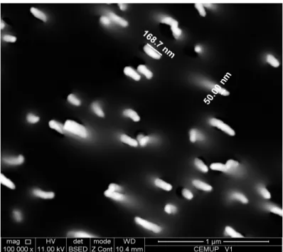
Figure 2.2 Autolab PSTAT 12 Potentiostat /Galvanostat 41 Figure 3.1 SEM image of 3D GNEEs created using a 50:50 DCM/EtOH mixture
applied to a Au-filled polycarbonate membrane (50 nm- diameter
pores) 45
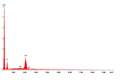
Figure 3.2 EDX spectrum of Au filled PCTE membrane before etching (2D) 46 Figure 3.3 EDX spectrum of Au filled PCTE membrane before etching (3D) 46 Figure 3.4 Cyclic voltammograms obtained at 3D and 2D GNEEs in 0.001M
K3[Fe(CN)6] in 0.1 M KNO3 at a scan rate of 50 mV/s 47 Figure 3.5 Cyclic voltammograms obtained at different scan rates for 2D GNEEs
in 0.01M K3[Fe(CN)6] and PBS, pH 6.5 at scan rates ranging from 10
xi
Figure 3.6 Cyclic voltammograms obtained at different scan rates for 3D GNEEs in 0.01M K3[Fe(CN)6] and PBS with pH 6.5 at scan rates ranging from
10 to 100 mV/s 49
Figure 3.7 log Ipc vs. log v for cyclic voltammogram of 0.01M K3[Fe(CN)6] and 0.1M PBS with pH 6.5 obtained used 3D GNEEs 50 Figure 3.8 log Ipc vs. log v for cyclic voltammogram of 0.01M K3[Fe(CN)6] and
0.1M PBS with pH 6.5 obtained used 3D GNEEs 52 Figure 3.9 Cyclic voltammograms of TyrE-GNEE (Solid line) and GNEE (Dotted
line) 0.1M PBS, pH 6.5, Scan Rate 50 mV/s 53
Figure 3.10 Cyclic voltammograms of the enzyme electrode in 0.1M PBS (pH 6.5) without (a) and with 1 x10⁻⁴ M catechol (b). Potential scan range
covers from -200 to 500 mV 54
Figure 3.11 Hydrodynamic voltammogram of L-dopa on GNEE at PBS, pH 6.5,
scan rate 50 mV/s 55
Figure 3.12 Effect of the flow rate on the oxidation of L-dopa on GNEE in 0.1 M at
PBS, pH 6.5 at constant potential -0.100 V 56
Figure 3.13 FIA responses of L-dopa (a) 10-3, (b)10-4, (c) 10-5 and (d) 10-6 M in 0.1 M PBS pH 6.5 at -0.100 V vs Ag/AgCl for five continuous injections 58 Figure 3.14 Calibration plot and curve fitting equation for L-dopa under optimized
conditions 58
Figure 3.15 FIA responses of dopamine (a) 10-3, (b)10-4, (c) 10-5 and (d) 10-6 M in 0.1 M PBS pH 6.5 at -0.100 V vs Ag/AgCl for five continuous injections 59 Figure 3.16 Calibration plot and curve fitting equation for dopamine under
optimized conditions 59
Figure 3.17 FIA responses of catechol (a) 10-3, (b)10-4, (c) 10-5 and (d) 10-6 M in
0.1 M PBS pH 6.5 at -0.100 V vs Ag/AgCl for five continuous injections 60 Figure 3.18 Calibration plot and curve fitting equation for catechol under optimized
conditions 60
Figure 3.19 FIA responses of phenol (a) 10-3, (b)10-4, (c) 10-5 and (d) 10-6 M in 0.1 M PBS ph 6.5 at -0.100 V vs Ag/AgCl for five continuous injections 61
xii
Figure 3.20 Calibration plot and curve fitting equation for phenol under optimized
conditions 61
Figure 3.21 FIA responses of L-dopa 10-4 M spiked in serum samples in 0.1 M PBS, pH 6.5 at -0.100 V vs Ag/AgCl for five continuous injections,
83% recovery was observed 64
Figure 3.22 FIA responses of Dopamine 10-4 M spiked in serum samples in 0.1 M PBS, pH 6.5 at -0.100 V vs Ag/AgCl for five continuous injections,
xiii
Index of Schemes
Scheme 2.1 Step 1- Electroless Au deposition, Step 2- Partial etching and exposing gold nanoarrays , Step 3- Aminoethnalthiol self assembled
40 Scheme 3.1 Mechanism of electroless deposition of gold on the PCTE membrane
pores
43
Index of Table
Table 1.1 Enzyme immobilization methods 15
Table 1.2 Role of nanomaterials in tyrosinase based electrochemical biosensors 34 Table 3.1 ΔEpk values as a functionof scan rates for 2D and 3D GNEEs 49 Table 3.2 FIA responses of L-dopa with interference 20mM ascorbic acid, 100mM
glucose and 100mM urea in 0.1M PBS pH6.5 at -0.1V vs Ag/AgCl 62 Table 3.3 FIA responses of dopamine with interference 20mM ascorbic acid,
100mM glucose and 100mM urea in 0.1M PBS pH6.5 at -0.1V vs
Ag/AgCl 63
Table 3.4 FIA responses of catechol with interference 20mM ascorbic acid, 100mM glucose and 100mM urea in 0.1M PBS pH6.5 at -0.1V vs
Ag/AgCl 63
Table 3.5 FIA responses of phenol with interference 20mM ascorbic acid, 100mM glucose and 100mM urea in 0.1M PBS pH6.5 at -0.1V vs Ag/AgCl 64
xiv
Abbreviations
DA Dopamine
DOPAC 3,4-dihydroxyphenylacetic acid
L-dopa Levedopa
Ag Antigene
Ab Antibody
DNA Deoxyribonucleic acid
EIAs Enzyme immunoassays
HRP Horseradish peroxidase
ALP Alkaline phosphatase
GOD Glucose oxidase
Z Impedance
R Resistance
C Capacitance
O2 Oxygen
H2O2 Hydrogen peroxide
SAMs Self assembled monolayers
NADH Nicotinamide adenine dinucleotide
ZnO Zinc oxide
BLMs Bi-layer lipid membranes
PVC Polyvinyl chloride
1D One-dimensional
CNT Carbon nanotubes
SWCNT Single- walled carbon nanotubes
NEAs Nanoelectrode arrays
NEEs Nanoelectrode ensembles
xv
HAuCl4 Chloroauric acid
GCE Glassy electrode carbon
GNEEs Gold nanoelectrode ensembles
2D Two-dimensional
3D Three-dimensional
FIA Flow injection analysis
D Dispersion coefficient
C0 Concentration of a pure dye
Cmax Concentration of injected dye as it
passes through the detector
C Measured concentration of the injection
R Reagent S Sample h height W Width A Area T Residence time
tb Peak width at the baseline
PPOs Polyphenoloxidases
Au-S Gold-sulfur
DOPA 3-(3,4-dihydroxyphenyl) alanine
SEM Scanning electron micrographs
PCTE Polycarbonate Track-etched
membranes
PBS Phosphate buffered saline
TFA Trifluoroacetic acid
Au Gold
xvi
EtOH Ethanol
GA Glutaraldehyde
EDX Energy dispersive X-ray
[Fe(CN)6]3- Ferricyanide
ΔEpk Peak separation
Ipc Cathodic peak
v Scan rate
NH2 Amino
TyrE Tyrosinase enzyme
1
2
1.1 IntroductionQuantification of extracellular levels of neurotransmitters in the brain with a high degree of quality and reliability has been a fundamental challenge for analytical chemists for years. With the ability to characterize how neurotransmitter levels change in response to the administration of different pharmacological agents, it is possible to learn about the mechanisms by which drugs elicit their effect. Characterizing neurotransmitter levels in diseased states enables mapping of a disease or treatment, and may guide the development of novel therapies.
1.2 Neurotransmitters
Neurotransmitters are brain chemicals that communicate information throughout the brain and body, relaying signals between neurons. Catecholamines originate from a wide range of neural pathways by employing biogenic amines as neurotransmitters [1]. The neurotransmitter metabolites released into the cerebrospinal fluid can be a sensitive indicator of neuronal functioning in nearby diencephalon structures [2]. Therefore, it is of great clinical importance to measure neurotransmitters and their metabolites level in the extracellular fluid in order to monitor neurotransmission process [3] (Fig.1.1). Functioning in dynamic balance are two kinds of neurotransmitters: the excitatory (such as nor-epinephrine), which stimulate, and the inhibitory (such as serotonin), which calm the brain to balance mood.
Dopamine (DA), which is the most important neurotransmitter among the catecholamines, plays an important role in the function of central nervous, renal, hormonal and cardiovascular systems. DA has also been associated with the reward system; the circuitry in the brain is responsible for the motivation to seek out stimuli as well as the emotions for feeling satisfied and satiated in one’s environment [4]. From the view of point of physiological importance, it is a challenge to monitor DA and its metabolite of 3,4-dihydroxyphenylacetic acid (DOPAC), because DA level control is vital in the treatment of Parkinson’s disease. Levodopa (L-dopa) is the medication of choice for the treatment of Parkinson’s disease, which is principally metabolized by L-dopa decarboxylase to dopamine, compensating for the deficiency of dopamine in the brain.
3
Figure 1.1 Structure of a typical chemical synapse
The analysis of neurotransmitters is of substantial interest for the rapid and early detection of neural disorders. Several electrochemical and optical methods for the analysis of neurotransmitters are available in recent literature. Other reports have addressed the analysis of neurotransmitters by capillary electrophoresis and by mass spectrometry. These conventional techniques are expensive, complicated and slow, whereas virtually inexhaustible development opportunity and immense market potential are giving the development of biosensor an edge over the others. Electrochemical techniques care an attractive method for the determination of DA because of their high sensitivity as well as their applicability to real-time detection of DA in brain tissues. Tyrosinase based electrochemical biosensor is a promising and effective tool for the determination of neurotransmitters such as DA and L-Dopa.
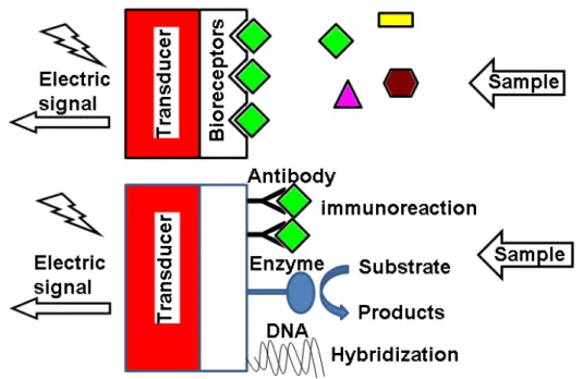
1.3 Biosensor
Biosensor-related research has shown tremendous growth over the last two decades. A biosensor is generally defined as an analytical device which converts a biological
4
response into a quantifiable and processable signal [5]. The general scheme of amperometric biosensors is shown in Fig.1.2. The interaction of the analyte with the bioreceptor is designed to produce an effect measured by the transducer, which converts the information into a measurable effect, for example, an electrical signal.
Figure 1.2 The general scheme of amperometric biosensors
Biosensors can be applied to a large variety of samples including body fluids, food samples, cell cultures and be used to analyze environmental samples. Designed for the purpose, biosensors are generally highly selective due to the possibility to tailor the specific interaction of compounds by immobilizing biological recognition elements on the sensor substrate that have a specific binding affinity to the desired molecule [6]. Typical recognition elements used in biosensors are: enzymes, nucleic acids, antibodies, whole cells, and receptors. Of these, enzymes are among the most common [7]. To fully exploit the specific interaction through biorecognition, the surface architecture of the sensor also must suppress any non-specific interaction. A tremendous research effort has been invested to find surface modifications with specific interaction capabilities over prolonged periods of time in biological fluids [8]. In particular, the ability to tailor the size and
5
structure and hence the properties of nanomaterials offers excellent prospects for designing novel sensing systems and enhancing the performance of the bioanalytical assay [8, 9]. Intense efforts have been devoted to the development of so-called second- and third generation biosensors [10].
1.3.1 Classifications of Biosensor
From the definition of biosensors, they can be classified either by their biological recognition element or their signal transduction mechanism. However, additional biosensor features could be analyzed.
1.3.2 Classification based on bioreceptors
Recent developments in biosensors research have centred on bioreceptors with improved biosensor design. Bioreceptors are used because they are important elements to specificity for biosensor technologies. They are biological molecular species (e.g., an antibody, an enzyme, a protein, or a nucleic acid) or a living biological system (e.g., cells, tissue, or whole organisms) that utilizes a biochemical mechanism for recognition. They allow binding the specific analyte of interest to the sensor for the measurement with minimum interference from other components in complex mixtures. According to bio receptor, biosensors can be classified into the following categories such as enzymatic biosensors, immunosensor, DNA (Deoxyribonucleic acid) biosensors, aptasensors and cells based biosensors etc.
1.3.2.1 Enzymatic biosensors
This class of biosensors employs enzymes as biocatalysts. Enzymes react with the analyte or the substrate producing a detectable signal through this biorecognition process [11]. An example of these types of biosensors is the use of an enzyme acting specifically to convert a reactant molecule into a product. Some enzymes show a specific sensitivity to a particular molecule (or substrate). Many enzymatic reactions involve cofactors. These cofactors are other molecules or ions that assist in the reaction. During the catalysis, the cofactors may be chemically changed, and as a consequence, the resulting physicochemical effects can monitor or detect the enzymatic process. The most famous
6
practical device for determination of blood glucose content is an enzymatic biosensor and it was developed by Yellow Springs Instruments in the early 1970s [12].
1.3.2.2 Immunosensors
Immunosensors are based on the antibody-antigen interaction and the transduction of the biorecognition event into a physical signal. The antigen is recognized as a foreign body. A specific antibody is generated to act against it by binding to it and operating to remove the antigen. By this specific recognition and interaction performed on the molecular level, antibodies and antigens can be exploited as a means for diagnostic testing. Antibodies can be raised in vitro to detect specific molecules. In this way, antibodies may serve as the basis for the biosensor detection system. The binding of an antigen (Ag) to the appropriate antibody (Ab) is accompanied by only small physicochemical changes. Lack of sufficient sensitivity for detecting analytes at low concentrations is a major impediment to development of label-free immunosensors. The utility of biosensing immunosensors would be greater if there was a proper strategy to amplify the immunological interactions so as to result in more pronounced changes [13]. The design and preparation of an optimum interface between the biological element and the detector material is the key part for this kind of sensors [14].
1.3.2.3 Enzyme immunoassays
Enzyme immunoassays (EIAs) based on electrochemical detection offer several potential advantages and have been applied in clinical, medical, biotechnological, food and environmental analysis. Among the enzyme labels employed, horseradish peroxidase (HRP), alkaline phosphatase (ALP) and glucose oxidase (GOD) are the most common. Recently, Ricci et al. [15] reviewed about recent advances, challenges, and trends of electrochemical EIAs focusing on HRP, ALP or GOD as labels over the past five years. Recently, label-free electrochemical immunoassay for detection of proteins has become an important topic in bioanalysis [16].
1.3.2.4 DNA based biosensors
DNA biosensors are commonly employed to detect specific sequences of DNA. They can reach high levels of selectivity and affinity based on the hybridization between a DNA.
7
Each type of cell has within it a unique signature in its DNA. All of the information contained in the DNA appears encoded in a series of amino acids and, as such, forms the identifying backbone of that structure. The recognition of these sequences is of fundamental importance to the control, reading, and detection of these molecular structures. The basic principle of a DNA biosensor is to detect the molecular recognition provided by the DNA probes and to transform it into the signal using a transducer.
1.3.2.5 Aptasensors
Aptamers are artificial nucleic acid ligands that can be generated against amino acids, drugs, proteins and other molecules. They are isolated from combinatorial libraries of synthetic nucleic acid by an iterative process of adsorption, recovery and reamplification. Aptamers, first reported in 1990, are attracting interest in the areas of therapeutics and diagnostics and offer themselves as ideal candidates for use as biocomponents in biosensors (aptasensors), possessing many advantages over state of the art affinity sensors. In general, aptamers are small (i.e., 40 to 100 bases), synthetic oligonucleotides that can specifically recognize and bind to virtually any kind of target, including ions, whole cells, drugs, toxins, low-molecular-weight ligands, peptides, and proteins. Aptamers can function as the biorecognition elements in biosensor applications [17]. Aptamers can be defined as in vitro selected functional oligonucleotides that bind a specific target molecule. Due to their inherent selectivity, affinity, and their advantages over traditional recognition elements, they represent an interesting alternative for biosensing. Aptamers are small in size in comparison to other biorecognition molecules such as antibodies, protein and enzymes. This allows efficient immobilization at high density. Therefore, production, miniaturization, integration, and automation of biosensors can be accomplished more easily with aptamers than with antibodies [18]. As for the protein-based biosensors, the significant conformational change of most aptamers upon target binding offers great flexibility in the design of biosensors.
1.3.2.6 Whole-Cells or Organelles based biosensors
Whole-cell bacterial biosensors are bacteria engineered to recognize a specific analyte. The signal-transduction is performed by the production of an easily quantifiable marker protein. In most cases, an existing regulatory system in the bacterial cell is exploited to
8
drive expression of a specific reporter gene, such as bacterial green fluorescent protein, beta-galactosidase and others [19].
1.3.3 Classification based on the transduction method
The advances in transduction are closely linked to the accelerated technological breakthroughs related to electronics, informatics, data mining, and computer technologies. Signal transduction and data analysis research, oriented to lowering the cost and portability of biosensor analysis, are areas of high activity in electrical and electronic engineering, and analytical chemistry and lead in accelerated pace to more reliable and easy to use biosensors. Biosensor technologies include transduction platforms based on four major types of transducers: electrochemical (electrodes), optical (optrodes), mass (piezoelectric crystals or surface acoustic wave devices), and thermal (thermistors or heat-sensitive sensors). These techniques have been adapted to detect analytes of interest based on the interaction with or functionality modification of biological targets. The specificity of the detection is determined by the biological component of the method.
1.3.3.1 Electrochemical Transducers
The biochemical signals can be used to generate a current/charge or may change conductivity between two electrodes. The corresponding transduction device can be described as potentiometric, amperometric and conductometric/impedimetric. Demands of high sensitivity, specificity, rapid analysis with accuracy of the analytical measurements have brought considerable thrust in the developing electrochemical biosensor as novel diagnostic tools in technology [20].
1.3.3.2 Optical Transducers
These have taken a new lease of life with the development of fibre optics, thus allowing greater flexibility and miniaturization. The techniques used include absorption spectroscopy, fluorescence spectroscopy, luminescence spectroscopy, internal reflection spectroscopy, surface plasmon spectroscopy and light scattering etc.
9
1.3.3.3 Electrical Transducers: Filed Effect Transistor based sensorsMiniaturization can sometimes be achieved by constructing one of the above types of electrochemical transducers on a silicon chip- based field-effect transistor.
1.3.3.4 Piezo-Electric Sensors
These devices involve the generation of electric currents from a vibrating crystal. The frequency of vibration is affected by the mass of material adsorbed on its surface, which could be related to changes in a reaction. Surjiuce acoustic wave devices are a related system.
1.3.3.5 Thermal Sensors
All chemical and biochemical processes involve the production or absorption of heat. This heat can be measured by sensitive thermistors and hence be related to the amount of substance to be analysed.
1.4 Electrochemical Biosensors
Among the various types of biosensors, the electrochemical biosensors are the most common as a result of numerous advances leading to their well understood biointeraction and detection process.
1.4.1 Classifications of electrochemical biosensors
The basic principle of electrochemical sensors is that the electroactive analyte is undergoes oxidized or reduced on the working electrode surface which is subjected to some predefined pattern of fixed or varying potential, and the variation on electron fluxes leads to the generation of an electrochemical signal, which is measured by the electrochemical detector. The two most important subclasses of electrochemical sensors include the voltammetric and potentiometric biosensors.
10
1.4.1.1 Voltammetric sensorsVoltammetric sensors investigate the concentration effect of the detecting species on the current potential characteristics of the reduction or oxidation of a specific reaction [21]. Amperometric sensors are a subclass of the voltammetric sensors. The principle of functioning for the amperometric sensors is based on the application of a fixed potential to the electrochemical cell, resulting in a current because of an oxidation or reduction reaction. The current is, then, used to quantify the species involved in the reaction [22, 23]. The versatility of amperometric biosensors is also apparent from their direct or indirect measurement capability. As Chaubey and Malhotra [24] describes, direct amperometry makes use of the intimate relationship between the products of the redox reaction and the measured current, whereas indirect amperometry uses conventional detectors to measure the metabolic substrate or product of the analyte of interest [25]. The amperometric biosensors are often used on a large scale for analytes such as glucose, lactate [26, 27], and sialic acid [28, 29].
1.4.1.2 Potentiometric biosensors
Potentiometric biosensors examine the potential difference measurement between the working electrode and the reference electrode as it relates to the redox reaction of the species of interest. The potentiometric biosensors monitor the accumulation of charge at zero current created by selective binding at the electrode surface [30]. A disadvantage of these sensors compared with the amperometric counterparts is the extended time period required for the potentiometric sensor to reach equilibrium required for data collection.
1.4.1.3 Impedimetric biosensors
Such devices follow either impedance (Z) or its components resistance (R) and capacitance (C); inductance typically has only a minimal influence in a typical electrochemical setup. Thus, the expression of impedance is as follows:
11
The inverse value of resistance is called conductance and for this reason some investigators name such systems as conductometric. Impedance biosensors include two electrodes with applied alternating voltage; amplitudes from a few to 100 mV are used. The impedance biosensor is commonly a functional part of the Wheatstone bridge. The enzymatically produced ions are able to provide a significant increase of impedance. Alternatively, impedance biosensors have been successfully used for microorganism growth monitoring due to the production of conductive metabolites [31, 32]. False positive results due to electrolytes from the samples are the main disadvantage of impedance biosensors. Impedimetric biosensors are less frequent compared to potentiometric and amperometric biosensors; nevertheless, there have been some promising approaches. Hybridization of DNA fragments previously amplified by a polymerase chain reaction has been monitored by an impedance assay [33]. A model impedance immunosensor containing electrodeposited polypyrrole film with captured avidin connected through biotin to anti-human IgG was able to detect antibodies as low as 10 pg/mL present in a sample [34].
1.4.1.4 Enzyme based electrochemical biosensors
Enzyme is a biological catalyst with extremely high specificity and efficacy. It must be remembered that a catalyst permit to reach easily the equilibrium without modifying its position. However, most of the enzymatic reactions take place in a short time at normal temperature, without using dramatic value of pressure and pH, and work at much higher rates in comparison to the common chemical organic and inorganic catalysts. Enzyme-catalyzed reactions are normally from 103 to 107 faster than the same noncatalyzed reactions. They are extremely specific and selective for the substrate which they interact with. Enzymes generally have a variable specificity degree, catalysing either a group of substrates that have correlated structures or a single molecule. Some kind of them assesses a good degree of stereo-specificity, as they catalyze only one of two substrate stereoisomer. The foremost general feature of enzymes is the reaction specificity. In fact they do not generate useless by-products of the reaction and give as high yield in the enzymatic reactions as almost 100%.Enzymes are generally bigger than the substrate they bind, so that only a little portion of substrate is effectively involved in the enzymatic reaction. The molecular recognition of the substrate is achieved by the well known lock
12
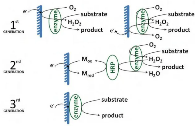
1.4.2 Amperometric enzyme sensorsThree different styles of amperometric enzyme sensors have been developed over the last 50 years. They are often referred to as first-, second-, and third-generation amperometric enzyme sensors (Fig. 3). They all require the enzymes be in close proximity to the electrode surface, but differ in the mechanism by which signal transduction occurs.
1.4.2.1 First generation amperometric enzyme sensors
First generation amperometric enzyme sensors were first proposed by Clark and Lyons in 1962[35], and later implemented by Updike and Hicks in 1967 [36]. Updike and Hicks coined the term enzyme sensors [35]. These sensors possess oxidase enzymes in close proximity to the electrode surface, which, upon interacting with substrate, consume oxygen (O2) and produce hydrogen peroxide (H2O2) (Fig. 1.3). Because O2 and H2O2 are electroactive and diffusible, the amount of O2 consumed or H2O2 produced by the reaction of the oxidase enzyme with substrate is used as a measure of substrate present [35-37].
13
1.4.2.2 Second generation amperometric enzyme sensorsSecond generation sensors also rely on a mediated electron-transfer mechanism for signal transduction to occur. These sensors typically incorporate horseradish peroxidase (HRP) and an oxidase enzyme to oxidize the substrate. These sensors operate on the principle that H2O2 is produced from the reaction of the oxidase enzyme with substrate, and HRP, an oxidoreductase, can both reduce H2O2 , and oxidize a mediator to initiate the electron transfer process (Fig. 1.3). The amount of oxidized mediator detected amperometrically at the electrode is used as a measure of the amount of substrate present. Redox mediators may be diffusible or non-diffusible [38- 41]. In the case that mediators are diffusible, some redox equivalents may be lost to diffusion, (i.e., diffuse out of the sensor and never be detected at the electrode). Because a loss of redox equivalents corresponds to ‘lost signal’, polymeric hydrogels that incorporate non-diffusible redox components have been prepared. The non-non-diffusible redox mediators utilize an electron hopping mechanism to facilitate electron transfer between redox sites. This prevents a loss of redox equivalents because electrons will hop from one redox site to another until they are detected at the electrode surface.
1.4.2.3 Third generation amperometric enzyme sensors
Third generation enzyme sensors rely on a direct, rather than mediated, electron transfer mechanism. The amperometric current measured is the result of oxidation or reduction of the enzyme’s prosthetic group, which serves as temporary trap of electrons or electron vacancies (Fig. 1.3). Third generation enzyme sensors frequently use self assembled monolayers (SAMs) to align the enzymes in a proper orientation, and connect the enzymes’ prosthetic groups to the electrode. As explained by Marcus theory, electron transfer decays exponentially with distance; hence, minimizing the distance between the enzymes’ prosthetic groups and the electrode is essential for the success of these sensors. In the event that an oxidase enzyme is attached to the self-assembled monolayer, electron transfer is not affected by the amount of O2 present. O2 must be present to withdraw electrons from oxidase enzymes in first and second generation amperometric enzyme sensors. However, in the case of third generation enzyme sensors, this can be accomplished by controlling the voltage applied to the electrode. The choice of the sensing electrode depends primarily upon the enzymatic system employed. Amperometric probes are highly suitable when oxidase or dehydrogenase enzymes,
14
generating electro-oxidizable peroxide or nicotinamide adenine dinucleotide (NADH) species, are employed.
1.5 Enzyme immobilization methods
The success of an enzyme biosensor ultimately relies on how well the enzyme bonds to the sensor surface and remains there during use. Immobilization between matrix and bioreceptor has been showed as the topical procedure for improving the enzyme stability and thus general biosensor performances. On the following sections different physical and chemical methods to immobilize the enzyme onto the electrode is discussed.
In order to make a viable biosensor, the biological component has to be properly attached to the transducer with maintained enzyme activity. This process is known as enzyme immobilization. Biosensors are usually designed with high enzyme loading to insure sufficient biocatalyst activities, and the enzymes are provided with an appropriate environment to sustain their activities. The local chemical and thermal environment can have profound effects on the enzyme stability. The choice of immobilization method depends on many factors, such as the nature of the biological element, the type of transducer used, the physicochemical properties of the analyte and the operating conditions in which the biosensor is to function, and overriding all these considerations is necessary for the biological element to exhibit maximum activity in its immobilized microenvironment [42]. Generally, there are four regular methods for enzyme immobilization and they are briefly described as described below (Table 1.1).
15
Table 1.1. Enzyme immobilization methods1.5.1 Adsorption
It is the simplest and fastest way to prepare immobilized enzymes. Adsorption can roughly be divided into two classes: physical adsorption and chemical adsorption. Physical adsorption is weak and occurs mainly via Vander Waals forces, ionic and hydrogen bonds, although sometimes hydrophobic bonding can become significant. These forces are weak, but sufficiently large in number to enable reasonable binding. Chemical adsorption is stronger and involves the formation of covalent bonds. Many substances adsorb enzymes on their surfaces, eg. alumina, charcoal, clay, cellulose, kaolin, silica gel, glass and collagen. Physical adsorption is mostly used for enzyme immobilization in ZnO-based glucose biosensors [43].
1.5.2 Entrapment
It refers to mixture of the biomaterial with monomer solution and then polymerised to a gel, trapping the biomaterial. However, this method can give rise to barriers to the diffusion of substrate, leading to the reaction delay. Besides, loss of bioactivity may occur
16
through pores in the gel. The gels commonly used include polyacrylamide, starch gels, nylon, silastic gels, conducting polymers, etc.
It is possible to distinguish between three general methods:
(i)- Entrapment behind a membrane: a solution or suspension of enzymes, cells, a slice of tissue is confined by an analyte permeable membrane as a thin film covering the detector;
(ii)- Entrapment of biological receptors within self assembled monolayers (SAMs) or bi-layer lipid membranes (BLMs);
(iii)- Entrapment of biomolecules within a polymeric matrix membranes (such as polyacrylonitrile, agar gel, polyurethane, or polyvinyl-alcohol), redox gels, sol-gels with redox centres [44].
1.5.3 Covalent bonding
In this method, the bond occurs between a functional group in the biomaterial to the support matrix. Some functional groups which are not essential for the catalytic activity of an enzyme can be covalently bonded to the support matrix. It requires mild conditions under which reactions are performed, such as low temperature, low ionic strength and pH in the physiological range.
1.5.4 Cross-linking
For this method, usually, biomaterial is chemically bonded to solid supports or to another supporting material such as cross-linking agent to significantly increase the attachment. It is a useful method to stabilize adsorbed biomaterials. Glutaraldehyde is the mostly used bifunctional agent. The agents can also interfere with the enzyme activity, especially at higher concentrations. Crosslinking is rarely used alone ass technique of immobilization, because the absence of mechanical properties and poor stability are severe limitations for biosensor development. This one is often used to enhance other methods of immobilization, normally in order to reduce cell leakage in other systems.
17
The immobilization of the receptor molecule on the sensor surface is a key point for the final performance of the sensor. The immobilization procedure must be stable and reproducible, and must retain the stability and activity of the receptor. One of the most promising strategies is immobilization based on nanotechnology. Nature of biosensing surface is very important namely the prolonged use of the sensor and an anticipated extended storage and working stability.
1.6 Importance of working electrode
In electrochemical sensing, choice of working electrode material is fundamental to the success of electrochemical measurement. In recent years solid electrodes of gold, platinum, silver, nickel, copper, various doped or undoped forms of carbon, dimensionally stable anions, etc. have replaced the conventional mercury electrodes, on the ground of toxicity. These materials can be either bare or chemically modified for improved selectivity, sensitivity and stability [45], mostly by using polymers of varied characteristics. Miniaturization of electrodes, as proposed by Wightman for the first time [46], with the advancement of micromachining, photolithography, microcontact printing, etc. has led to the development of microelectrodes (<2 mm dimension), and has opened the horizon of in vivo and in vitro applicability of electrochemical sensor systems requiring only microliter volumes of analyte and reagent [47]. But the demand for a low-cost, disposable, biosensor strips or sticks for easy commercialization was realized with recent technologies such as screen-printed electrodes [48, 49], which involves deposition of electrode material, mainly carbon and noble metals, on inert PVC or ceramic backing. Cheap, miniaturized, easy-to-use, disposable chips for electrochemical analysis of bio-analytes are very essential and many groups are working in this direction [50].
1.7 Role of nanotechnology in biosensors developments
Nanotechnology involves the study, manipulation, creation and use of materials, devices and systems typically with dimensions smaller than 100 nm. Nanotechnology is playing an increasingly important role in the development of biosensors [51, 52, 53]. Sensitivity and other attributes of biosensors can be improved by using nanomaterials in their construction. Nanomaterials, or matrices with at least one of their dimensions ranging in scale from 1 to 100 nm, display unique physical and chemical features because of effects such as the quantum size effect, mini size effect, surface effect and macro-quantum
18
tunnel effect. Use of nanomaterials in biosensors allows the use of many new signal transduction technologies in their manufacture. Because of their submicron size, nanosensors, nanoprobes and other nanosystems are revolutionizing the fields of chemical and biological analysis, to enable rapid analysis of multiple substances in vivo analysis.
1.7.1 Nanotechnology in electrochemical biosensors
Nanotechnology brings new possibilities for biosensor construction and for developing novel electrochemical bioassays. Nanoscale materials have been used to achieve direct wiring of enzymes to electrode surface, to promote electrochemical reaction, to impose barcode for biomaterials and to amplify signal of biorecognition event. The resulting electrochemical nanobiosensors have been applied in areas of cancer diagnostics and detection of infectious organisms. Nanomaterials, an emerging sub-discipline in chemistry has enabled the development of ultrasensitive electrochemical biosensors due to their high surface area, favourable electronic properties and electrocatalytic activity as well as good biocompatibility induced by nanometer size and specific physicochemical characteristics.
The sampling component of a biosensor contains a bio-sensitive layer that can either contain bioreceptors or be made of bio-receptors covalently attached to the transducer. For biosensing purposes, a layer of receptor molecules that are capable of binding the analyte molecules in a selective way must be previously immobilized on the transducer surface. The immobilization of the receptor molecule on the sensor surface is a key point for the final performance of the sensor. The immobilization procedure must be stable and reproducible, and must retain the stability and activity of the receptor. One of the most promising strategies is immobilization based on nanotechnology [54, 55]. It is essential to create a biosensing surface in which the sensing mechanism is immobilized. The biosensing surface may contain enzymes, antibodies, antigens, microorganisms, mammalian cells, tissues, or receptors. Nature of biosensing surface is very important namely the prolonged use of the sensor and an anticipated extended storage and working stability.
19
One-dimensional (1-D) nanostructures, such as carbon nanotubes (CNT) and semiconductor- or conducting polymer nanowires, are particularly attractive materials for working electrode in biosensors. Because of the high surface-to-volume ratio and novel electron transport properties of these nanostructures, their electronic conductance is strongly influenced by minor surface perturbations (such as those associated with the binding of macromolecules). Such 1-D materials thus offer the prospect of rapid (real time) and sensitive label-free bioelectronic detection, and massive redundancy in nanosensor arrays. These nanomaterials would allow packing a huge number of sensing elements onto a small footprint of an array device. The remarkable properties of CNT suggest the possibility of developing superior electrochemical sensing devices, ranging from amperometric enzyme electrodes to label-free DNA hybridization biosensors [55]. An extremely important challenge in amperometric enzyme electrodes is the establishment of satisfactory electrical communication between the active site of the enzyme and the electrode surface. The redox center of most oxidoreductases is electrically insulated by a protein shell. Because of this shell, the enzyme cannot be oxidized or reduced at an electrode at any potential. The possibility of direct electron-transfer between enzymes and electrode surfaces could pave the way for superior reagentless biosensing devices, as it obviates the need for co-substrates or mediators and allows efficient transduction of the biorecognition event. ‘‘Trees’’ of aligned CNT in the nanoforest, prepared by self assembly, can act as molecular wires to allow electrical communication between the underlying electrode and redox proteins which is covalently attached to the ends of the single- walled carbon nanotubes (SWCNT) [56,57] . Willner’s group demonstrated that aligned reconstituted glucose oxidase (GOD) on the edge of SWCNT can be linked to an electrode surface [58]. Such enzyme reconstitution on the end of CNT represents an extremely efficient approach for ‘plugging’ an electrode into GOD. Arrays of nanoscopic gold tubes or wires have been prepared by electroless deposition of the metal within the pores of polycarbonate particle track-etched membranes [59]. Glucose oxidase was immobilized onto the preformed self-assembled monolayers (SAMs) (mercaptoethylamine or mercaptopropionic acid) of gold tubes, via cross-linking with glutaraldehyde or covalent attachment by carbodiimide coupling. Glucose responses as large as 400 nA /mM cm2 were obtained. Based on a slimier method of template synthesis,[60] immobilized glucose oxidase in the polypyrrole nanotubes and produced a biosensor. Compared to conventional biosensor, immobilization on nanomaterials enhanced the amount of the enzyme loading, the retention of the immobilized activity and the sensitivity of the biosensor [9].
20
1.7.2 Nanoarrays, nanotubes, nanoparticules electrodesNano-structured materials have proven as one of the most powerful tool in new technologies and research, due to their absolutely peculiar properties at nanometer size scale. Many studies have shown that optical, mechanical, photo-catalytic and transport properties drastically changes, depending on quantum size effect, as the mean diameter of the particles is in the exaction size regime (i.e. 10 nm) [61–69]
Various nanostructures have been investigated to determine their properties and possible applications in biosensors. These structures include nanotubes, nanofibers, nanorods, nanoparticles and thin films.
Nanoparticles have numerous possible applications in biosensors. For example, functional nanoparticles (electronic, optical and magnetic) bound to biological molecules (e.g. peptides, proteins, nucleic acids) have been developed for use in biosensors to detect and amplify various signals. Some of the nanoparticle-based sensors include the acoustic wave biosensors, optical biosensors, magnetic and electrochemical biosensors, as discussed next. [70]
Nanoelectrodes can be categorized as individual nanoelectrodes, nanoelectrode arrays (NEAs), or nanoelectrode ensembles (NEEs). [71, 72] Individual nanoelectrodes have been produced from carbon fibers and metals, such as platinum and gold wires, produced by flame- or electrochemical-etching methods [71, 72]. The active radii of electrode tips are from a few tens to several hundreds of nanometers, with varying surface roughness. Carbon fiber nanoelectrode can be produced with very smooth surfaces, as verified by scanning electron microscopy. Carbon nanotubes (CNTs) and metal nanowires can be assembled in densely packed arrays of NEAs and NEEs. The alignment of individual nanoelectrodes can vary from well-ordered periodic arrays to random collections of nanostructured materials. These platforms have been produced from CNTs, pure metallic nanowires, metal oxide nanowires, and magnetic nanoparticles [71, 72]. The geometric order of the platforms differentiates arrays from ensembles; NEAs consist of periodic arrangement of individual nanoelectrodes, whereas NEEs are more random collections of nanoelectrodes.
21
1.8 Self Assembled Monolayer Modified ElectrodesIn the last years, chemical modified electrodes by self assembled monolayer of alkanothiols have been extensively used because their simplicity and efficiency [73]. The functionalization of such self-assembled monolayer is important issue today because molecular architecture can be build which confer new surface properties [74, 75]. The design and construction of such novel molecular devices is opening a huge number of applications in areas such as corrosion protection, wetting, microelectronics, optics, chemical, biochemical and electrochemical sensors [76-80]. Despite the great versatility and perspectives show by electrochemical sensors, the utility of an electrode is limited by gradual degradation of its surface. From this point of view, the gold surface is a preferred one, because has the third best electrical conductivity of all metals at room temperature and its inertness prevent the formation of insulating surface oxides [77, 79].
1.9 Gold electrodes in biosensor fabrication
Gold electrodes have been increasingly used in designing electrochemical biosensors because they allow durable immobilization of biomolecules to the electrode surface while controlling the molecular architecture of the recognition layer, most often via binding to self-assembled monolayers (SAMs) [80, 84]. The gold surface modification with organic thiols compounds opens numerous opportunities for the construction of modified electrodes by using a well known and efficient reaction. In generally, such modified electrodes are obtained in two ways [85-87]: (i) adsorption of thiols on gold surface, followed by the adsorption of specific receptor on thiols monolayer (embedment procedure), or (ii) adsorption of functionalized thiols on gold surface, where the functional groups of thiols have the receptor role (Figure 1.4). In both cases, the performances of the obtained modifications are significant dependent by the gold surface quality. If the gold surface is the smooth-faced one, its cover degree is higher, the obtained monolayer is well ordered, and the electrodes have great analytical performances.
22
Figure 1.4 Formation of self assembled monolayer on goldEnzyme immobilization on flat gold surfaces, however, often suffers from low amounts of biomolecules and poor electrical contact to the transducer. Actually, most efforts are directed to two new directions: (1) production of composite electrodes made of gold nanoclusters and immobilized enzymes, which exploit the enhanced catalytic activity of the gold nanoparticles [88-90], and (2) the three-dimensional structuration of gold electrodes with nanometer- sized dimensions for biosensor applications (e.g., microporous gold electrodes [91], nanopatterning of porous gold films [92] or gold nanoelectrode ensembles [93]).
1.9.1 Gold nanomaterials in enzyme biosensors
The electrodes are usually modified by gold nanomaterials in different ways to improve the performance of the biosensor. The electrode surface could be roughened by gold nanoparticles to enhance the interaction of enzyme or biomolecules with the electrode.
1.9.1.1 Gold nanoparticles
Gold nanoparticles could provide a stable immobilization for biomolecules retaining their bioactivity. Moreover, electron transfer between redox proteins and electron surfaces is facilitated, which is induced by many factors, such as the high surface-to-volume ratio, high surface energy, decreased proteins-metal particles distance and the functioning as electronconducting pathways between prosthetic groups and the electrode surface from the gold nanoparticles. Gold nanoparticles are normally synthesized by chemical route and electrodeposition. Pingarron et al. recently reported a review on gold nanoparticle-based electrochemical biosensors, in which gold-nanoparticle-based enzyme biosensor are summarized [94]. An example is the construction of acetylcholinesterase biosensor in
23
which electrode was modified by electrodeposited gold nanoparticles at the electrode surface after hydrolysis of acethlthiocholine by the immobilization enzyme [95]. This method is valuable for the development of new devices for the sensitive detection of potentially dangerous and deadly neurotoxins. Carbon paste electrode could be modified by the colloidal gold consisting of pretreated graphite power with colloid gold solution and paraffin oil [96]. GOD was immobilized onto the modified electrode via physical adsorption. Such kind of GOD biosensor can efficiently exclude the interference of commonly coexisted uric and ascorbic acid [97]. The similar methodology is also favored for other substrate detection, such as phenol and hydrogen peroxide [96-98]. Gold electrode can be modified by attachement of gold nanoparticles via covalent bond. These gold nanoparticles by chemical route were self-assembled on gold electrode by dithiol via Au-S bond, where dithiol was physically absorbed on the electrode surface by putting gold electrode immersed into a dithiol ethanol solution [99]. A cystamine monolayer was then chemisorbed onto those gold nanoparticles and exposed to an array of amino groups, after that GOD was immobilized by covalently attached to the cystamine modified electrode [99]. Zhang et al., reported that the biosensor provided a linear response to glucose from 20 µM – 5, 7 mM with a sensitivity of 88 µA/cm2 mM. The sensor had a good reproducibility and remained stable over 30 days [99].
1.9.1.2 Gold nanocomposites
A wide variety of matrices, including inorganic materials, organic polymers, and other commercially available solid supports, have been used for enzyme immobilization. Chitosan, as mentioned in pervious part, is one of the most promising immobilization matrices due to its excellent properties. Colloidal gold nanoparticles have been also used as the matrix for the enzyme immobilization to retain the macromolecules’ bioactivity. The adsorption of colloidal gold nanoparticles on the chitosan membrane could provide an assembly of gold nanoparticle mulilayers and a suitable microenvironment similar to the native environment of biomolecules. Based on this approach, a disposal biosensor was fabricated for the rapid detection of H2O2 by entrapping HRP in colloidal gold nanoparticle modified chitosan membrane [96, 97]. The biosensor was characterized with good detection precision and storage stability. Based on a similar methodology, glucose [100] and HRP [101] biosensors were prepared by self-assembling gold nanoparticles on chitosan hydrogel modified Au electrodes. Nanocomposites by combination of gold nanoparticles with inorganic or organic nanomaterials have shown to possess interesting properties, which can be profited for the development of electrochemical biosensors. An
24
example of such nanocomposites is a colloidal gold-CNT composite electrode using Teflon as the non-conducting binding material [102]. The constructed biosensor showed significantly improved responses to H2O2, and the incorporation of GOD into the new composite matrix allowed the preparation of a mediator less glucose biosensor with a remarkably higher sensitivity than that from other GOD-CNT bioelectrodes [102]. Hybrid nanocomposites of gold nanoparticles and organic materials are proposed, in which gold and polypirrole (PPy) are fabricated by wet chemical route using chloroauric acid (HAuCl4) and pyrrole as the reaction reagents [103]. The reaction occurs in mild aqueous conditions and doesn’t involve application of an electrical potential, surfactants or solvents that could affect the biological activity. A stable nanocomposite strongly adhered to the surface of glassy electrode carbon (GCE) electrode and could be employed for electrochemical characterization without loss of the immobilized material.
1.9.1.3 Gold nanoarrays
Many different metals have been electrodeposited using anodic alumina and nuclear track-etched polycarbonate. Electrodeposition inside nanoporous membrane templates [104, 105] has proved to be a versatile approach to fabrication of freestanding metallic nanowires. In general, nanoporous templates are widely available and relatively inexpensive: templates permit the preparation of materials with a high degree of homogeneity and reproducibility. Gold nanoarrays are a widely used nanoelectrode platform, which can be facilely prepared through the templated synthesis methods and other approaches. [71, 72] An apparent advantage of gold nanoelectrode ensembles (GNEEs) is an enhanced signal-to-background current ratio, leading to enhanced detection limits. [71, 72] Recently, three dimensional (3D) structured NEEs have aroused considerable interest for electrochemical studies and application because of their unique configurations and large surface areas. [71, 72] The 3D structured NEEs are typically produced by etching upper layers of the template membrane from a flat two-dimensional (2D) array, creating additional surfaces and chemistries for detection. The ability to devise catalytic properties in gold NEEs make these very attractive for fabrication of electrochemical sensors for use in clinical diagnostic applications. These results indicate that GNEE is a good immobilization matrix, providing a large well-defined surface area with the capacity to modify the nanowire surfaces by linking to proteins. The prospect of spatial patterning with biomolecules is limited by maintaining the active and functional state of the protein. Approaches to orient and retain the electroactive states of these biomolecules can lead to enhanced sensitivities. The use of spacers has led to increased
25
activities by decreasing deleterious surface effects, such as denaturation and unfolding of proteins onto electrodes or assemblies. [106, 107, 108].
1.10 Flow Injection Analyses
Flow injection analysis (FIA) is one of the most powerful analytical tools to monitor automatically a large variety of on-line processes for multi-components systems. The application of this methodology in biosensing systems has been showed very useful to investigate the main sensor parameters. Theoretical considerations about the dependence of the signal upon the experimental parameters, i.e. the diffusion flow rate and the geometric configuration, have been also performed compared to continuous flow techniques.
1.10.1 Basic Components:
The basic scheme of an FIA system is showed in Fig. 1.5 and consists of four essential components:
Figure 1.5 Flow Injection Analysis System
a) A propelling unit which produces the flow of one or more solutions, either containing reagents or merely acting as the carrier(s).The propelling system must force the carrier stream through the units, in a perfectly reproducible, pulse-free and as constant as possible flow rate. This function can be performed by a peristaltic pump (the most used in FIA applications, up to now), a gas-pressure system or even gravity-based units;


![Figure 3.4 Cyclic voltammograms obtained at 3D and 2D GNEEs in 0.001M K 3 [Fe(CN) 6 ] in 0.1 M KNO 3 at a scan rate of 50 mV/s](https://thumb-eu.123doks.com/thumbv2/123dok_br/15938612.1095983/63.892.166.658.685.1060/figure-cyclic-voltammograms-obtained-gnees-kno-scan-rate.webp)
![Figure 3.5 Cyclic voltammograms obtained at different scan rates for 2D GNEEs in 0.01M K 3 [Fe(CN) 6 ] and PBS, pH 6.5 at scan rates ranging from 10 to 100 mV/s](https://thumb-eu.123doks.com/thumbv2/123dok_br/15938612.1095983/64.892.173.787.660.1071/figure-cyclic-voltammograms-obtained-different-rates-gnees-ranging.webp)
