ARTICLE
Liposomal nanotheranostics for multimode
targeted in vivo bioimaging and near
‐infrared
light mediated cancer therapy
Rajendra Prasad
1
, Nishant K. Jain
1
, Amit S. Yadav
2,3
, Deepak S. Chauhan
1
, Janhavi Devrukhkar
1
,
Mukesh K. Kumawat
1
, Shweta Shinde
1
, Mahadeo Gorain
2
, Avnesh S. Thakor
4
, Gopal C. Kundu
2,3
,
João Conde
5
✉
& Rohit Srivastava
1
✉
Developing a nanotheranostic agent with better image resolution and high accumulation into
solid tumor microenvironment is a challenging task. Herein, we established a light mediated
phototriggered strategy for enhanced tumor accumulation of nanohybrids. A multifunctional
liposome based nanotheranostics loaded with gold nanoparticles (AuNPs) and emissive
graphene quantum dots (GQDs) were engineered named as NFGL. Further, doxorubicin
hydrochloride was encapsulated in NFGL to exhibit phototriggered chemotherapy and
functionalized with folic acid targeting ligands. Encapsulated agents showed imaging
bimodality for in vivo tumor diagnosis due to their high contrast and emissive nature.
Tar-geted NFGL nanohybrids demonstrated near infrared light (NIR, 750 nm) mediated tumor
reduction because of generated heat and Reactive Oxygen Species (ROS). Moreover, NFGL
nanohybrids exhibited remarkable ROS scavenging ability as compared to GQDs loaded
liposomes validated by antitumor study. Hence, this approach and engineered system could
open new direction for targeted imaging and cancer therapy.
https://doi.org/10.1038/s42003-020-1016-z
OPEN
1Department of Biosciences and Bioengineering, Indian Institute of Technology Bombay, Powai, Mumbai, Maharashtra 400076, India.2Laboratory of Tumor
Biology, Angiogenesis and Nanomedicine Research, National Center for Cell Science, Pune 411008, India.3School of Biotechnology and Kalinga Institute of Medical Sciences (KIMS), KIIT Deemed to be University, Institute of Eminence, Bhubaneswar 751024, India.4Interventional Regenerative Medicine and
Imaging Laboratory, Department of Radiology, Stanford University, Palo Alto, CA 94304, USA.5Centre for Toxicogenomics and Human Health, Genetics,
Oncology and Human Toxicology, NOVA Medical School, Faculdade de Ciências Médicas, Universidade Nova de Lisboa, 1169-056 Lisboa, Portugal. ✉email:[email protected];[email protected]
123456789
imaging, therapeutics, and targeting agents in a single platform,
the so-called targeted theranostics agents, have been proposed for
better outcomes
6,7,14. However, integrating an
“all in one
plat-form” at nanoscale may reduce its diagnostic and therapeutic
efficacy due to premature release of loaded diagnostic and
ther-apeutic agents
24–29. These pre-leaked cargo molecules may cause
numerous side effects for healthy cells/tissues
30,31. Further, the
integration of all agents in one system can result in complicated
synthesis
routes
with
low
product
yield
and
low
reproducibility
7,32–34.
In addition, the potential impact of enhanced permeability and
retention effect, improving the uptake of nanohybrids into solid
tumor microenvironment is a critical concern
7,14,20,35–37. Second,
targeting biomolecules improve the circulation and accumulation
of injected nanoprobes for selective targeting to cancer cells, but
are not yet convincing
6,38–40. Now, the question about how to
improve the accumulation of nanosized particles into solid tumor
environment is the main focus of the present work. Further, it has
been noticed that the external
field or stimuli, such as
near-infrared (NIR) light, temperature, magnetic
field, etc., may
improve the uptake of nanoparticles; however, these are tested
only up to cellular level, and very few reports are available at the
in vivo level
7,41,42. Presently, computerized tomography (CT) and
emissive contrast imaging have been realized as the most
con-vincing imaging approaches among all imaging modalities due to
high electron and radiodensity of the used contrast agent (only
for heavy metal contrast agents) in CT and the strong
fluores-cence ability of emissive probes (especially for photostable
emissive agents) that exhibits ineffective tissue penetration in NIR
range
6,7,21,43.
In terms of therapeutic strategy, several treatment plans have
been applied for cancer cell ablation. Among them, NIR
light-responsive phototriggered strategy (known as photothermal
therapy, PTT) has emerged as a dynamic therapy for localized
treatments using a variety of nanotheranostic agents
6,7,38,44. In
PTT, photothermal agents produce heat through surface plasmon
resonance and electron–hole delocalization under NIR light
irradiation
45–48. Recently, apart from heat, the generation of
reactive _cioxygen species (ROS) has been noticed as a side
product of PTT
7,42,49. Hence, the combined effect of generated
heat and ROS from a single system shows oxidative and thermal
damage of treated cancer cells/or tumor
7,42. However, the
pro-duced nonspecific and uncontrolled ROS, and heat affect the
surrounding healthy tissues that may cause inflammation,
eschars, mutation, protein denaturation, cell apoptosis or
necro-sis, and mitochondrial dysfunctions
42. To avoid the above
lim-itations, and to improve the potential effect of PTT, the ROS
scavengers
50must be delivered precisely in the heterogeneous
tumor environment that is specific for the target site/or
ROS-enriched area. With this concept, few nanosized scavenger
sys-tems, especially platinum-coated gold nanorods (Pt-GNRs), have
the combination effect of chemotherapy-photothermal therapy
(chemo-PTT). Remarkably, ROS has been observed during NIR
light exposure that is scavenged by targeted NFGL nanohybrids
ensuring the catalytic effect of loaded plasmonic AuNPs.
Results
Stepwise assembly of multimode liposomal nanotheranostics.
Two different imaging probes, viz., AuNPs and GQDs, were
integrated with liposomes (named as NFGL) using
solvent-evaporated thin-film hydration method. The surface of liposomal
nanohybrids was decorated with FA-targeting ligand that was
further investigated for site-selective tumor imaging (contrast and
fluorescent imaging modality) and NIR light-mediated tumor
reduction (see Fig.
1
a, b). First, the engineered liposomal
nano-hybrids were analyzed by low-beam voltage (100 kV)
transmis-sion electron microscopic (TEM) measurements exhibiting about
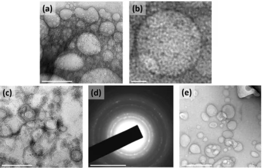
50 nm in size (see Fig.
2
). Very few particles were observed with
particle sizes between 60 and 100 nm. Further, better dispersion
and particle-size distribution of prepared NFGL, FA attached
NFGL, and drug-loaded FA attached NFGL nanohybrids were
measured with dynamic light scattering (DLS), microscopic, and
aqueous dispersion measurements shown in Supplementary
Fig. 1. Prior to a detailed description of NFGL nanohybrids,
herein, highly aqueous dispersible
fluorescent and NIR-active
GQDs were obtained from ethanolic extracts of Mangifera indica
(mango) leaves through microwave-assisted synthesis route
51.
Obtained GQDs were with better size distribution and clear
fringes as shown in Supplementary Fig. 2a, b. GQDs showed
thickness of about 0.9–1 nm as measured through AFM (see
Supplementary Fig. 2c). Next, the polymer-stabilized AuNPs as
ROS scavengers were synthesized by using an earlier reported
procedure with some modifications
52. Controlled size distribution
of synthesized AuNPs was seen through TEM imaging
mea-surement as shown in Supplementary Fig. 2d. The encapsulation
of tiny particles (GQDs, ~25% and AuNPs, ~33%) in the
lipo-somal cavity were clearly observed from TEM images (see
Fig.
2
a–c). These loading capacities were analyzed using optical
density measurements (see Supplementary Eq. 1). The
selected-area electron diffraction (SAED) pattern of NFGL indicated the
successful entrapment of the AuNPs (see Fig.
2
d). Parent
lipo-somal nanostructures were shown in Fig.
2
e. Interestingly, the
drastic contrast difference between empty and cavity-filled
lipo-somes was clearly observed from TEM images that indicated the
success of nanohybrids preparation.
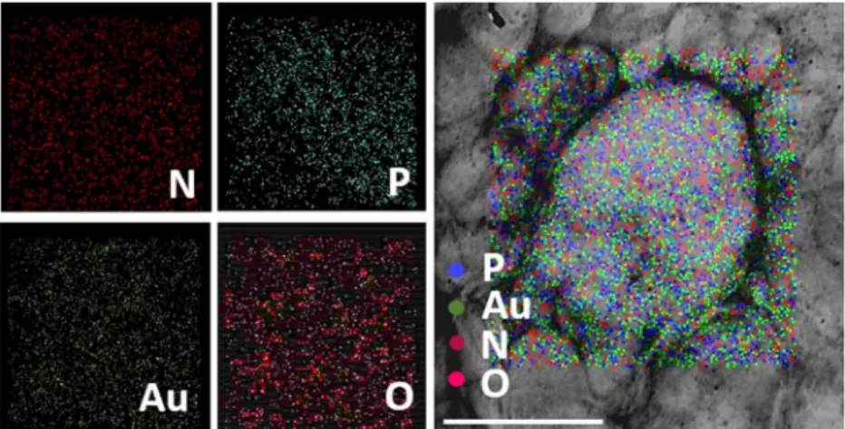
Moreover, the elemental components, such as nitrogen (N),
phosphorous (P), oxygen (O), and gold (Au) of NFGL, were
analyzed through elemental analysis (elemental mapping and
energy dispersive X-ray analysis, EDAX), confirming the
encapsulation of AuNPs as shown in Fig.
3
and Supplementary
Fig. 3. Further, surfactant directed process that described the
Fig. 1 Schematic representation of liposomal nanotheranostics for multimode-targeted bioimaging and phototriggered cancer therapy. a Folic acid targeting ligand decorated self-assembled liposomal nanohybrid loaded multimode imaging probes, viz., gold nanoparticles (AuNPs as radiocontrast for X-ray computed tomography and reactive oxygen species scavenger) and graphene quantum dots (GQDs asfluorescent contrast for near-infrared fluorescence imaging and photothermal agent). Designed functional liposomal nanohybrids demonstrating photothermal response/heat and the generation of reactive oxygen species (ROS, considered as the side product of photothermal therapy) under near-infrared (NIR) light exposure.b NIR light mediated cancer therapeutic representation with tumor-bearing mice model using engineered liposomal nanotheranostic agents and targeted imaging bimodality of breast cancer through X-ray computed tomography (X-ray CT) and in vivo imaging system (IVIS). Liposomal nanotheranostics treated cancer cells displaying the production of ROS (green emission represents the presence of ROS captured by DCFDA (2′,7′-dichlorofluorescin diacetate) dye staining) during NIR light exposure, scale bar= 20 µm.
Fig. 2 Transmission electron microscopic (TEM) images of engineered liposomal nanotheranostics showing spherical morphology. a, b TEM images showing the successful encapsulation of graphene quantum dots (GQDs) in liposomal cavity, scale bar= 100 nm and 10 nm. c TEM imaging observation of gold nanoparticles (AuNPs) and graphene quantum dots (GQDs) encapsulated with liposomal nanohybrids named as NFGL, scale bar= 500 nm. d Selected area electron diffraction (SAED) pattern of NFGL nanohybrids, scale bar= 100 nm. e Microscopic image showing the distribution of parent liposomes (loaded with AuNPs and GQDs) with maintained spherical morphology, scale bar= 200 nm.
mixing of silver nitrate (AgNO
3) with HAuCl
4(gold precursor) in
the presence of ascorbic acid (reducing agent) produced
monodispersed AuNPs that were characterized by TEM and
EDAX. Negative
ζ potentials of −44 mV and −38 mV were
obtained for GQDs and AuNPs due to the presence of
–OH and
–SH functional groups of quantum dots and AuNPs. Liposomes
showed the positive
ζ potential of 28 mV due to a cationic choline
head group of lipid molecules, whereas NFGL showed
ζ potential
of 10 mV, ensuring the interaction of GQDs and AuNPs with
lipid self-assembly (see Supplementary Fig. 4). From RAMAN
spectra, D and G bands of carbon framework confirmed the
presence of GQDs (1328 and 1590 cm
−1) in the NFGL (1323 and
1587 cm
−1) as shown in Supplementary Fig. 5.
Optical properties of liposomal nanotheranostics. The optical
properties of NFGL were analyzed through absorption and
photoluminescence spectroscopic measurements (see Fig.
4
). The
absorption spectra of NFGL showed a sharp band at ~524 nm,
indicating the transverse peak of loaded AuNPs that was further
stable even after 24 h, demonstrating the better photostability of
encapsulated AuNPs (see Fig.
4
a). The peak sharpness revealed
the high dispersion of AuNPs. Moreover, loaded AuNPs in NFGL
were confirmed through TEM images and elemental analysis (as
shown in Figs.
2
c and
3
). Contrasting ability of NFGL
nanohy-brids was evaluated and discussed in the present work due to high
electron density and atomic number of these encapsulated AuNPs
(discussed below). In addition, the hydrophilic cavity of
lipo-somes was loaded with emissive GQDs that demonstrated broad
absorption in the NIR region (700–900 nm), indicating their
applicability for phototherapy. Peaks of unsaturated carbonic
frameworks in GQDs (C=C and C=O) were noticed between 200
and 300 nm (see Fig.
4
a). Emission spectra of NFGL, GQDs
loa-ded parent liposomes, and purified GQDs, indicated the
orange-yellow emissive nature of fabricated nanohybrids (570 nm
emission at 500 nm of excitation) due to the surface defects and
functional groups of encapsulated GQDs as shown in Fig.
4
b. In
addition, the liposomal cavity of nanohybrids was also loaded
with anticancer drug doxorubicin hydrochloride that was
con-firmed through spectroscopic analysis where the absorption of
doxorubicin hydrochloride was observed at around 493 nm
(shown in Supplementary Fig. 6).
Multimodal characteristics of liposomal nanotheranostics.
Entrapped AuNPs induced the contrast modality of the fabricated
NFGL nanohybrids due to high electron density and high atomic
number of Au. Radiodensity of NFGL was measured at various
concentrations (5–100 µg/mL) using clinical X-ray computed
tomography (X-ray CT, TOSHIBA 64 CT scanner, 120 kVp tube
voltage, 250 mA tube current, 5 mm slice thickness, and 1 s
rotation time) scanner showing a linear correlation between
contrast and concentration of nanohybrids (see Fig.
5
a).
Radio-density and brightness were analyzed by recording the Hounsfield
units (HU) for the selected area of interest followed by RadiAnt
DICOM Viewer software. The second imaging modality, viz., NIR
fluorescence (NIRF) of engineered NFGL nanohybrids, was
measured by using the in vivo imaging system (IVIS) at 500 nm
of excitation wavelength. The aqueous suspension (100 µg/mL) of
NFGL demonstrated the promising emission property due to the
photoluminescence ability of loaded photostable GQDs as shown
in Fig.
5
b (emission from sample contained tubes shown in the
inset). Phosphate-buffered saline (PBS) and parent liposomes
were used as control during imaging measurements.
Importantly, the unsaturated carbonic framework in GQDs
made them photothermally active under NIR light irradiation.
About 750 nm wavelength of the NIR laser (1 W/cm
2) was
applied to conduct the phototransduction of GQDs, and NFGL
Fig. 3 Microscopic elemental mapping of engineered liposomal nanotheranostics. Elemental composition of gold nanoparticles (AuNPs) and graphene quantum dots (GQDs) encapsulated with liposomal nanohybrids (NFGL) analyzed through transmission electron microscopic (TEM) images showing the presence of nitrogen (N in maroon color), phosphorous (P in blue color), gold (Au in emerald color), and oxygen (O in pink color) elements with individual and merged imaging, scale bar= 300 nm.Fig. 4 Optical properties of designed NFGL nanohybrids validated through spectroscopic measurements. a Absorption spectra of parent liposome (loaded with AuNPs and GQDs), prepared graphene quantum dots (GQDs), and gold nanoparticles (AuNPs) and graphene quantum dots (GQDs) loaded liposomal nanohybrids named as NFGL at two different time points, viz., 0.5 h and 24 h, indicating the presence of multimode probes (GQDs and AuNPs) within liposomal particles. b Photoluminescence spectra of prepared graphene quantum dots, GQDs encapsulated liposomes, and engineered NFGL nanohybrids using 500 nm excitation wavelength, demonstrating better emissive property of fabricated nanotheranostics.
nanohybrids showed a regular rise in temperature with respect to
NIR exposure time. Hyperthermia temperature (43 °C) was
noticed in 3 min, whereas ablation temperature (~49.9 °C) was
recorded within 10 min of light irradiation using 0.5 mg/mL
concentration of NFGL that was compared with parent
liposomes, indicating the better NIR activity of encapsulated
GQDs (see Fig.
5
c). Moreover, at 0.2 mg/mL concentration of
NFGL, the temperature reached ~43.3 °C after 3 min of NIR
exposure that was further raised up to 55 °C after 10 min of light
exposure. About 1 mg/mL concentration of NFGL showed
maximum temperature of about 60 °C after 10 min of NIR light
exposure (see Supplementary Fig. 7). Similarly, GQDs loaded
liposomes showed good photothermal response that was due to
the loaded GQDs. In light of heat-response measurements, the
temperature reached 43.2 °C in 4 min at 0.2 mg/mL
concentra-tion, and that was further recorded to about 54 °C in 10 min of
exposure. Quick hyperthermia temperature (~43.3 °C in 2 min)
was recorded at 0.5 mg/mL concentration of nanohybrids, and
the maximum temperature of about 59 °C in 10 min was recorded
at 1 mg/mL concentration of nanohybrids. The temperature was
stabilized at 37 °C for negative control (PBS and liposomes) as
shown in Supplementary Fig. 7. Next, the phototriggered drug
release performance of DOX-loaded NFGL was examined in
various environments. Before NIR exposure, negligible drug
release (about 3.5%) was obtained in physiological conditions (pH
7.4), whereas more than 70% was observed after NIR irradiation
during 24 h of incubation time due to produced heat by loaded
GQDs. In case of acidic environment (cancer cell interior
environment, pH 3.0), about 77% of drug released was noticed
within 6 h even without NIR light exposure that was further
achieved ~98% after NIR light exposure (only one shot of 10-min
NIR exposure), indicating the combined effect of chemo-PTT (see
Supplementary Fig. 8). In the disintegration test, controlled
morphology of nanohybrids was seen in the physiological
condition before NIR irradiation, whereas disintegrated particles
were noticed after NIR exposure due to generated heat from
encapsulated GQDs as shown in Supplementary Fig. 9. Further,
in acidic environment (pH 3.0), the degradation of the designed
system was evidently noticed due to protonation of lipid and
GQDs that revealed the destabilization of lipid self-assembly.
Interestingly, the complete disintegration of nanohybrids was
observed in acidic environment (pH 3.0) under the NIR light
exposure. The colloidal stability of designed nanohybrids was
ensured by dispersion examinations before and after laser
exposure (750 nm) at 1 h and 24 h of incubation time, showing
high dispersion without any turbidity (see Fig.
5
d). In addition,
the aqueous dispersion of designed NFGL, DOX-loaded NFGL,
and GQDs loaded liposomal nanohybrids was tested up to 1 week
of time period as shown in Supplementary Fig. 1. Hence, these
examinations confirmed the multifunctionality of designed
liposomal nanohybrids.
In vitro evaluation of liposomal nanotheranostics. To evaluate
the intracellular localization and targeted cancer therapeutics,
FA targeting ligand was attached to the NFGL nanohybrids using
polyethylene glycol (NH
2–PEG) linker
53. FTIR spectra revealed
Fig. 5 Multimodal characteristics of liposomal nanotheranostics. a Contrast measurements (also known as radiodensity) of designed gold nanoparticles (AuNPs) and graphene quantum dots (GQDs)-loaded liposomal nanohybrids named as NFGL at various concentrations (5–100 µg/mL) using a clinical TOSHIBA 64 CT clinical scanner with 5 mm slice thickness and 1 s rotation time compared with parent liposome (loaded with AuNPs and GQDs), revealing the presence of AuNPs (high electron coefficient and density) within the liposomal framework. b Emission performance of NFGL and compared with parent liposomes and PBS using the in vivo imaging system (IVIS) showing the better contrast ability for deep tissue penetration, and indicating the presence of GQDs within liposomal assembly.c Time-dependent photothermal transduction performance of NFGL nanohybrids at 0.5 mg/mL concentration using 750 nm of NIR light irradiation (1 W/cm2) compared with parent liposome (n = 3), ensuring the potential impact of phototriggered therapy. d Digital
photographs showing dispersion of NFGL at ambient conditions, and during laser exposure after 1 h and 24 h of time periods.
that the O–H stretching vibrations at 3376 cm
−1, C=O stretching
vibrations, and N–H bending at 1650 and 1587 cm
−1were
assigned to the CONH group. Peaks between 1640 and 1655 cm
−1were attributed to the bending of NH
2, and stretching vibrations
of CH
2obtained at 2947 cm
−1confirmed the presence of
NH
2–PEG. Further, the bands between 1390 and 1550 cm
−1indicated aromatic ring stretch of pteridine and p-amino benzoic
acid moieties of FA
6,7. Thus, FTIR spectra confirmed the presence
of oxygen rich functional groups on GQDs and surface
functio-nalization of NFGL nanohybrids (see Supplementary Fig. 10a, b).
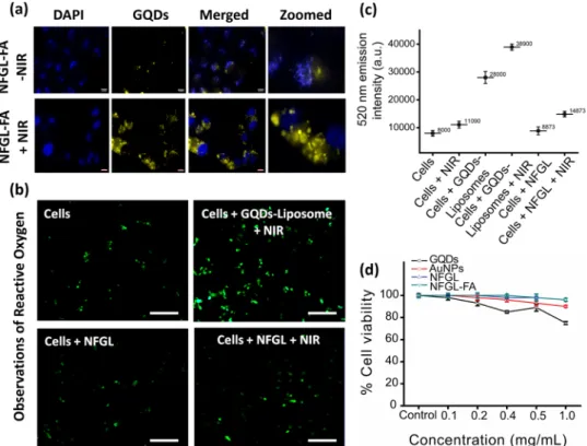
Next, 4T1 breast cancer cell lines were treated with emissive
nanohybrids before and after NIR exposure. Enhanced
distribu-tion of nanohybrids in the cancer cell interior was observed after
NIR irradiation as compared with without NIR treatment,
may indicate the damage of cellular membrane by generated heat
(6 h of
fluorescence imaging, Fig.
6
a). On the other hand,
pro-mising cellular uptake (6 h of incubation time) of NFGL
nano-hybrids with 4T1 cells was achieved with the help of FA
functionalization (NFGL–FA) as shown in Supplementary Fig. 11.
Recently, ROS has been noticed as the side product of
light-mediated photothermal strategy that damages the healthy cells via
oxidizing the cellular matrix
7,42. Similarly, we observed the
presence of ROS along with photothermal heat during NIR light
exposure (750 nm) due to the hydroxyl and carboxyl groups of
loaded GQDs in liposomal nanohybrids (see Supplementary
Fig. 12). Remarkably, the NFGL nanohybrids showed minimal
appurtenance of ROS from cancer cell interior during NIR light
exposure, maybe due to the ROS scavenging effect
50of loaded
AuNPs. Further, the presence of ROS was detected by green
(2′,7′-dichlorofluorescin diacetate, DCFDA) dye staining that was
confirmed through fluorescence microscopic images as shown in
Fig.
6
b, c. The homogeneous and deep intracellular distribution of
ROS from cancer cell interior was clearly noticed during NIR
light exposure that allowed the easy uptake of nanoparticles as
shown in Supplementary Fig. 13. However, the clear mechanism
of ROS generation during NIR light irradiation experiments is
unknown and under investigation.
Next, 24 h MTT measurements in NIH-3T3 cell lines showed
more than 90% cell viability for GQDs, AuNPs, NFGL, and
FA-functionalized NFGL (0.1–1 mg/mL, Fig.
6
d). Further, in vitro
therapeutics examinations were carried out on 4T1 and MCF-7
cancer cell lines using different formulations of nanohybrids as
shown in Supplementary Figs. 14 and 15, respectively. In
4T1 cells, FA-attached GQDs (GQD-FA) and NFGL (NFGL–FA)
showed noteworthy cancer cell death (about 80%) under NIR
light treatment due to the photothermal and oxidative damage
that was about 89% in the case of DOX-loaded NFGL–FA under
NIR light exposure due to the combined chemo-PTT effect.
Before NIR light exposure, these systems showed negligible cell
death (18–25%). Whereas, non-targeted NFGL demonstrated
minimum cell death (32%) due to poor cellular uptake.
Interestingly, only NIR light and NFGL nanohybrids showed
~98% cell viability that was comparable with untreated cancer
cells (about 100% considered as control group). In MCF-7 cells,
nanohybrids showed more than 95% cell viability, whereas about
45 and 35% cell viabilities were observed after NIR light exposure
due to the photothermal and ROS effect. DOX–NFGL showed
~70% cell viabilities before FA attachment, and dropped to 42%
Fig. 6 In vitro validations of formulated gold nanoparticles (AuNPs) and graphene quantum dots (GQDs) loaded liposomal nanotheranostics. a Cancer cell imaging and cellular uptake of folic acid functionalized NFGL nanotheranostic agents (NFGL–FA) with 4T1 breast cancer cell lines with and without NIR light exposure (750 nm, 1 W/cm2for 10 min of exposure), scale bar= 10 µm. b Observations of produced reactive oxygen species (ROS, as a side productof photothermal therapy) during NIR light irradiation using various formulations of NFGL nanohybrids treated with 4T1 cancer cell lines; green emissive ROS are noticed by (2′,7′-dichlorofluorescin diacetate, DCFDA) dye staining. c Quantitative analysis of produced ROS from nanohybrids treated with breast cancer cells with and without NIR light irradiation (n = 3). d Percentage cell viability of NFGL nanohybrids before and after folic acid functionalization and its major components (GQDs and AuNPs) using 24 h MTT assay at different concentrations (0.1–1 mg/mL, n = 3).
after FA attachment, due to the targeted chemotherapeutic effect
of NFGL nanohybrids. Moreover, only 10% cell viability was
observed for targeted combined chemo-PTT (DOX–NFGL–FA
under NIR light exposure). Hence, we believed that the NIR light
irradiations improve the efficacy of heat and ROS that enhance
the uptake of nanoparticles through photothermal and oxidative
rupture of cancer cell membrane.
Imaging bimodality for tumor diagnosis and biodistribution.
In the present work, we established a NIR light triggered strategy
for enhanced accumulation of injected NFGL based
nanother-anostic agents into 4T1 breast tumor bearing mice models as
shown in Fig.
7
a. FA attached NFGL (NFGL–FA) was tested as a
safe multimode contrast agent (X-ray CT and NIRF imaging) for
localized tumor diagnosis (see Fig.
7
b). After 2 weeks of tumor
growth, a single dose of nanohybrids was subcutaneously
admi-nistered (100 µl) at the tumor site. After 1 h post injection of
NFGL–FA, the tumor area was exposed with 10 min of NIR light
(750 nm, 1 W/cm
2), and examined for X-ray CT and NIRF
imaging as shown in Fig.
7
c, d, respectively. In vivo X-ray CT
images of NIR treated animals exhibited higher brightness and
contrast from tumor area that were much better as compared
with without NIR treated and pre-injected mice. The enhanced
contrast from tumor area was maintained even after 48 h of post
injection, indicating the strong binding and higher accumulation
ability of injected liposome based nanocontrast agents (see
Fig.
7
c). These obtained outcomes were corroborated with NIRF
imaging modality that revealed the improved emission intensity
from NIR light exposed tumor area as compared with without
NIR treated and pre-injected mice as shown in Fig.
7
d.
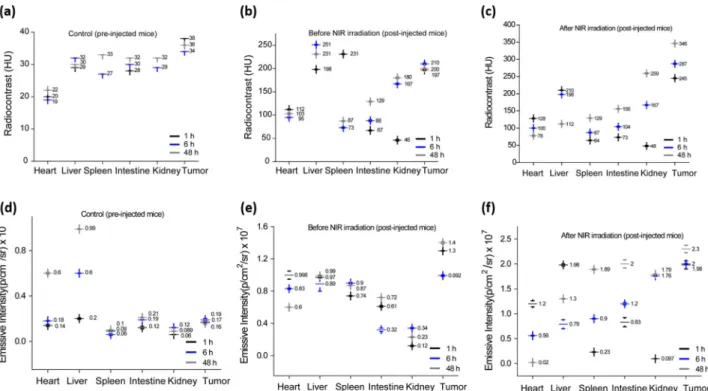
Further, the designed targeted nanohybrids were tested for
biodistribution and tumor accumulation measurements at various
time points (1, 6, and 48 h) followed by subcutaneous post
injection as shown in Fig.
8
and Supplementary Fig. 16.
Whole body scans of NFGL–FA-injected animals were captured
by using imaging bimodalities (CT and IVIS) that were compared
with the control group (untreated/pre-injected animals). In
biodistribution measurements of NIR treated animals, the coronal
and axial CT slices of mice body showed the higher radiodensity
and contrast (HU values) from tumor (~243 HU), heart (~140
HU), liver (about 210 HU), spleen (68 HU), intestine (~79 HU),
and kidneys (~42 HU) that were considerably different from the
radiodensity of NIR exposed animal’s organs like heart (~110
HU), liver (about 198 HU), spleen (50 HU), intestine (~52 HU),
kidneys (~39 HU), and tumor (~200 HU) and pre-injected mice
as shown in Fig.
8
a–c. Importantly, the contrast and brightness
decreased gradually from the upper abdominal organs and
accumulated in the lower abdomen organs with higher contrast
and brightness with respect to post-injection time (6 h and 48 h)
in all cases as shown in Supplementary Table 1. However,
pre-injected animals demonstrated negligible contrast (in the range of
20–40 HU) from tumor area (38–40 HU for 6–48 h of time) and
other major organs in all the cases. The drastic change in
Fig. 7 Site-selective multimode tumor imaging. a Planned NIR light mediated phototriggered strategy for post-injected 4T1 tumor bearing mice showing enhanced tumor uptake of injected gold nanoparticles (AuNPs) and graphene quantum dots (GQDs) loaded liposomal nanotheranostics with folic acid functionalization (NFGL–FA). b Experimental flowchart from day 0 (cell culture) to tissue collection (36 days) followed by multimodal tumor diagnosis and biodistribution experiment setup.c Localized tumor diagnosis and specific biodistribution measurements after 48 h of time before and after NIR light exposure (750 nm, 1 W/cm2for 10 min) followed by whole body X-ray computed tomography scans with coronal and axial CT slices of mice body usingTOSHIBA 64 CT scanner at 120 kVp tube voltage and 250 mA tube current with 5 mm slice thickness and 1 s rotation time.d Targeted deep tumor localization in mice body before and after NIR light exposure (750 nm, 1 W/cm2for 10 min) using the in vivo imaging system (IVIS). In both imaging
modalities, pre-injected mice were considered as control groups.
radiodensity and brightness between pre-injected and without
NIR exposure, and NIR treated post-injected tumor bearing mice,
demonstrated the better accumulation and strong binding ability
of NFGL–FA nanohybrids.
On the other hand,
fluorescent based imaging measurements
demonstrated the promising tumor accumulation and binding
ability that were measured up to 48 h before and after NIR
exposure (see Fig.
8
d–f). After NIR light exposure, the
momentous emission from the tumor site was noticed within
an hour of NFGL–FA post injection that was observed up to 48 h,
which was much better from pre-injected and without NIR treated
animals. Biodistribution measurements of post-injected animals
(1, 6, and 48 h) confirmed the smooth circulation and specific
distribution of injected nanohybrids. Within 1 h of post injection,
NIR treated mice demonstrated maximum emissive intensity
from tumor (1.98 × 10
7p/s/cm
2/sr), heart (1.2 × 10
7p/s/cm
2/sr),
liver (1.98 × 10
7p/s/cm
2/sr), spleen (0.28 × 10
7p/s/cm
2/sr),
intes-tine (0.64 × 10
7p/s/cm
2/sr), and kidneys (0.18 × 10
7p/s/cm
2/sr)
that was further increased in lower abdominal organs and on the
tumor site (2.0 × 10
7p/s/cm
2/sr and 2.3 × 10
7p/s/cm
2/sr) with
respect to the course of time (6 h and 48 h) as given in
Supplementary Table 2. We noticed a drastic difference in
emission intensities between the pre-injected, without NIR
exposure, and NIR treated animals that demonstrated the
potential role of NIR light for enhanced tumor uptake of
nanohybrids. Importantly, the contrast and emission
enhance-ment in lower abdominal organs may reveal the clearance of
small sized imaging agents.
To evaluate site-selective tumor imaging and specific
biodis-tribution (1, 24, and 48 h), NFGL–FA was intravenously injected
into tumor bearing mice (10 mg/kg body weight, single-dose
administration of 100 µl). Maximum emission intensity from
tumor area was achieved after 24 h of post injection, specifying
the
highest
accumulation
of
nanohybrids
in
tumor
microenvironment that was further notable up to 48 h due to
the strong binding ability of NFGL–FA (see Fig.
9
a,
Supplemen-tary Fig. 17a). In biodistribution of injected nanohybrids, the
fluorescent intensity from the heart, lungs, liver, kidneys, spleen,
intestine, and tumor was measured after 48 h of post injection as
shown in Fig.
9
b. Moreover, remarkable emission intensity from
tumor, intestine, and kidneys was noticed compared with the liver
and lungs (see Supplementary Fig. 17b). Remarkable emission
from tumor area corroborated the large uptake of nanohybrids in
tumor microenvironment. Stimulatingly, there was no emission
from the heart and spleen, and emission from the liver, intestine,
and kidneys may indicate the easy excretion of injected
nanohybrids.
In vivo therapeutic evaluation for localized tumor reduction. A
comprehensive investigation of light to heat transduction, ROS
scavenging performance, and in vitro anticancer efficacy ensured
the suitability of prepared liposomal nanotheranostics for in vivo
therapeutic studies. Next,
five groups of tumor-bearing animals
(three mice per group) were treated with various therapeutic
conditions to evaluate the tumor ablation. In detail, a minimal
dose (10 mg/kg body weight) of engineered nanohybrids, such as
FA
attached
NFGL
(NFGL–FA), doxorubicin
hydro-chloride loaded NFGL–FA (DOX–NFGL–FA), and GQDs loaded
liposomes with FA attachment, was intravenously administered
for phototriggered antitumor therapy and tumor ablation. In vivo
therapeutic ability of designed nanohybrids was demonstrated on
4T1
tumor
bearing
mice
for
targeted
chemotherapy
(DOX–NFGL–FA), NIR light mediated site-specific phototherapy
(NFGL–FA under NIR light exposure), and localized NIR
light triggered combined chemophototherapy (DOX–NFGL–FA
during 750 nm of NIR light irradiation) as shown in Fig.
9
c–f.
ROS scavenging performance at the in vivo level was tested by
using NFGL–FA and FA attached GQDs loaded liposome
Fig. 8 Multimode image analysis using in vivo X-ray computed tomography (X-ray CT) and near-infraredfluorescence imaging showing specific biodistribution with promising contrasting ability of post-injected gold nanoparticles (AuNPs) and graphene quantum dots (GQDs) loaded liposomal nanotheranostics with folic acid functionalization (NFGL–FA) in major organs and tumor. a–c Quantitative analysis from X-ray CT imaging (n = 3 mice per group) andd–f near-infrared fluorescence imaging using the in vivo imaging system (n = 3 mice per group) with and without NIR exposure experiments and compared with pre-injected mice (considered as control groups).(GQDs–liposome–FA) nanohybrids in photothermal conditions.
Especially, GQDs liposome–FA treated mice showed better tumor
reduction compared with NFGL–FA treated mice during NIR
light exposure (see Fig.
9
c–e). The combined effect of produced
ROS (a side product of PTT) and generated heat causes the tumor
regression in the case of GQDs–liposome–FA, whereas the
pro-duced ROS was catalyzed by loaded AuNPs in the NFGL–FA
nanohybrids during NIR light mediated antitumor activity
resulting in low tumor reduction (see Fig.
9
d, e). During in vivo
therapeutic course, various qualitative and quantitative analyses
of tumor volume, weight, animal health, and body weight were
evaluated. A drastic reduction in tumor volume and weight was
measured in the case of DOX–NFGL–FA (39.04 mm
3and 0.041
g) during NIR light treatment due to the combined effect of the
released
anticancer
drug
and
generated
heat.
Whereas,
GQDs–liposome–FA (82.07 mm
3and 0.085 g) treated mice
showed promising tumor reduction due to the combined effect of
produced ROS and heat (a key factor of PTT).
From in vivo ROS scavenging measurements during NIR light
irradiation, the designed NFGL–FA nanohybrids showed better
tumor shrinking (120.35 mm
3and 0.070 g) compared with the
control group (142.07 mm
3and 0.119 g, pre-treated animals) due
to the promising effect of generated heat as validated with the
obtained data given in Fig.
9
d, e. In addition, there were no
symptoms observed of eschars and inflammation on the animal’s
body during NIR light exposure, indicating the ROS scavenging
ability of treated NFGL–FA nanohybrids (see Fig.
9
c). Besides,
DOX–NFGL–FA showed favorable tumor reduction (61.59 mm
3and 0.070 g) without NIR light irradiation due to the impactful
effect of targeted chemotherapy. Likewise, multiple therapeutic
observations were described in the present work, demonstrating
the reduction of 4T1 breast tumor (see Fig.
9
f). Hence, multimode
diagnostics and therapeutic observations indicated the potential
impact of designed liposomal nanotheranostics for cancer
treatments.
In vivo toxicity of liposomal nanotheranostics. In vivo toxicities
were measured through histopathology analysis, hemolysis study,
mice health, and body weight observations. The effect of injected
nanohybrids on major organs like heart, liver, spleen, and
kid-neys, was evaluated through histopathology analysis at different
time points (6 h and 48 h) as shown in Supplementary Fig. 18a.
After 2 weeks, treated animals were sacrificed, and major organs
were collected to examine the pathological changes in organs by
hematoxylin and eosin (H&E) staining. H&E examinations
demonstrated (1) healthy myofibers and muscle bundles in the
Fig. 9 Localized tumor diagnosis and phototriggered tumor reduction measurements. a Whole body in vivo imaging for site-selective 4T1 tumor diagnosis at various time points (1, 24, and 48 h) of intravenously injected gold nanoparticles (AuNPs) and graphene quantum dots (GQDs) loaded liposomal nanotheranostics with folic acid functionalization (NFGL–FA). b Ex vivo imaging of collected major organs and 4T1 tumor after 48 h from intravenously nanotheranostics injected animals.c Digital photographs of 4T1 tumor bearing mice during their therapeutic conditions after intravenous injection of NFGL–FA nanotheranostics (n = 3 mice per group) showing the successive tumor regression in various therapeutic conditions with good health of treated mice.d, e Measurements of tumor reduction by tumor volume (mm3,*p < 0.05) and tumor weight (gram,*p < 0.05,**p < 0.01) analysis (n = 3mice per group) during various therapeutic conditions using different formulations of NFGL–FA nanotheranostics with and without NIR light exposure (750 nm, 1 W/cm2for 10 min), and compared with the control group of animals (pre-injected and untreated mice).f Digital photograph of collected
tumors after various therapeutic assessments using different formulations of NFGL–FA nanotheranostics representing the promising tumor reduction and potential impact of phototriggered cancer therapy.
heart, (2) normal portal triad, hepatocyte, and central vein in the
liver and (3) no acute changes in glomeruli and tubules of the
kidney that confirmed the biocompatibility of the injected
nanotheranostics agent. Further, hemocompatibility
examina-tions (10–200 µg/mL concentration of nanohybrids) showed
negligible hemolysis (below 5% at 100 µg/mL concentration),
whereas maximum hemolysis (6–10%) was calculated at the
highest concentration (200 µg/mL) of nanohybrids shown in
Fig.
10
a. Hemolysis procedure was adopted from previously
reported methods
6using DI water and PBS as positive and
negative controls. The surface functionalization and functional
group masking on the nanohybrid’s surface helped in reducing
the hemotoxicity. Moreover, the in vivo toxicity/or safety of
post-injected nanotheranostic agents was examined by animal health
and body weight measurements. The obtained results showed
normal health of all animals (pre-injected and post injected) with
the controlled body weight demonstrating the better
bio-compatibility of designed nanohybrids (see Fig.
10
b,
Supple-mentary Fig. 18b).
Discussion
Self-assembled liposomal targeted nanotheranostics platform
demonstrated site-specific 4T1 tumor diagnosis and
photo-triggered tumor ablation. Enhanced cellular and tumor
accumu-lation of designed nanotheranostics with better contrasting and
therapeutic ability were evaluated methodically. Good aqueous
dispersion, better colloidal stability (tested up to 1 week),
bio-compatibility, hemobio-compatibility, and easy degradation ensured
the in vivo investigation of nanotheranostics. Engineered
nano-hybrids showed imaging bimodality with spatial resolution of
solid tumor due to encapsulated dual imaging probes (AuNPs
and GQDs) in the liposomal cavity. Post-injected nanohybrids
(subcutaneously and intravenously) exhibited strong tumor
binding ability (tested up to 48 h) due to the potential targeting
effect of FA. Further, surface functionalization revealed the
spe-cific biodistribution and smooth circulation of injected
nano-theranostics. Enhanced contrast (brightness and
fluorescence) of
nanohybrids in lower abdomen organs may indicate their easy
excretion from the animal body. Moreover, single wavelength
NIR light exposure (750 nm, 1 W/cm
2) resulted in promising
cancer cell death and selective tumor reduction because of
gen-erated photothermal heat and ROS (as the side product of
PTT
50). The ROS inhibition was successfully achieved at in vitro
and in vivo levels, demonstrating noteworthy cancer cell death
and tumor reduction without affecting the surrounding healthy
tissue. From in vivo analysis, the surrounding area of tumor
(NIR exposed area) was noticed normally without any eschars
and inflammation during NIR light treatment that signposted the
ROS scavenging ability of liposomal nanohybrids. Importantly,
the combined chemophototherapy effect of nanohybrids
exhib-ited superior tumor reduction compared with stand-alone
ther-apy (chemotherther-apy and PTT) due to the synergistic effect of
generated heat and released anticancer drug. Overall, negligible
hemolysis, enhanced cell viability, normal animal health, and
controlled body weight confirmed the biocompatibility of
designed nanohybrids. Hence, the fabricated liposomal
nano-theranostics systems could be a safe and potential platform for
multimode tumor diagnostics and therapeutics.
Methods
Reagents. 1,2-Distearoyl-sn-glycero-3-phosphocholine (DSPC) was procured from Lipoid, Switzerland. GQDs were obtained from Mangifera indica (known as mango tree in the local campus of IIT Bombay, India). (2′,7′-dichlorofluorescin diacetate, DCFDA), Hydrogen tetrachloroaurate (HAuCl4),
1-ethyl-3-(3-dimethy-laminopropyl) carbodiimide (EDC), sodium citrate dehydrate, silver nitrate, cho-lesterol, trypsin-EDTA, 3-(4,5-dimethylthiazol-2-yl)-2,5-diphenyl-tetrazolium bromide (MTT), N-hydroxysuccinimide (NHS), FA, poly(ethylene glycol) 2-aminoethyl ether acetic acid (COOH–PEG-NH2, Mn3500), Thiol-PEG-Carboxyl,
doxorubicin hydrochloride (DOX.HCL), sodium borohydride (NaBH4, 99%),
ascorbic acid (99.5%), and N-cetyltrimethylammonium bromide (CTAB, 99%) were purchased from Sigma-Aldrich Pvt. Ltd., USA. Phosphate-buffered saline (PBS, pH 7.4), fetal bovine serum (FBS), Dulbecco’s Modified Eagle Medium (DMEM), dimethyl sulfoxide (DMSO), 4′,6-diamidino-2-phenylindole (DAPI), and antibiotic–antimycotic solution were obtained from HiMedia Pvt. Ltd., India. Millipore (>18.2 MΩ cm) was used for all experiments.
Characterization techniques. TEM images were performed with exposure of low-beam voltage (100 kV) using Cryo-mode (FEI Tecnai G2). Fourier transform-infrared spectroscopy (FTIR) was done using 3000 Hyperion Microscope with Vertex 80 FTIR System (Bruker, Germany). Raman spectra were recorded using a Jobin-Yvon Labram spectrometer. Samples were excited using 532 nm laser at 5 mW. Dynamic light scattering and zeta potential measurements were recorded by using (DLS)-BI200SM, Brookhaven Instruments Corporation, USA. Further, UV–vis spectroscopy was performed at a path length of 1 cm using Perkin Elmer Lamda-25. Fluorescence spectroscopy was performed using Shimadzu at a slit width of 5 nm (excitation and emission) in high-sensitivity mode. Atomic force microscopy measurements were recorded by using atomic force microscope (PSIA XE-100) using tapping mode. Fine clean silicon wafers as substrates were used for AFM measurements that were made by drop-casting process. Fluorescence microscopy was carried out by using 465–95, 525–45, and 540–80 nm filters from an invertedfluorescent microscope: Nikon Eclipse TE 2000S. ESR (electron spin resonance) analysis was performed using ESR spectrometer (JES-FA 200). The signals were recorded at room temperature at standard frequency. The ROS measurements were done on a microplate reader. In vivo emissive images were recorded using IVIS spectrum imaging system (IVIS spectrum Xenogen), and contrast images were recorded at clinical CT scanner (TOSHIBA 64 at 120 kVp tube voltage and 250 mA tube current with 5 mm slice thickness and 1 s rotation time). NIR light mediated photothermal transduction experiments were performed graphene quantum dots (GQDs) loaded liposomal nanotheranostics (NFGL) before and after FA attachment at various concentrations (10–200 µg/mL, n = 3). b Body weight measurements of post-injected various mice groups (n = 3). Both the analysis demonstrate the good biocompatibility and safety of engineered liposomal nanotheranostic agents.
by using 750 nm NIR laser source using 1 W/cm2power density. All digital pictures were captured by using mobile camera (One Plus 6 T).
Synthesis of GQDs. GQDs were synthesized using Mangifera indica leaves fol-lowed by an earlier reported recipe with some modifications51. Small pieces of
leaves were dipped in ethanol overnight. The acquired leaf’s extract was centrifuged andfiltered, that was further concentrated by ethanol evaporation through rotary evaporator. The obtained material was diluted with Milli-Q water and treated under a microwave oven for 5 min (800–900 W). Finally, the prepared residue was dispersed in ethanol andfiltered through the syringe filter to obtain GQDs. The preparedfinal product (GQDs) was dried at 65–70 °C overnight, and stored in dark conditions at room temperature for further usage.
Synthesis of AuNPs. Gold nanoparticles were prepared by using a previously reported method with various modifications47,52. The AuNPs were prepared by
mixing 80μL of 0.01 M NaBH4, 100μL of 0.01 M HAuCl4, 140μL of 0.01 M citrate
sodium, and 9.625 mL of ultrapure water under stirring at 2000 rpm. The prepared mixture was added to the solution of 800μL of 0.01 M HAuCl4, 100μL of 0.01 M
AgNO3, 100μL of 0.8 M HCl, 400 μL of 0.15 M ascorbic acid, and 50 mL of 0.09 M
CTAB solution. The reaction solution was left at 37 °C overnight. After completion of the reaction, the AuNPs were collected via centrifugation (15 K rpm) that were functionalized with the PEG dithiol through surfactant exchange process at room temperature overnight. To obtain the PEGylated AuNPs, 3 mL of CTAB-stabilized AuNPs were incubated with 20 mg of PEG dithiol overnight at room temperature. The surface-modified AuNPs were collected through centrifugation (15 K rpm for 10 min) and washed thoroughly with Milli-Q water, and were dispersed in Milli-Q water for further usage.
Synthesis of GQD-loaded liposomal nanohybrids. The preparation method of liposomes was adopted from our earlier reported procedure with some modifica-tions7. A mixture of DSPC (80 mg) and cholesterol (20 mg) was dissolved in 15 mL
of chloroform at room temperature conditions. After making the complete mix-ture, the solvent was completely removed at 41 °C. After 1 h of solvent evaporation, 30 ml of PBS containing 1 mg offluorescent GQDs was added to obtain a phos-pholipid thinfilm and kept for further film hydration that was hydrated at 45–47 °C for 60 min using rota evaporator. The hydrated suspension was kept overnight and the next day, having taken to room temperature (37 °C), that was further sonicated (probe sonication of 5 cycles with 40% intensity with 2 s on/off pulse) for 10 min to obtain the small vesicles. The obtained liposomal nanohybrids were characterized through microscopic analysis and used for nanomedicine applications.
Synthesis of liposomal nanotheranostics (NFGL). To design NFGL, a mixture of DSPC (80 mg) and cholesterol (20 mg) was dissolved in 15 mL of chloroform at room temperature, and the solvent was completely removed at 41 °C followed by evaporation process. After obtaining the thinfilm, 30 mL of PBS containing 1 mg of emissive GQDs and 1 mL of surface-modified AuNPs was added to phospholipid thinfilm during film hydration process and further hydrated at 45–47 °C for 60 min. The above hydrated suspension was kept overnight and then sonicated for 10 min to obtain the small size vesicles followed by probe sonication (5 cycles with 40% intensity with 2 s on/off pulse). For the synthesis of doxorubicin hydrochloride (DOX.HCL)-loaded NFGL (DOX–NFGL), 0.5 mg/mL DOX was prepared in PBS (pH 7.4). The aqueous solution of DOX was added during thin-film hydration. Further, the remaining procedure was the same as described earlier. The mass of drug loaded in NFGL was calculated by subtracting the mass of drug in the supernatant from the total mass of drug used. Percentage loading efficiency was calculated according to the following equation:
% Loading ¼Mass of drug in NFGL
Mass of NFGL ´ 100 ð1Þ
Surface functionalization of NFGL with FA. FA (150 mg) was activated by 1-ethyl-3-(3-dimethylaminopropyl) carbodiimide (EDC, 60 mg) and
N-hydroxysuccinimide (NHS, 40 mg) in 45 mL of Milli-Q water for 24 h. The reaction mixture was protected from light. In all, 5 mL of 0.5 mg/mL aqueous solution of amine functionalized polyethylene glycol (COOH–PEG–NH2) was mixed with
activated FA and allowed to react for 12 h. The PEGylated FA (1 mg/mL) as targeting ligand was attached on the surface of NFGL (5 mg/mL) through incu-bation process at room temperature. After completion of the reaction, the products were dialyzed for 24 h.
Aqueous dispersibility, degradation, and photostability. Synthesized nanohy-brids (1 mg/mL) were mixed in aqueous suspension that was observed for 24 h of incubation time at ambient conditions and laser exposure (750 nm, 1 W/cm2for 10 min). During dispersibility test, the digital photographs were captured at 1 h and 24 h of time periods. Further, the photostability of NFGL (1 mg/mL) was checked up to 2 days under 30 min of intermittent exposure of NIR (750 nm) and UV light (365 nm). The prepared NFGL–FA nanotheranostics was treated in various
conditions, such as NIR light exposure, mixing in acidic condition, and exposed with NIR light (combination therapeutic condition) to evaluate the degradation. In addition, the aqueous suspension of NFGL, DOX–NFGL, and GQDs–liposomes was prepared. These prepared suspensions were kept at room temperature for up to 1 week time period to observe the turbidity and aggregation. Digital photographs of treated NFGL were captured at various time points (1st, 2nd, 3rd, and 7th day). Photothermal transduction assessment. Various concentrations (0.2–1.0 mg/mL) of NFGL in PBS were prepared for photothermal transduction experiments. The surrounding temperature (water bath) was stabilized to 37 °C. About 100 µL of NFGL and GQDs-loaded liposomes were added into 96-well plates and exposed to 750-nm (1 W/cm2power) continuous wave (CW) NIR laser source for 10 min. Time-dependent photothermal response was recorded by a digital thermometer. PBS and parent liposomes were used as controls.
ROS analysis. ROS production from NFGL was analyzed using DCFDA (2′,7′-dichlorofluorescin diacetate) dye staining. The designed nanohybrids were treated with NIR light, and ESR (electron spin resonance) analysis was performed using ESR spectrometer (JES-FA 200). About 750 nm light was irradiated over NFGL dispersed in water, and signals were recorded at standard frequency (8.75–9.65 GHz) at room temperature. To examine the ROS from cancer cell lines in the therapeutic conditions, 4T1 and MCF-7 cells were seeded at the density of 2 × 10⁴ in 96-well plates, and 100 µL of 1 mg/mL NFGL was added and left for 24 h of incubation. The next day, these cells were irradiated with 750 nm light for 10 min. After treatment, thefluorescence (excitation: 495 nm, emission: 520 nm) was measured at different time intervals after adding DCFDA (10 µM) dye. In these experiments, various phases of NFGL, such as NFGL treated cells before NIR exposure, GQDs loaded liposomes with NIR irradiation, and NFGL treated cancer cells after NIR light exposure, were tested to ensure the ROS generation during PTT measurements, and scavenging the ROS by using engineered NFGL nano-hybrids in a real situation of phototherapy.
Stimuli-triggered drug release. To check the kinetics of drug release in response to NIR light, 2 mL of DOX–NFGL was added in a dialysis bag immersed in 200 mL of PBS at various pH (3 and 7.4). At various time intervals, and before and after NIR light irradiation, 2 mL of solution was collected and replaced with the same volume of fresh PBS solution. The amount of DOX released was measured with the help of the following equation:
% Release ¼Mass of drug at time tð Þ
Initial mass of drug ´ 100 ð2Þ
In vitro biocompatibility. To check the biocompatibility of NFGL and its variants, NIH-3T3 cells were seeded at a density of 2 × 10⁴cells per well in 96-well plates and incubated for 24 h in 5% CO2atmosphere at 37 °C using Dulbecco’s Modified
Eagle’s Medium (DMEM, Gibco, Carlsbad, CA, USA) supplemented with 10% FBS and penicillin/streptomycin. In MTT assay, after 24 h of incubation, 100 µL of different concentrations (0.1–1 mg/mL) of NFGL, FA attached NFGL, GQDs, and surface modified AuNPs were added into the wells. Following 24 h of incubation, wells were washed off with PBS, and 20 µL of MTT dye was added. Formazan crystals formed after 4 h were dissolved using 200μL of DMSO. Optical absorbance was recorded at 570 and 690 nm using microplate reader (Tecan Infinite 200 PRO). The percentage of cell viability was calculated in reference to untreated cells (control).
In vitro cellular uptake and ROS measurements. Breast cancer cells were cul-tured in DMEM culture media that was supplemented with 10% FBS and peni-cillin/streptomycin. To check the targeting ability of NFGL, 4T1 cells were seeded into 12-well plates at a density of 2 × 10³cells/well and incubated for 24 h in 5% CO2atmosphere at 37 °C. After being rinsed with PBS, 100 µg/mL of NFGL–FA
nanoparticles were added and treated in the conditions of with and without NIR light (750 nm NIR light exposure was for 10 min). After 6 h of incubation, cells were washed with PBS three times to get rid of all the unbound particles. About 4% paraformaldehyde solution was added to the cells, left for 10 min, and then nuclei were stained with 4,6-diamidino-2-phenylindole (DAPI, 1 µg/mL in PBS). At the end of the incubation period, the staining solution was repeatedly washed with PBS. The coverslip was mounted on a drop of 70% glycerol on a glass slide, and fluorescence images were taken using fluorescence microscope. To evaluate the in vitro ROS from cancer cellular environment, 4T1 cells were treated with NFGL and GQDs loaded liposomal nanohybrids in the presence and absence of NIR light irradiation. After 10 min of NIR light exposure and additional 6 h of incubation, these cells were stained with DCFDA (2′,7′-dichlorofluorescin diacetate) dye. The green emission intensity from the cells were recorded after adding DCFDA (10 µM).
In vitro therapeutic and ROS scavenging performance. 4T1 breast cancer cells were seeded into 96-well plates at a density of 2 × 10⁴cells/well and left overnight in 5% CO2atmosphere at 37 °C. After rinsing the wells with PBS, cells were
group 10: DOX–NFGL–FA treated cells (targeted chemotherapy), and group 11: cells treated with DOX–NFGL–FA along with 10 min of NIR light exposure (tar-geted combined chemo-PTT). After treatment, the plates were incubated for another 24 h; thereafter, MTT assay was performed as described above. In vivo tumor growth measurements. Experimental protocols on Balb/c mice were approved by the Institutional Animal Ethical Committee (IAEC) of the National Centre for Cell Science, Pune, India (NCCS, Pune). The IAEC allows us to conduct the in vivo experiments as per institute guidelines according to the National Centre for Cell Science, Pune, India’s laws. IAEC’s laws approved that all experiments were performed in completion with the guidelines of the IAEC research program under B318 project. In vivo examinations were followed by using our earlier reported methodology7. Six-week old female Balb/c mice were used for
the present study. In total, 2 × 1054T1 breast cancer cells were injected sub-cutaneously into the mammary fat pad of Balb/c mice, and tumor growth evaluated.
Image-guided tumor uptake and biodistribution studies. Due to multimode-contrasting probes (emissive GQDs and multimode-contrasting AuNPs) in NFGL, the designed system was able to demonstrate its imaging bimodality with breast tumor model. A one-time dose (10 mg/kg body weight) of targeted NFGL–FA nanohy-brids was subcutaneously injected into 4T1 tumor site of female Balb/c mice. After 1 h of post injection, mice were exposed for 10 min of NIR light (750 nm, 1 W/ cm2); then the in vivo NIRF images of treated animals were captured at various time points (1, 6, and 48 h) in the anesthesized condition using IVIS. These mice were compared with non-NIR light exposed and pre-injected mice group (control). The emissive intensity (xenogen for NIRF at 500 nm of excitation) from whole body luminescence and major organs was measured to examine the in vivo biodistribution of post-injected animals at various time points (1, 6, and 48 h). Similarly, time-dependent X-ray computed tomography (X-ray CT) images were captured using clinical scanner (TOSHIBA 64 CT scanner) at 120 kVp tube voltage and 250 mA tube current with 5 mm slice thickness and 1 s rotation time. A single dose (10 mg/kg) of NFGL–FA was subcutaneously injected into 4T1 tumor site of female Balb/c mice. Post-injected mice were exposed for 10 min of NIR light (750 nm, 1 W/cm2); then the in vivo CT scans of treated animals were captured at various time points (1, 6, and 48 h) in the anesthesized condition. These scans were compared with non-NIR light exposed and pre-injected mice group (control). The brightness from the whole body and major organs was measured to examine the in vivo biodistribution of post-injected animals at various time points (1, 6, and 48 h). On the other hand, a minimal dose (10 mg/kg body weight) of NFGL–FA nanohybrids was intravenously injected into 4T1 tumor bearing female Balb/c mice. In vivo NIRF images of NFGL–FA nanohybrid injected animals were cap-tured at various time points (1, 24, and 48 h) using IVIS. The biodistribution of injectedfluorescent nanohybrids was measured with major organs of tumor bear-ing mice at different time points of post injection, as mentioned above. Further, after 48 h of post administration, treated animals were sacrificed, and all major organs were collected and used for ex vivo NIRF imaging.
Near‐infrared light mediated tumor reduction. To observe the therapeutic outcomes (antitumor activity), different formulations of designed nanohybrids, such as NFGL–FA, DOX–NFGL–FA, and GQDs–liposome–FA, were tested on 4T1 tumor-bearing mice with and without NIR light exposure. A total offive sets of animal groups were prepared (3 mice per group) as follows: (1) control animal group (only pre-injected and untreated animals), (2) DOX–NFGL–FA injected animals (tumor reduction by targeted chemotherapeutic effect that was without NIR light treatment), (3) DOX–NFGL–FA injected animals (tumor reduction by targeted chemo–photothermal therapeutic effect that was under NIR light treat-ment), (4) GQD–liposome–FA injected animals (tumor reduction by produced ROS and generated heat under NIR light irradiation), and (5) NFGL–FA injected animals (tumor reduction by generated heat under NIR light irradiation where produced ROS was scavenged by the nanoparticles). Once the tumor size was
culture day (considered as day 0), post-administered animals were sacrificed, and all major organs were collected for hematoxylin and eosin validations, and to investigate the tissue injury. During whole treatment planning, the body weight of all treated animals was observed and compared with the control animal group. In hemolysis study, we followed the previously reported method with some mod-ifications6. Healthy animals were used to collect 0.5 mL of blood in
ethylenedia-minetetraacetic acid-stabilized tubes that were further centrifuged and washed with PBS. Further, 0.1 mL of collected red blood cells were diluted with 4 mL of PBS. In all, 500 µL volumes of RBCs were treated with various concentrations (10–200 µg/ mL) of nanohybrids, such as liposomes, NFGL, and FA attached NFGL. Treated nanohybrids were incubated with RBCs for a period of 6 h at room temperature (RT), and then the mixtures were centrifuged at 5000 rpm for 10 min. UV–Vis absorption spectroscopy was used to calculate the hemolysis of treated nano-particles through observing the released hemoglobin into the solution from hemolyzed RBCs (absorbance of hemoglobin at 540 nm). The percentage of hemolysis was calculated using the equation: % hemolysis= [(absorbance of the used sample− absorbance of negative control)/(positive control absorbance − negative control absorbance)] × 100.
Statistics and reproducibility. Statistical measurements of all experiments were demonstrated in triplicate. Graphs were plotted by using OriginPro 8 and sigma plot 10.0 software. For in vivo studies, experimental protocols with samples of animals (n= 3 mice per group and total n = 5 treatment groups) on 6-week-old Balb/c female mice were approved by the Institutional Animal Ethical Committee (IAEC) of the National Centre for Cell Science, Pune, India (NCCS, Pune). The IAEC allows us to conduct the animal experiments as per institute guidelines according to the National Centre for Cell Science, Pune, India’s laws. IAEC’s laws approved that all experiments were performed in completion with the guidelines of the IAEC research program under B318 project. All cell lines were from American Type Culture Collection (ATCC) that were authenticated and cultured at the National Centre for Cell Science, Pune. Remarkable observations between different groups were assessed by t test.
Reporting summary. Further information on research design is available in the Nature Research Reporting Summary linked to this article.
Data availability
Supporting data for the present study are available within this article or in the Supplementary Informationfile, and other data information are available from the authors on their reasonable request.
Received: 21 November 2019; Accepted: 7 May 2020;
References
1. Barreto, J. et al. Nanomaterials: applications in cancer imaging and therapy. Adv. Mater. 23, H18–H40. (2011).
2. Kim, J., Piao, Y. & Hyeon, T. Multifunctional nanostructured materials for multimodal imaging, and simultaneous imaging and therapy. Chem. Soc. Rev. 38, 372–390 (2009).
3. Kwon, O. S. et al. Dual-color emissive upconversion nanocapsules for differential cancer bioimaging in vivo. ACS Nano 10, 1512–1521 (2016). 4. Patra, J. K. et al. Nano based drug delivery systems: recent developments and
future prospects. J. Nanobiotechnol. 16, 71 (2018).
5. Park, S., Aalipour, A., Vermesh, O., Yu, J. H. & Gambhir, S. S. Towards clinically translatable in vivo nanodiagnostics. Nat. Rev. Mater. 2, 17014 (2017).