Radiol Bras. 2016 Mai/Jun;49(3):150–157 150
Estimating
131
I biokinetics and radiation doses to the red
marrow and whole body in thyroid cancer patients: probe
detection
versus
image quantification
*
Estimativa da biocinética do 131
I e das doses de radiação na medula óssea vermelha e no corpo inteiro em pacientes com câncer de tireoide: comparação entre o método de detecção de sonda e a quantificação de imagens cintilográficas
Willegaignon J, Pelissoni RA, Lima BCGD, Sapienza MT, Coura-Filho GB, Queiroz MA, Buchpiguel CA. Estimating 131I biokinetics and radiation doses to the red marrow and whole body in thyroid cancer patients: probe detection versus image quantification. Radiol Bras. 2016 Mai/Jun;49(3):150–157.
Abstract
R e s u m o
Objective: To compare the probe detection method with the image quantification method when estimating 131I biokinetics and radiation
doses to the red marrow and whole body in the treatment of thyroid cancer patients.
Materials and Methods: Fourteen patients with metastatic thyroid cancer, without metastatic bone involvement, were submitted to therapy planning in order to tailor the therapeutic amount of 131I to each individual. Whole-body scans and probe measurements were
performed at 4, 24, 48, 72, and 96 h after 131I administration in order to estimate the effective half-life (T
eff) and residence time of 131I
in the body.
Results: The mean values for Teff and residence time, respectively, were 19 ± 9 h and 28 ± 12 h for probe detection, compared with
20 ± 13 h and 29 ± 18 h for image quantification. The average dose to the red marrow and whole body, respectively, was 0.061 ± 0.041 mGy/MBq and 0.073 ± 0.040 mGy/MBq for probe detection, compared with 0.066 ± 0.055 mGy/MBq and 0.078 ± 0.056 mGy/MBq for image quantification. Statistical analysis proved that there were no significant differences between the two methods for estimating the Teff (p = 0.801), residence time (p = 0.801), dose to the red marrow (p = 0.708), and dose to the whole body (p =
0.811), even when we considered an optimized approach for calculating doses only at 4 h and 96 h after 131I administration (p >
0.914).
Conclusion: There is full agreement as to the feasibility of using probe detection and image quantification when estimating 131I biokinetics
and red-marrow/whole-body doses. However, because the probe detection method is inefficacious in identifying tumor sites and critical organs during radionuclide therapy and therefore liable to skew adjustment of the amount of 131I to be administered to patients under such
therapy, it should be used with caution.
Keywords: Thyroid neoplasms; Iodine radioisotopes/therapeutic use; Radioisotopes/pharmacokinetics; Dosimetry; Radiotherapy.
Objetivo: Comparar o desempenho dos métodos de detecção de sonda e quantificação de imagens na estimativa da biocinética do radioisótopo 131I e das doses de radiação na medula óssea vermelha e no corpo inteiro durante a radioiodoterapia em pacientes com
câncer de tireoide.
Materiais e Métodos: Catorze pacientes portadores de câncer metastático de tireoide, sem acometimento ósseo, foram submetidos ao planejamento terapêutico visando estabelecer a melhor atividade de 131I a ser empregada na radioiodoterapia. Imagens cintilográficas
e captações de corpo inteiro foram adquiridas 4, 24, 48, 72 e 96 h após a administração de atividades traçadoras de 131I, visando
estimar a meia-vida efetiva (T½ef) e o tempo de residência do 131I no organismo dos pacientes.
Resultados: Os valores médios de T½ef e tempo de residência foram, respectivamente, 19 ± 9 h e 28 ± 12 h pelo método de detecção
de sonda e 20 ± 13 h e 29 ± 18 h pela quantificação de imagens. As doses médias na medula óssea vermelha e no corpo inteiro foram, respectivamente, 0,061 ± 0,041 mGy/MBq e 0,073 ± 0,040 mGy/MBq pelo método de detecção de sonda e 0,066 ± 0,055 mGy/MBq e 0,078 ± 0,056 mGy/MBq pela quantificação de imagens. A análise estatística demonstrou que os dois métodos apresen-tam desempenho semelhante no tocante à estimativa de T½ef (p = 0,801), tempo de residência (p = 0,801) e doses, tanto na medula
óssea vermelha (p = 0,708) como no corpo inteiro (p = 0,811), mesmo com métodos otimizados de dosimetria que levam em consideração somente dois pontos de medida (4 h e 96 h) após a administração de 131I (p > 0,914).
Conclusão: Existe excelente concordância entre o método de detecção de sonda e a quantificação de imagens quanto à estimativa da biocinética do 131I e das doses absorvidas de radiação. Contudo, o método de detecção de sonda deve ser usado com cuidado por ser
incapaz de identificar regiões metastáticas e órgãos críticos durante a terapia com radionuclídeos, podendo distorcer ajustes da atividade de 131I a ser administrada durante a radioiodoterapia.
Unitermos: Neoplasias da tireoide; Radioisótopos do iodo/uso terapêutico; Radioisótopos/farmacocinética; Dosimetria; Radioterapia. José Willegaignon1, Rogério Alexandre Pelissoni2, Beatriz Christine de Godoy Diniz Lima2, Marcelo Tatit
INTRODUCTION
The management of patients with differentiated thyroid cancer involves radioiodine therapy to ablate remnants of thyroid tissues after surgical resection of the gland or in the treatment of metastases(1). A large amount of 131
I is gener-ally administered to patients during the treatment of meta-static diseases. Under these circumstances, it would be more appropriate to tailor the amount according to individual needs and to the radiotoxic effect on healthy organs, such as the red marrow, lungs, kidneys, and salivary glands(2,3).
Several of the dosimetric methods used for adjusting the amount of 131
I to be administered in therapy are based on delivering a maximum radiation dose of 2–3 Gy to red-mar-row tissues, while abiding by the rules for radioiodine-avid lung metastases(2,4). When determining the radiation dose
per unit of 131
I activity (mGy/MBq) to be received by the red marrow and whole body, sequential measurements of the circulating levels of 131
I are generally required. Typically, the circulating level of 131
I is either estimated through inva-sive procedures, such as the collection and analysis of blood samples, or inferred from whole-body radiation measure-ments with a radiation detection probe or by image quanti-fication(5,6).
Considering the radiation detection probe and image quantification methods as two different procedures, to be used as alternatives for determining radiometric data, our aim here was to compare their performance when estimat-ing 131
I biokinetics and radiation doses to the whole body and to the red marrow.
MATERIALS AND METHODS
Patient characteristics and radiometric data acquisition
Fourteen patients with metastatic differentiated thyroid cancer were submitted to a dosimetric protocol in order to tailor the amount of 131
I to be administered in individual therapy. None of the patients showed evidence of distant metastasis. The study was approved by the local research ethics committee.
Prior to 131
I administration, patients underwent 4–6 weeks of thyroid hormone withdrawal and were maintained on an iodine-poor diet, in order to raise the endogenous thyroid-stimulating hormone level (to > 30 mIU/L) and stimulate 131
I uptake by thyroid tissue remnants and me-tastases. In the present study, exogenous recombinant hu-man thyroid-stimulating hormone was not administered.
The proportional whole-body 131
I retention was calcu-lated at 4, 24, 48, 72, and 96 h after 131
I administration with a radiation detection probe and a gamma camera. Patient data and measurement times were documented.
Radiation detection probe measurements
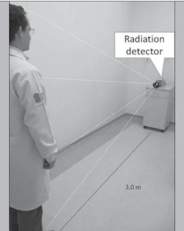
An NaI radiation detector (identiFINDER™ Digital Spectrometer; Thermo Fisher Scientific Inc., Erlangen, Germany), with a 35 mm × 51 mm crystal, was used. The individuals were placed in a standing position 3.0 m from the detector, as illustrated in Figure 1. Each measurement was taken three times, for 3 min each time, in the same lo-cation (an area with a low background radiation level) every day. The net radioactivity (counts/min) was determined only for the anterior acquisition. Before each patient measurement, the background radiation was measured (also for 3 min) in the absence of the patient. Although partial shielding of the detector was considered propitious, the shielding between the patient and the NaI crystal was removed in order to re-duce background radiation. The solid angle of the detector
* Study conducted at the Instituto do Câncer do Estado de São Paulo Octavio Frias de Oliveira (Icesp) and at Faculdade de Medicina da Universidade de São Paulo (FMUSP), São Paulo, SP, Brazil.
1. PhD, Chief Medical Physicist, Instituto do Câncer do Estado de São Paulo Octavio Frias de Oliveira (Icesp), São Paulo, SP, Brazil.
2. Technologist, Instituto do Câncer do Estado de São Paulo Octavio Frias de Oliveira (Icesp), São Paulo, SP, Brazil.
3. PhD, Assistant Professor, Radiology Department, Faculdade de Medicina da Universidade de São Paulo (FMUSP), São Paulo, SP, Brazil.
4. MD, Nuclear Physician, Instituto do Câncer do Estado de São Paulo Octavio Frias de Oliveira (Icesp), São Paulo, SP, Brazil.
5. MD, Radiologist Physician, Instituto do Câncer do Estado de São Paulo Octavio Frias de Oliveira (Icesp), São Paulo, SP, Brazil.
6. PhD, Full Professor, Radiology Department, Faculdade de Medicina da Univer-sidade de São Paulo (FMUSP), São Paulo, SP, Brazil.
Mailing address: Dr. José Willegaignon. Serviço de Medicina Nuclear, Instituto de Radiologia –Hospital das Clínicas da Faculdade de Medicina da Universidade de São Paulo. Travessa da Rua Doutor Ovídio Pires de Campos, s/nº, Cerqueira César. São Paulo, SP, Brazil, 05403-010.E-mail: j.willegaignon@gmail.com.
Received May 4, 2015. Accepted after revision July 6, 2015.
was sufficient to receive photons from the entire body of the patient. The duration of the measurement was sufficient to providing net counts > 105
at each measuring point, even when the background radiation was subtracted.
Image acquisition
A dual-head gamma camera—for single-photon emission computed tomography/computed tomography (SPECT/ CT)—with a high-energy collimator was used in order to estimate the 131
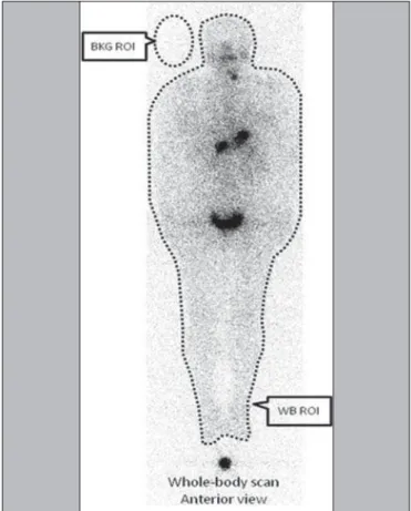
I activity within the body, by whole-body image quantification and as a function of time. Only planar nuclear medicine images were used for quantification. As depicted in Figure 2, the activity was estimated on the basis of the radiation counts for designated regions of interest (ROIs) around the whole body, minus the background ra-diation, as follows:
WBnet = WBpixels × (WBcts/pixel – BKGcts/pixel) where WBnet is the net whole-body count, WBpixels is the total number of pixels for the whole-body ROI in question,
WBcts/pixel is the average count per pixel for the whole-body ROI in question, and BKGcts/pixel is the average count per pixel of background radiation.
A copy of the ROIs (WB and BKG) from the first images was replaced in later images. The same calculation method was applied to each of the five images acquired from each patient. The SPECT/CT was provided with the software
Syngo MI Applications 2009A®
(Siemens Medical Solutions USA, Inc.; Malvern, PA, USA). Whole-body planar images were acquired in a 256 × 1024 matrix with a scan speed of 8 cm/min. All acquired images were analyzed using the free software Image J, version 1.45s (Wayne Rasband, National Institutes of Health; Bethesda, MD, USA).
One patient was used as a standard source for evaluating the remaining whole-body activity as a function of time af-ter 131
I administration, net whole-body counts being normal-ized to the first data point (taken as 100%). After 131
I admin-istration, the patients were allowed to void only after the first probe measurement and image acquisition. When estimating 131
I biokinetics and radiation doses, we considered only the anterior view (for probe measurement and image acquisition), given that our experience has shown that there is little differ-ence between the data estimated with this methodology and those estimated by considering conjugate views (anterior and posterior acquisition). Based on whole-body scanning, the net count rate from anterior acquisition only is, on average, approximately 5% superior to that of conjugate acquisition, indicating that determining only the anterior count rate is a practical and easy method for evaluating radioiodine reten-tion as a funcreten-tion of elapsed time after 131
I administration.
Red-marrow and whole-body absorbed dose calculation
For each patient, the cumulative whole-body activity (Ãwb) was calculated by applying the following equation:
Ãwb = (1.443 × A0 × Teff)
where A0 is the amount of radioactivity administered, and
Teff is the effective whole-body half-life of 131
I.
To describe the radiometric data plotted on the graph “estimated whole-body activity versus time of measurement”, the Teff was determined by a simple exponential function ad-justment:
y = a + be–λeff t
where λeff = 0.693 / Teff.
According to the radiometric data acquired by probe de-tection or image quantification, Ãwb, Teff, and residence time were calculated in duplicate for each patient. The residence time was estimated by dividing the Ãwb by the total amount of 131
I administered (A0).
The absorbed dose of radiation per unit of 131
I activity (mGy/MBq), estimated for the whole body and the red mar-row, was calculated with OLINDA/EXM computer soft-ware(7). We estimated the absorbed dose by introducing the 131
I residence time into the computer program, choosing a specific adult anthropomorphic phantom (adult male or adult female) 131
I radionuclide, and adjusting the parameters of the software according to patient body mass and thyroid tis-sue mass. The last was estimated by calculating the total 131
I uptake by remnant tissue after thyroidectomy and consider-ing 1% of the 131
I uptake per gram of tissue. As described in a previous study(8), the red-marrow dose was adjusted accord-ing to patient weight.
RESULTS
Of the 14 patients evaluated, two were male and 12 were female. Patient ages ranged from 25 to 63 years. All of the patients had previously undergone total or near-total thy-roidectomy. The mean value ± 1 standard deviation for pa-tient weight was 77 ± 19 kg, and the mean height was 1.63 ± 0.06 m. The activity of the tracer 131
I ranged from 74 MBq to 126 MBq, with a mean value of 87 ± 14 MBq, adminis-tered to patients (orally in liquid form) according to a dosi-metric protocol and as an aid in staging the disease. Patient characteristics are presented in Table 1.
Table 2 presents the effective half-life and residence time of 131
I for each patient, determined by considering radiomet-ric data acquired by means of probe detection and image
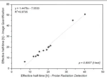
quantification. The mean half-life and residence time values were 19 ± 9 h and 28 ± 12 h, respectively, for probe detec-tion, compared with 20 ± 13 h and 29 ± 18 h, respectively, for image quantification. The effective half-life calculated from image quantification was, on average, 9.38% higher than that calculated from probe measurements, the differ-ence ranging from 0.09% to 34.53% among the patients. A similar difference was observed in residence times. As can be seen in Figures 3 and 4, no significant difference was found between the probe detection and image quantification methods in terms of the calculated effective half-lives or resi-dence times (p = 0.801 for both).
Radiation doses to the red marrow and whole body per unit of 131
I activity administered are presented in Table 3. Mutual correlations, when estimated by probe detection or image quantification, are shown in Figures 5 and 6. When estimated by probe detection, the mean red-marrow and whole-body doses were 0.061 ± 0.041 mGy/MBq and 0.073 ± 0.040 mGy/MBq, respectively, compared with 0.066 ± 0.055 mGy/MBq and 0.078 ± 0.056 mGy/MBq, respectively, Table 1—Patient characteristics and units of 131I activity administered to patients
for dosimetric purposes.
Patient P1 P2 P3 P4 P5 P6 P7 P8 P9 P10 P11 P12 P13 P14 Mean value Gender Female Female Male Female Female Female Female Female Female Female Female Male Female Female Age (years) 63 31 59 49 53 27 37 50 25 51 45 47 26 32 42.50 ± 12.68 Weight (kg) 66.6 124.0 65.0 72.1 103.0 81.8 73.4 76.5 43.0 67.5 72.9 67.6 77.2 88.9 77.02 ± 19.04 Height (m) 1.62 1.58 1.69 1.66 1.57 1.66 1.55 1.56 1.69 1.66 1.61 1.58 1.72 1.69 1.63 ± 0.06
131I activity (MBq) 74.00 82.51 93.24 86.95 126.17 85.10 85.84 74.74 78.07 74.37 77.33 103.23 86.95 92.50 87.21 ± 13.99
Table 2—Estimated effective half-life and residence time of 131I when
consider-ing individually based radiometric data acquired with probe detection and planar image quantification. Patient P1 P2 P3 P4 P5 P6 P7 P8 P9 P10 P11 P12 P13 P14 Mean value
Probe detection Image quantification Effective half-life (h) 20.50 15.18 40.20 19.22 21.30 14.54 13.20 20.09 20.12 7.20 17.43 33.54 11.98 14.05 19.18 ± 8.57 Residence time (h) 29.58 21.90 58.01 27.73 30.73 20.98 19.05 28.99 29.03 10.39 25.15 48.40 17.29 20.27 27.68 ± 12.36 Effective half-life (h) 20.53 15.99 50.84 17.62 21.49 12.41 11.50 19.90 20.14 6.48 16.90 45.12 11.44 12.71 20.22 ± 12.57 Residence time (h) 29.62 23.07 73.36 25.43 31.01 17.91 16.59 28.72 29.06 9.35 24.39 65.11 16.51 18.34 29.18 ±
18.14 Figure 4. Correlations between residence times calculated from radiometric data acquired with the probe detection and image quantification methods.
Table 3—Estimation of red-marrow and whole-body radiation doses when con-sidering radiometric data from probe detection and planar image quantification.
Patient P1 P2 P3 P4 P5 P6 P7 P8 P9 P10 P11 P12 P13 P14 Mean value
Probe detection Image quantification Red-marrow
(mGy/MBq) 0.067 0.025 0.155 0.057 0.042 0.037 0.038 0.056 0.119 0.023 0.051 0.123 0.033 0.033 0.061 ±
0.041
Whole-body (mGy/MBq) 0.084 0.036 0.167 0.073 0.060 0.050 0.050 0.073 0.120 0.029 0.066 0.134 0.043 0.045 0.073 ±
0.040
Red-marrow (mGy/MBq)
0.067 0.026 0.196 0.052 0.043 0.032 0.033 0.055 0.119 0.021 0.050 0.165 0.031 0.030 0.066 ±
0.055
Whole-body (mGy/MBq) 0.084 0.038 0.211 0.067 0.060 0.042 0.043 0.072 0.120 0.026 0.064 0.181 0.041 0.040 0.078 ±
0.056
Figure 6. Correlations between radiation doses to the whole body, as calculated from radiometric data acquired with the probe detection and image quantification methods.
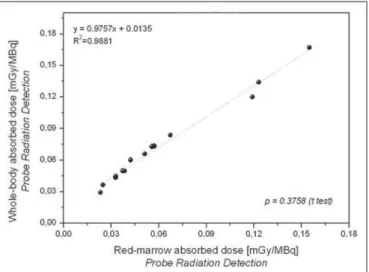
when estimated by image quantification. The mean differ-ences between the probe detection and image quantification methods in terms of the radiation doses calculated was simi-lar to those found for the effective half-lives and residence times. The statistical analysis revealed no significant differ-ence between the two methods, in terms of the estimated radiation doses to the red marrow (p = 0.708) and to the whole body (p = 0.811).
As can be seen in Figures 7 and 8, there were a good correlation between the estimated doses delivered to the red marrow and whole body for the same patient, as calculated by probe detection (p = 0.376; R2 = 0.988) and image quan-tification (p = 0.490; R2 = 0.993). Overall (as calculated by both methods), the mean radiation dose per unit of 131
I ac-tivity administered received by the whole body was approxi-mately 27% higher than that received by the red marrow (range, 0.84–44.0%).
Figure 7. Correlations between radiation doses to the red marrow and radiation doses to the whole body when only radiometric data acquired with probe detection were considered.
Figure 8. Correlations between radiation doses to the red marrow and radiation doses to the whole body when only radiometric data acquired with image quanti-fication were considered.
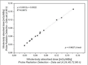
The radiation doses presented in Table 3 were calcu-lated by considering measurements obtained at five time points (4, 24, 48, 72, and 96 h). However, an optimized approach can be applied, given that similar dose estimation results are obtainable when using only the measurements obtained at the two extremes (4 h and 96 h). Correlations between the optimized and the non-optimized method can be seen in Figures 9, 10, 11 and 12. Statistical analysis with a t-test showed that there was no significant difference be-tween the two methods for estimating doses (p > 0.914).
It is important to note that, when estimating the radia-tion doses to the red marrow and whole body in this study, we did not take into consideration doses received by the patients less than 4 h after 131
I administration. Soon after oral administration, 131
I is mainly accumulated in the stom-ach, and requires a certain time before entering into blood circulation. It is assumed that immediate whole-body 131
I
dispersion would lead to incorrect radiation dose estimation. However, if one considers immediate 131
I dispersion in the first 4 h, the dose to the red marrow or whole body would account for only approximately 10% of the total dose received by the two. However, this assumption was not made in the present study, because the main aim here was to compare the performance of probe detection and image quantifica-tion in radiaquantifica-tion dose estimaquantifica-tion.
DISCUSSION
Since 1962, when Benua et al.(9) presented a study on
radioiodine dosimetry for metastatic thyroid cancer patients, many papers on the same topic have been published(8).
However, there is still controversy regarding this point, due to a scarcity of studies comparing dose-response correlations according to the dosimetric method employed. In most dosi-metric methods, consideration (for dose calculation) is given
Figure 9. Correlations between radiation doses to the red marrow calculated by considering measurements obtained at five time points (4, 24, 48, 72, and 96 h) and those calculated by considering measurements obtained at only two (4 h and 96 h), using only radiometric data acquired with the probe detection method.
Figure 11. Correlations between radiation doses to the red marrow calculated by considering measurements obtained at five time points (4, 24, 48, 72, and 96 h) and those calculated by considering measurements obtained at only two (4h and 96 h), using only radiometric data acquired with the image quantification method.
Figure 10. Correlations between radiation doses to the whole body calculated by considering measurements obtained at five time points (4, 24, 48, 72, and 96 h) and those calculated by considering measurements obtained at only two (4 h and 96 h), using only radiometric data acquired with the probe detection method.
only to the use of extensive radiometric data from patients undergoing diagnostic or therapeutic procedures. Neverthe-less, these data can be acquired by collecting and measuring radiometric data from body fluids or even inferred from body radiation measurement.
According to the European Association of Nuclear Medi-cine Dosimetry Committee blood and bone marrow dosim-etry guidelines for differentiated thyroid cancer(5), the probe
and image quantification methods are considered as alter-native procedures for estimating the amounts of radioactive iodine inside the body and provide similar absorbed-dose results when those data are used for internal dose calcula-tion. However, from our point of view, there have been no studies presenting dosimetric data in a satisfactory way to confirm this assumption. In addition, the limitations of us-ing only radiometric data acquired by probe detection to tailor the 131
I amount to be administered to metastatic thyroid cancer patients have not been well addressed. In this con-text, the present study makes a contribution to internal do-simetry by furnishing comparisons of dosimetric data esti-mated by probe detection and image quantification.
On the basis of the results of the current study, and by applying the methodology described here, we can state that the probe detection and image quantification methods pro-vided similar results when estimating 131
I effective half-life and residence time within the body. Therefore, both meth-ods can be considered valid for determining these param-eters. The differences between the two methods when esti-mating radiation doses per unit of 131
I activity administered, either to the red marrow or to the whole body were not sta-tistically significant. The mean radiation dose to per unit of 131
I activity administered was considerably higher for the whole-body doses than for the red-marrow doses, and the difference was similar to that reported in other studies(8). It is important to note that, depending on the physical charac-teristics of each patient (e.g., weight and height), that dif-ference could reach values even higher than the 27% observed in the present study, which is a representative value for a standard patient (70 kg in weight and 1.70 m tall), hence the impropriety of using that value for every patient without some kind of adjustment according to patient biotype, espe-cially in the case of patients presenting bone metastases.
When determining appropriate doses, good correlations were found between the absorbed doses determined at five time points (4, 24, 48, 72, and 96 h) and those determined at only two (4 h and 96 h), when either probe detection or image quantification was used. Reducing the number of data points necessary for estimating radiation doses implies a general reduction in the costs involved in nuclear medicine therapy planning, while working in favor of implementing this procedure in the daily routine of a nuclear medicine department. A similar study of optimizing the amount of radiometric data necessary for dosimetry in Graves’ disease therapy was previously conducted by our group using the probe detection method(10,11). Another group of authors used
positron emission tomography images to estimate the ra-diation doses given to patients with thyroid cancer(12). Both studies were in agreement, in that it is possible to achieve a considerable reduction in the amount of radiometric data required for providing adequate dosimetry. However, it is possible that more data are needed in the case of tumor dosimetry.
The probe detection and image quantification methods provide similar results in determining radiometric data for planning red-marrow and whole-body dosimetry for thyroid cancer patients without metastatic bone involvement. How-ever, the probe detection method should be used with a cer-tain degree of caution, given that, in some clinical cases, the red-marrow is not the first organ at risk during therapy, es-pecially in patients who present with regional or dissemi-nated diseases. Unlike image quantification, probe detection is unable to identify tumor sites or evaluate the 131
I bioki-netics within specific organs, such as the kidneys, lungs, and brain, or in tumors surrounding the spinal cord. At our fa-cility, several patients presented to therapy planning with metastases involving or adjacent to the spinal cord with high 131
I uptake. Such metastases merit special attention, with restrictions of the absorbed dose being determined accord-ing to the dimension of the irradiated area duraccord-ing radionu-clide therapy procedures, such as external radiotherapy, such analysis having been the modus operandi for decades. It is also quite common to see patients presenting with lung metastases, a situation in which the lung, rather than the red marrow, is the limiting organ for determining the total amount of 131
I to be applied in therapy. Therefore, the evalu-ation of tumor sites and dose restriction based on 131
I bioki-netics in a specific organ or tumor cannot be determined only by probe detection, in which the analysis and quantification of nuclear medicine images is the rule. Tailoring the amount of 131
I to be administered to a patient based on biokinetics data obtained from probe detection alone could lead to the use of incorrect procedures, with the possibility of causing severe damage to the patient.
From our perspective, there is no doubt that the future of nuclear medicine dosimetry will involve image quantifi-cation, given the various advantages of its use in therapy planning, such as restoring the bases of radionuclide therapy, thus avoiding cell damage caused by the delivery of inap-propriate radiation doses to a specific organ or tumor.
CONCLUSIONS
There is strong positive agreement between the probe detection and image quantification methods for estimating 131
I biokinetics and radiation doses to the red marrow and whole body for patients with metastatic thyroid cancer with-out metastatic bone involvement. Nevertheless, care should be taken when using the probe detection method, because it is unable to identify tumor sites and critical organs during therapy planning, which could have a negative impact on the adjustment of 131
REFERENCES
1. Luster M, Clarke SE, Dietlein M, et al. Guidelines for radioiodine therapy of differentiated thyroid cancer. Eur J Nucl Med Mol Im-aging. 2008;35:1941–59.
2. Dorn R, Kopp J, Vogt H, et al. Dosimetry-guided radioactive io-dine treatment in patients with metastatic differentiated thyroid cancer: largest safe dose using a risk-adapted approach. J Nucl Med. 2003;44:451–6.
3. Song H, He B, Prideaux A, et al. Lung dosimetry for radioiodine treatment planning in the case of diffuse lung metastases. J Nucl Med. 2006;47:1985–94.
4. Sgouros G, Song H, Ladenson PW, et al. Lung toxicity in radioio-dine therapy of thyroid carcinoma: development of a dose-rate method and dosimetric implications of the 80-mCi rule. J Nucl Med. 2006;47:1977–84.
5. Lassmann M, Hänscheid H, Chiesa C, et al. EANM Dosimetry Com-mittee series on standard operational procedures for pre-therapeu-tic dosimetry I: blood and bone marrow dosimetry in differentiated thyroid cancer therapy. Eur J Nucl Med Mol Imaging. 2008;35: 1405–12.
6. Hindorf C, Glatting G, Chiesa C, et al. EANM Dosimetry
Com-mittee guidelines for bone marrow and whole-body dosimetry. Eur J Nucl Med Mol Imaging. 2010;37:1238–50.
7. Stabin MG, Sparks RB, Crowe E. OLINDA/EXM: the second-gen-eration personal computer software for internal dose assessment in nuclear medicine. J Nucl Med. 2005;46:1023–7.
8. Willegaignon J, Sapienza MT, Buchpiguel CA. Comparison of dif-ferent dosimetric methods for red marrow absorbed dose calcula-tion in thyroid cancer therapy. Radiat Prot Dosimetry. 2012;149: 138–46.
9. Benua RS, Cicale NR, Sonenberg M, et al. The relation of radioio-dine dosimetry to results and complications in the treatment of meta-static thyroid cancer. Am J Roentgenol Radium Ther Nucl Med. 1962;87:171–82.
10. Willegaignon J, Sapienza MT, Coura Filho GB, et al. Determining thyroid (131)I effective half-life for the treatment planning of Graves’ disease. Med Phys. 2013;40:022502.
11. Willegaignon J, Sapienza MT, Coura-Filho GB, et al. Graves’ dis-ease radioiodine-therapy: choosing target absorbed doses for therapy planning. Med Phys. 2014;41:012503.