online | memorias.ioc.fiocruz.br
An experimental protocol for the establishment of dogs
with long-term cellular immune reactions to
Leishmania
antigens
Márcia Cristina Aquino Teixeira1/+, Geraldo Gileno de Sá Oliveira2, Patrícia Oliveira Meira Santos2,3,
Thiago Campanharo Bahiense1,2, Virginia Maria Goes da Silva2,4, Márcio Silva Rodrigues2,
Daniela Farias Larangeira1,2, Washington Luis Conrado dos-Santos2,5,
Lain Carlos Pontes-de-Carvalho2,5
1Universidade Federal da Bahia, Salvador, BA, Brasil 2Centro de Pesquisa Gonçalo Moniz-Fiocruz, Salvador, BA, Brasil 3Universidade Federal do Ceará, Fortaleza, CE, Brasil 4Universidade Estadual do Sudoeste da Bahia, Jequié, BA, Brasil
5Escola Bahiana de Medicina e Saúde Publica, Salvador, BA, Brasil
Domestic dogs are considered to be the main reservoirs of zoonotic visceral leishmaniasis. In this work, we evaluated a protocol to induce Leishmania infantum/Leishmania chagasi-specific cellular and humoral immune responses in dogs, which consisted of two injections of Leishmania promastigote lysate followed by a subcutaneous inoculation of viable promastigotes. The primary objective was to establish a canine experimental model to provide positive controls for testing immune responses to Leishmania in laboratory conditions. After inoculation of viable promastigotes, specific proliferative responses of peripheral blood mononuclear cells (PBMCs) to either Leishmania lysate or recombinant proteins, the in vitro production of interferon-γ by antigen-stimulated PBMCs and a signifi -cant increase in circulating levels of anti-Leishmania antibodies were observed. The immunized dogs also displayed positive delayed-type hypersensitivity reactions to Leishmania crude antigens and to purified recombinant proteins. An important finding that supports the suitability of the dogs as positive controls is that they remained healthy for the entire observation period, i.e., more than seven years after infection. Following the Leishmania antigen lysate injections, the infection of dogs by the subcutaneous route appears to induce a sustained cellular immune response, leading to an asymptomatic infection. This provides a useful model for both the selection of immunogenic Leishma-nia antigens and for immunobiological studies on their possible immunoprotective activities.
Key words: Leishmania infantum - Leishmania chagasi - dogs - immunization - cellular immune response
Visceral leishmaniasis (VL) is increasingly becom-ing a major public health concern due to its dispersal to previously non-endemic rural areas, the emergence of disease foci in urban areas and, more recently, its op-portunistic association with human immunodeficiency virus infection (Mestre & Fontes 2007, Alvar et al. 2008, Maroli et al. 2008, Cerbino Neto et al. 2009). In the New World, VL is caused by Leishmania chagasi,which is be-lieved to be identical to Leishmania infantum (reviewed by Maurício et al. 2000). Therefore, in the present paper, the parasite will be referred to as L. infantum.
Several pieces of evidence have been reported that support a role for dogs as domestic reservoirs of L. in-fantum: first, Leishmania-infected dogs are frequently present in domestic and peridomestic environments in VL-endemic areas, second, dogs from endemic areas ex-hibit high seropositivity rates and third, sick dogs suffer from intense skin parasitism, which, theoretically, more readily exposes Leishmania to the sand fly (Travi et al. 2001, Mestre & Fontes 2007, Maroli et al. 2008, Verçosa
Financial support: PRONEX, PADCT, RENORBIO, CNPq, INO-VABIO, CADCT/FAPESB
+ Corresponding author: marciat@uf ba.br Received 15 August 2010
Accepted 1 December 2010
et al. 2008). The main control measures adopted in Brazil are vector control using insecticides, treatment of human cases and serological surveys, followed by culling of se-ropositive canines. However, the massive elimination of dogs between 1990-1997 did not reduce the incidence of the human disease (Costa & Vieira 2001), indicating that the implementation of this measure alone was ineffective. Accordingly, the World Health Organization considers the deployment of an effective vaccine to be an important al-ternative for the control of leishmaniasis (Desjeux 2002). Studies on natural and experimental L. infantum infections in dogs indicate that many animals develop asymptomatic infections, probably due to an efficient cellular immune response against the parasite (Cabral et al. 1992, Pinelli et al. 1994, Dye 1996, Dos-Santos et al. 2008), suggesting that a canine vaccine is feasible. In fact, the availability of purified and recombinant Leish-mania proteins, together with the increasing understand-ing of the immunological mechanisms involved in the control of leishmaniasis, have enhanced studies of anti-Leishmania vaccines in the canine model (Borja-Cabre-ra et al. 2002, 2008, 2010, Fujiwa(Borja-Cabre-ra et al. 2005, Moreno et al. 2007, Rodríguez-Cortés et al. 2007b, de Lima et al. 2010). However, the number of Leishmania immuniza-tion/infection studies in canine models is still limited in comparison to those reported in murine models.
monitor and quantify certain biological parameters, such as (i) the development of clinical signs of disease, (ii) the levels and isotypic profiles of anti-Leishmania antibod-ies, (iii) the presence, nature and intensity of antigen-specific proliferative responses and/or the production of cytokines in antigen-stimulated peripheral blood mono-nuclear cells (PBMCs), (iv) the presence of skin hyper-sensitivity in response to inoculated antigens and (v) the determination of the capacity of immunized dogs to transmit the parasites to sand flies. Concerning the mea-surement of these parameters, one of the major problems in studying canine immune responses to Leishmania an-tigens is the lack of a convenient source of antigen-spe-cific canine lymphocytes that may be used as positive controls in biological assays for cell-mediated immunity, which appears to be protective against Leishmania infec-tion in dogs (Leandro et al. 2001, Rodrigues et al. 2007, Araújo et al. 2009, Bourdoiseau et al. 2009). In dogs, as in human beings, cell-mediated immune responses are assessed either by injecting adequately diluted antigenic preparations into the skin and subsequently measuring a delayed-type hypersensitivity response or by stimulat-ing PBMCs with antigens in vitro and measurstimulat-ing cell proliferation or cytokine production (Santos-Gomes et al. 2000, Chamizo et al. 2005, Carrillo et al. 2008, Dos-Santos et al. 2008, Bourdoiseau et al. 2009). Immortal-ized T cell lines (Barsov 2009, Bartelt et al. 2009), which are exhaustively selected in vitrofor antigen reactivity, do not represent an ideal substitute for freshly-collected PBMCs as control cells for in vitroimmunological re-actions because it is difficult to guarantee that the cell lines reflect the unselected PBMC population, in terms of both their repertoire of antigen recognition and their adaptation to in vitro conditions. It is operationally com-plex to obtain Leishmania-infected asymptomatic dogs in endemic areas to serve as positive controls for cell-mediated immune responses and there is no guarantee that the asymptomatic dogs will not become sick with the passage of time.
With the aim of obtaining a source of Leishmania antigen-responder canine lymphocytes, L. infantum -specific cellular and humoral immune responses were evaluated in vivo and in vitro at various time points of an immunization protocol (after injection of a Leishma-nia promastigote lysate and the subsequent inoculation of viable promastigotes). The results indicate that, fol-lowing the immunization with parasite crude antigens, the induction of a sub-clinical L. infantum infection is more efficient than immunization with parasite antigens alone in terms of eliciting both humoral and cellular im-mune responses against either native or recombinant L. infantum antigens.
SUBJECTS, MATERIALS AND METHODS
Parasite and antigens-The Leishmania used in this study was isolated from the bone marrow of a human patient and identified as L. infantum (MHOM/BR2000/ Merivaldo2 strain). The parasite was maintained by pas-sage in golden hamsters and cultivation in Schneider’s Drosophila medium (Sigma Aldrich, St. Louis, MO, USA), supplemented with 20% foetal bovine serum
(FBS) (Gibco BRL, Gaithersburg, MD, USA), at 22ºC and at pH 7.2. For the immunization of dogs and the de-tection of antibodies in serum samples by enzyme linked immunosorbent assay (ELISA), promastigotes in the sta-tionary phase of growth were washed by centrifugation with 0.15 M phosphate-buffered saline (PBS), pH 7.2 and lysed by ultrasonic exposure (5 cycles of 30 s at 80% output with intermediate cooling cycles of 1 min) (Bran-son Sonifier 450W, Bran(Bran-son Instruments, Danbury, CT, USA). The lysate was stored at -20ºC until use. The Lci1 (an Hsp70 heat shock protein polypeptide segment) and Lci2 (a kinesin polypeptide segment) L. infantum amas-tigote recombinant antigens (patent PI0900961-2, INPI, Brazil, 2009), encoded by plasmids constructed with L. infantum amastigote cDNA, were expressed in Escheri-chia coli and purified either by an inclusion body (in-soluble aggregate of over-expressed proteins) isolation protocol (Babu et al. 2000) or by immobilized metal-ion affinity chromatography using nickel-chelating Sepha-rose Fast Flow columns (GE Healthcare, Uppsala, Swe-den), according to the directions of Jedrzejas et al. (1998). The degree of purity of the recombinant antigens was an-alyzed by 10% polyacrylamide gel electrophoresis in the presence of sodium dodecyl sulphate, followed by Coo-massie blue staining, as described previously (Laemmli 1970). The protein content of each antigen preparation was determined by protein reaction with fluorescamine (Lorenzen & Kennedy 1993). For the lymphoproliferative assays, crude and purified antigens were dialysed against Roswell Park Memorial Institute (RPMI) medium and sterilised by gamma-ray irradiation.
Animals and experimental design - Twenty-nine mixed-bred dogs, two to four-years old, were obtained from the Salvador City Zoonosis Control Center and kept in a kennel at the Gonçalo Moniz Research Center, Oswaldo Cruz Foundation (Fiocruz), Salvador, Bahia, with daily recreation in a dedicated area. All the dogs were examined and treated when necessary for anaemia, intestinal parasitosis and ectoparasitosis and all received routine vaccination against leptospirosis, distemper, ad-enoviruses, parainfluenza and parvoviruses during an acclimatization period. All the dogs were confirmed to be free of Leishmania based on the absence of parasites by microscopic examination of spleen aspirate cultures and the absence of detectable specific antibodies by ELI-SA (detailed in Determination of anti-Leishmania anti-body levels) and immunofluorescence, as described by Paranhos-Silva et al. (1996). Periodic surveys of the ken-nel for the presence of vectors of Leishmania, performed with the use of light traps, produced negative results. A group of four animals (dogs 1-4) was subjected to the following subcutaneous immunization protocol: (i) two biweekly injections of a lysate derived from 108 Leish-mania promastigotes in incomplete Freund adjuvant and (ii) an inoculation of 108 live stationary-phase Leishma-nia promastigotes four weeks later. A control group of three animals (dogs 5-7) was treated at the same time intervals with saline and adjuvant and then saline alone instead of viable promastigotes. For the assessment of
-ter seven and four years of immunization, respectively, other control groups consisted of untreated animals and animals injected subcutaneously with 200 µg of the Lci1 or Lci2 L. infantum recombinant antigens (Santos 2007). Cellular and humoral immune responses against Leishmania were evaluated three weeks after the second lysate injection, six and 45 weeks after the live parasite inoculation and four-seven years after the immuniza-tion. Spleen aspirates were collected every three months for the first two years after the inoculation of viable pro-mastigotes and were cultured in biphasic medium (blood agar-Schneider’s medium). The animals were clinically monitored by a veterinarian throughout the experiment, which was conducted in accordance with the Fiocruz guidelines for the use of experimental animals.
Determination of anti-Leishmania antibody lev-els - ELISAs were performed as described elsewhere (Paranhos-Silva et al. 1996). Microtitre plate (Corning Laboratory Science Co, NY, USA) wells were coated overnight at 4ºC with 20 µg.mL-1 (protein content) of Leishmania stationary-phase promastigote lysate or 0.5 µg.mL-1 of Leishmania recombinant protein in
100 µL volumes of 0.1 M carbonate buffer (pH 9.6). The wells were washed three times with PBS contain-ing 0.05% Tween 20 (Sigma Aldrich, St. Louis, MO, USA) and blocked with PBS containing 5% dried skim milk for 1 h at 37ºC. After three washing procedures, the wells were incubated with 100 µL of 1/400 dilutions of test sera in PBS containing 3% (dry w/v) skim milk and 0.05% Tween 20 for 1 h at 37ºC. The wells were washed again as before and 100-µL volumes of an anti-dog IgG-peroxidase conjugate (Sigma Aldrich, St. Louis, MO, USA), diluted at 1/2,000, were added and incubated for 1 h at 37ºC. The wells were then washed again before the addition of 0.4 µL.mL-1 of 30% hydrogen peroxide and
0.4 mg.mL-1 o-phenylenediamine (Sigma Aldrich, St.
Louis, MO, USA) in 0.05 M phosphate-citrate buffer, pH 5.0. After 20 min, the reaction was stopped by the addi-tion of 25 µL of 2 N sulphuric acid and the optical den-sity (OD) for 492-nm wave-length light was determined using a microtitre reader. Positive control sera from dogs with parasitologically proven infections and negative control sera from dogs with negative spleen cultures tak-en from a leishmaniasis non-tak-endemic area were includ-ed in all plates. ODs above the cut-off were considerinclud-ed positive. To calculate the cut-off, the mean OD and the standard deviation (SD) of the mean were calculated based on sera from 53 dogs taken from Leishmania -free areas. Then the obtained SD value was divided by the mean OD. This provided the SD/mean ratio of the 53 negative controls. The cut-off value was different for each ELISA plate (correcting for the intrinsic inter-assay variations found in any enzymatic-based inter-assay) and corresponded to the mean OD obtained with three negative control sera included in each ELISA plate plus two times the product of the mean OD of the three negative control sera and the SD/mean ratio of the 53 negative controls (Paranhos-Silva et al. 1996).
Lymphocyte proliferation assay - PBMCs were iso-lated by centrifugation of blood and diluted with
ster-ile Hanks’ balanced salt solution (Gibco-Invitrogen, Carlsbad, CA, USA) at a 1:1 (v/v) ratio with Histopaque-1077 (Sigma Aldrich, St. Louis, MO, USA) solution. Purified PBMCs were washed three times at 4ºC with RPMI medium and viable cells were adjusted to 2 x 10⁶
in RPMI supplemented with 10% heat-inactivated FBS,
50 μg.mL-1 gentamicin and 2 mM L-glutamine (com
-plete medium). Two hundred microlitres containing 2 x 10⁵ cells was then cultured in complete medium either
in the presence of 4 µg.mL-1 concanavalin A (Con A)
(Sigma Aldrich, St. Louis, MO, USA) for three days or with 20 µg.mL-1 (protein content) Leishmania lysate,
0.5 µg.mL-1 Lci1 or 16 µg.mL-1 Lci2, for five days. These
antigen and mitogen concentrations were found to be op-timal in previous assays utilizing PBMCs of naturally infected dogs. All cultures were carried out in flat-bot-tomed microtitre plates (Corning Incorporated, Corning, NY, USA) in triplicate in a 100% humidified atmosphere with 5% CO2 at 37ºC. Cells were pulsed during the last 18 h of culture with 1 µCi of [3H]thymidine (GE
Health-care UK Ltd, Little Chalfont, England) and harvested onto glass fibre filters utilising a cell harvester. Radioactivity uptake was measured in a liquid scintillation beta counter (Matrix 9600 Direct Beta Counter; Packard). Prolifera-tive responses were expressed as a stimulation index (SI), which was calculated by dividing the mean counts of stimulated cultures by the mean counts of non-stimulated cultures. Stimulation indices equal to or higher than 2.5 were considered to be indicative of proliferation.
Quantification of interferon (IFN)-γ levels - PB-MCs were obtained as described above. Isolated PBPB-MCs (107 cells/mL) were resuspended in complete RPMI
me-dium and then plated onto wells of 6-well tissue culture plates (Costar Corning Inc, USA). Cells were incubated with 20 µg.mL-1 L. infantum lysate, 10 µg.mL-1 of Con
A (positive control) or medium alone (negative control) at 37ºC with 5% CO2. Supernatants were collected from the cultures at 48 h and stored at -20ºC until use. PBMCs from dogs that were injected with saline (2 animals) or with a mixture of the Lci1 and Lci2 recombinant antigens
(5 animals) were used as controls. IFN-γ was measured
by sandwich ELISA, according to the manufacturer’s rec-ommendations (R&D Systems, Minneapolis, USA), using
specific anti-dog IFN-γ (1 µg.mL-1), biotinylated anti-dog
IFN-γ (100 ng.mL-1) and streptavidin conjugated to
horse-radish peroxidase (1:200). Absorbance values at 450 nm were read at OD450 in an automatic Microplate Reader EL 3311 (Boehring Mannheim, Germany). A standard curve
for IFN-γ was performed in successive dilutions from
32 ng.mL-1-.03125 ng.mL-1 of recombinant canine IFN-γ.
Data are expressed in nanograms per millilitre.
Delayed-type hypersensitivity reaction - The skin test was performed as described previously (Paranhos-Silva et al. 2001) using freeze-thawed, lysed L. infantum promastigotes diluted in sterile saline as the antigen. Dogs were injected intradermally at different sites of the abdomen with the following: (i) 100 µL of lysate con-taining 250 µg of protein, (ii) different concentrations of purified Lci1 (12.5, 25 and 50 µg.mL-1) and Lci2 (25,
alone. Intradermal indurations were measured 48 h and 72 h after injections. Reactions showing a diameter equal or larger than 5 mm were considered positive.
Statistical analyses - Results from measurements between two groups of dogs were statistically com-pared using the Mann-Whitney non-parametric U test. The statistical significance of the observed differences between three or more groups was determined by the Kruskal-Wallis test and Dunn’s test was used as a post test to determine the statistical significance between two
groups. A p value ≤ 0.05 was considered significant.
RESULTS
Specific antibody responses against total or recom-binant L. infantum antigens - All dogs exhibited rela-tively low (OD values below 0.4 in the ELISA) but sig-nificantly (p< 0.05) increased levels of anti-L. infantum IgG activity after two injections of promastigote lysate (Table I). Six weeks after the subcutaneous inoculation of virulent promastigotes, the antibody levels either remained constant (dogs 3 and 4) or increased (dogs 1 and 2) (Table I). A large and statistically significant (p < 0.05) increase in anti-L. infantum antibody levels was observed 45 weeks after infection compared to levels observed before immunization and at three weeks after the intradermal injection of Leishmania lysate. Reactiv-ity of the serum antibodies against the Lci1 recombi-nant protein was observed in two animals (dogs 2 and 3) 45 weeks after viable promastigote inoculation. None of the animals had detectable serum antibodies against the Lci2 protein during the follow-up period (data not shown). Sera from the control group had no detectable anti-Leishmania antibodies (Table I).
Lymphoproliferative response and IFN-γ production - Proliferative responses to crude Leishmania antigens were not observed after two subcutaneous injections of L. infantum lysate (Fig. 1A). Responses were observed
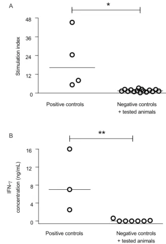
only after inoculation of live promastigotes and were considerably higher after 45 weeks than after six weeks of infection in three of the four dogs (Fig. 1A). After 45 weeks (10.5 months) of infection, all animals responded to Lci1, while two of the four dogs (animals 3 and 4) responded to Lci2 protein, with a mean SI of 6.6 and 4.5, respectively (Fig. 1B). No PBMCs from animals of the control, non-immunized group had lymphoproliferative responses to the L. infantum lysate or to the recombinant proteins (Fig. 1B). Seven years after immunization, PB-MCs from the animals still proliferated in response to L. infantum lysate (Fig. 2A).
Four years after the immunization, Leishmania an-tigen-stimulated PBMCs from the immunized animals
produced detectable levels of IFN-γ, which were sig
-nificantly different from the levels produced by PBMCs from seven animals that were injected with either saline (2 animals) or with a mixture of two recombinant anti-gens (5 animals) (Fig. 2B). PBMCs from immunized
ani-mals still produced IFN-γ in response to Leishmania
an-tigen seven years after immunization (data not shown).
Delayed-type hypersensitivity to crude and recom-binant Leishmania antigens - Forty-five weeks after im-munization, all dogs from the infected group had posi-tive skin test reactions at the site of injection of crude L. infantum antigens, varying from 10-30 mm at 48 h and from 7-26 mm at 72 h (Table II). Three immunized ani-mals had larger reactions at 48 h, while one dog had an increase of induration area after the next 24 h (Table II). Three of four dogs from the immunized group reacted to higher concentrations of Lci1 and one responded to both Lci1 and Lci2 (Table II). No positive reactions were detected in the three control dogs.
Clinical and parasitological evaluation - The pres-ence of parasites in spleen aspirate cultures was initially observed in two of the four immunized animals (dogs 2 and 3) six months after promastigote inoculation.
There-TABLE I
IgG anti-Leishmania antibodies in the sera of animals immunized with Leishmania infantum (dogs 1-4) or non-immunized (dogs 5-7) against L. infantum lysate and the Lci1 (an Hsp70 heat shock protein polypeptide segment) recombinant antigen
Dog
IgG anti-Leishmania lysate IgG anti-Lci1 recombinant protein
Before immunization
Three weeks after last of two lysate injections
Six weeks after infection
Forty-five weeks after
infection
Before immunization
Three weeks after last of two lysate injections
Six weeks after infection
Forty-five weeks after
infection
1 0.033a 0.202b 0.495b 2.013b 0.120 0.086 0.117 0.130
2 0.190 0.379 0.802 3.038 0.141 0.256 0.278 1.706
3 0.198 0.446 0.541 3.584 0.199 0.226 0.397 0.772
4 0.109 0.405 0.442 0.657 0.108 0.079 0.086 0.115
5 0.155 0.032 0.174 0.128 0.131 0.109 0.096 0.124
6 0.142 0.177 0.124 0.212 0.138 0.142 0.107 0.154
7 0.102 0.110 0.088 0.040 0.076 0.101 0.089 0.106
after, Leishmania parasites were isolated at least once from the spleen cultures of all infected dogs, despite the absence of clinical signs of VL, such as loss of weight, cachexia, alopecia and onychogryphosis. Dogs from the control group also remained healthy and all their spleen cultures were negative during the follow-up period.
DISCUSSION
A canine experimental model that can be used to study different immunological mechanisms involved in resistance and susceptibility to L. infantum infec-tion or to test potential vaccine candidates should ful-fil the following requirements: (i) provide Leishmania antigen-primed cells to test antigens for cellular immune
responses in vitro, (ii) allow the assessment of delayed hypersensitivity reactions in vivo and (iii) be long-last-ing. In this paper, we describe a protocol to develop an experimental model with these characteristics involving injections of a crude, easily obtained Leishmania lysate and the inoculation of live L. infantum amastigotes by the intradermal route. The animals treated with this protocol fulfil the requirements listed above based on the follow-ing results: (i) their PBMCs proliferated and/or produced
IFN-γ in response to Leishmania lysate or recombinant
antigens, (ii) they had clearly positive delayed-type skin hypersensitivity reactions to Leishmania lysates and (iii) their immune systems remained sensitized to Leishma-nia antigens for several years (at least 7 years). These
0 5 10 15 20 25
St
im
ula
ti
on
ind
ex
Three
weeks after last
of two
lysate injections
Six weeks
after infection
45 weeks
after infection
1 2 3 4
0 1 2 3 4 5 6 7
Dog identification Dog identification
St
im
ula
ti
on
ind
ex
Lci1 recombinant protein
Lci2 recombinant protein 1 2 3 4 1 2 3 4
5 6 7 5 6 7 5 6 7
1 2 3 4 5 6 7
A
B
Fig. 1: lymphoproliferative response to Leishmania antigens in Leish-maniainfantum immunized dogs. A: proliferative response of periph-eral blood mononuclear cells (PBMC) stimulated with L. infantum
promastigote lysate at different time points during the follow-up pe-riod; B: proliferative response of PBMC cultures stimulated with L. infantum Lci1 (an Hsp70 heat shock protein polypeptide segment) and Lci2 (a kinesin polypeptide segment) recombinant proteins after 45 weeks of infection. Each column represents the result obtained from an individual animal identified by the number under its base. Animals 1-4 were immunized with L. infantum as described in the Subjects, Materials and Methods; animals 5-7 were injected with saline only.
0 12 24 36 48
*
Negative controls + tested animals Positive controls
S
ti
m
u
la
ti
o
n
i
n
d
e
x
0 4 8 12 16
**
IFN-
c
o
n
c
e
n
tr
a
ti
o
n
(
n
g
/m
L
)
Negative controls + tested animals Positive controls
A
B
Fig. 2: lymphoproliferation and interferon-γ (IFN-γ) production by
peripheral blood mononuclear cells (PBMC) in response to Leishma-nia lysate in Leishmaniainfantum immunized dogs. A: proliferative response of PBMC, prepared from blood collected seven years after immunization, stimulated with L. infantum promastigote lysate; B:
IFN-γ levels in supernatants of cultures of PBMC (prepared from
results confirm previous reports that indicated that the inoculation of dogs with L. infantum by the intradermal route induces long pre-patent periods and stimulates cell-mediated immune responses (Killick-Kendrick et al. 1994, Santos-Gomes et al. 2000, Leandro et al. 2001, Paranhos-Silva et al. 2003, Travi et al. 2009), although the follow-up periods mentioned in these reports are not as long as those in the present work.
In this study, the elicitation of strong cellular and hu-moral immune responses was clearly not associated with the injection of promastigote lysate: three weeks after the second lysate injection, the cellular immune response was undetectable in a lymphoproliferative assay and only low levels of anti-L. infantum antibodies were produced by the four animals. This scenario completely changed after the inoculation of 108 viable promastigotes; the dogs
devel-oped increased cellular immune responses that persisted for at least seven years. These persistent immune respons-es likely reflect a long-lasting asymptomatic infection, as suggested by the transient detection of Leishmania amas-tigotes in spleen cultures. Thus, the increase in antibody production, the elicitation of lymphoproliferative response and the in vitroIFN-γ production of antigen-stimulated PBMCs suggests that parasite-host interactions during the course of subclinical infection favours the triggering of both cellular and humoral anti-Leishmania immune re-sponses in dogs. Moreover, the delayed skin hypersensi-tivity reaction found in the present work, together with the
production of IFN-γ, is indicative of a Th1-type immune
response (Dos-Santos et al. 2008).
It is possible that the dogs in the present study were found to remain consistently healthy after infection with stationary-phase promastigotes because of the previous
injections of promastigote extract. The experimental in-fection of dogs with Leishmania produces inconsistent results with regards to the development of disease and the establishment of sub-clinical infection (Abranches et al. 1991, Nieto et al. 1999, Rhalem et al. 1999, Cam-pino et al. 2000, Santos-Gomes et al. 2000, Leandro et al. 2001, Paranhos-Silva et al. 2003, Chamizo et al. 2005, Rodríguez-Cortés et al. 2007a, Carrillo et al. 2008).
This lack of consistency and reproducibility may be due to various factors, such as the route of inoculation, the size of the inoculum, the developmental stage of the parasite and the dog breed (reviewed by Moreno & Alvar 2002). Indeed, the genetic background of the dog may be an important factor in the outcome of experimental infection (Solano-Gallego et al. 2000, Dantas-Torres 2007). In Brazil, it may be more appropriate to use mixed-breed dogs in experimental models to more ac-curately represent the genetic diversity of the dog popu-lation in Leishmania-endemic areas. In the experiments described herein, mongrel dogs, similar to the ones found inhabiting the Brazilian Bahia state endemic areas, were used. A desirable canine model for the screening of anti-gens with immune-protective properties through the as-sessment of their reactivity with products of the immune response would include healthy animals that have cellu-lar immune responses to Leishmania antigens (disease-resistant dogs), as was achieved in the present work. Of course, the experimental canine model reported here would not be appropriate for the study of pathological and/or immunological features of canine VL. The obser-vation that the PBMCs from immunized dogs prolifer-ated when incubprolifer-ated in vitro with the Lci1 (4 dogs) and the Lci2 (2 dogs) recombinant antigens and that the dogs TABLE II
Skin reactions to a Leishmania infantum lysate and to the Lci1 (an Hsp70 heat shock protein polypeptide segment) and Lci2 (a kinesin polypeptide segment) L. infantum recombinant antigens in animals immunized with L. infantum (dogs 1-4)
or non-immunized (dogs 5-7), 45 weeks after infection with L. infantum
Diameter of skin indurations (mm) after injection of
Dog
Time of
reaction reading Saline
250 µg of L. infantum lysate
12.5 µg of Lci1 25 µg of Lci1 50 µg of Lci1 25 µg of Lci2 50 µg of Lci2 100 µg of Lci2
1 48a
72 0 0 10 7 0 0 5 8 7 7 0 0 0 0 0 0 2 48 72 0 3 24 20 0 0 9 8 4 3 0 0 4 7 6 8 3 48 72 3 0 30 22 0 0 0 0 4 10 0 0 0 0 0 0 4 48 72 0 0 22 26 0 0 0 0 0 0 0 0 0 0 0 0 5 48 72 2 0 4 0 0 0 0 0 0 0 0 0 0 0 4 4 6 48 72 0 0 3 0 0 0 0 0 0 0 0 0 0 0 3 0 7 48 72 0 0 0 0 0 0 0 0 0 0 0 0 0 0 0 0
had a delayed-type skin response to the Lci1 (3 dogs) and the Lci2 (1 dog) recombinant antigens indicates that the dog model described herein can be useful in attempts to identify antigens associated with immunity in resistant dogs. The fact that some dogs responded to the recombi-nant antigens and some did not is not surprising, given the heterogeneity of the immune response to Leishmania antigens in different dogs (Teixeira et al. 2007).
The availability of these immunized dogs that are ap-parently protected against the development of disease, as demonstrated by the lack of clinical symptoms and the presence of in vitro lymphoproliferative and delayed-type hypersensitivity responses to leishmanial antigens, provides a long-term source of Leishmania-sensitised animals for different experiments. Indeed, they have been previously used to show that subclinical infection with L. infantum in different animal species, including dogs, is an effective protocol to produce humoral im-mune responses against L. infantum amastigotes (Fróes et al. 2004). Additionally, supernatants from antigen-stimulated PBMCs from these dogs could stimulate macrophages from healthy animals to control in vitro infection by L. infantum (Rodrigues et al. 2007).
The present study does not, however, exclude a pos-sible suppressive or enhancing role for the initial injec-tions of parasite lysate on the immune response, which could have subsequently been elicited or greatly stimu-lated by the infection. To investigate this possibility, a group of infected, non-immunized animals would have to be included in a study such as the one reported herein. Both ethical and financial aspects should be considered before carrying out this proposed study because the model reported herein involving two subcutaneous in-jections of easily obtained Leishmania lysate, followed by the inoculation of dogs with live promastigotes by the subcutaneous route, seems to induce a sustained cellular immune response, leading to an asymptomatic infection. The model could therefore be useful for both the selection and immunobiological studies of immuno-genic Leishmania antigens, providing a useful source of responder animals with which to assess the potential of antigens as targets of cellular and humoral immune reac-tions and serving as a positive control for L. infantum -specific cellular immune responses in initial trials of canine vaccine candidates.
REFERENCES
Abranches P, Santos-Gomes G, Rachamim N, Campino L, Schnur LF, Jaffe CL 1991. An experimental model for canine visceral leish-maniasis. Parasite Immunol 13: 537-550.
Alvar J, Aparicio P, Aseffa A, Den Boer M, Cañavate C, Dedet JP, Gradoni L, Ter Horst R, López-Vélez R, Moreno J 2008. The re-The re-lationship between leishmaniasis and AIDS: the second 10 years.
Clin Microbiol Rev 21: 334-359.
Araújo MS, de Andrade RA, Sathler-Avelar R, Teixeira-Carvalho A, Andrade MC, Vianna LR, Mayrink W, Reis AB, Malaquias LC, Mello MN, Martins-Filho OA 2009. T-cell-derived cytokines, ni-T-cell-derived cytokines, ni-tric oxide production by peripheral blood monocytes and seric anti-Leishmania (Leishmania) chagasi IgG subclass patterns fol-lowing immunization against canine visceral leishmaniasis using Leishvaccine and Leishmune. Vaccine 27: 1008-1017.
Babu KR, Swaminathan S, Marten S, Khanna N, Rinas U 2000. Pro-duction of interferon-alpha in high cell density cultures of recombi-nant Escherichia coli and its single step purification from refolded inclusion body proteins. Appl Microbiol Biotechnol 53: 655-660.
Barsov EV 2009. Selective immortalization of tumor-specific T cells to establish long-term T-cell lines maintaining primary cell char-acteristics. Methods Mol Biol 511: 143-158.
Bartelt RR, Cruz-Orcutt N, Collins M, Houtman JC 2009. Com-parison of T cell receptor-induced proximal signaling and downstream functions in immortalized and primary T cells.
PLoS One 4: e5430.
Borja-Cabrera GP, Correia Pontes NN, da Silva VO, Paraguai de Sou-za E, Santos WR, Gomes EM, Luz KG, Palatnik M, Palatnik de Sousa CB 2002. Long lasting protection against canine kala-azar using the FML-QuilA saponin vaccine in an endemic area of Brazil (São Gonçalo do Amarante, RN). Vaccine 20: 3277-3284.
Borja-Cabrera GP, Santos FN, Bauer FS, Parra LE, Menz I, Morgado AA, Soares IS, Batista LM, Palatnik-de-Sousa CB 2008. Immu- Immu-nogenicity assay of the Leishmune vaccine against canine vis-ceral leishmaniasis in Brazil. Vaccine 26: 4991-4997.
Borja-Cabrera GP, Santos FN, Santos FB, Trivellato FA, Kawasaki JK, Costa AC, Castro T, Nogueira FS, Moreira MA, Luvizotto MC, Palatnik M, Palatnik-de-Sousa CB 2010. Immunotherapy with the saponin enriched-Leishmune vaccine versus immuno-chemotherapy in dogs with natural canine visceral leishmaniasis.
Vaccine28: 597-603.
Bourdoiseau G, Hugnet C, Gonçalves RB, Vézilier F, Petit-Didier E, Papierok G, Lemesre JL 2009. Effective humoral and cellular im-munoprotective responses in Li ESAp-MDP vaccinated protected dogs. Vet Immunol Immunopathol128: 71-78.
Cabral M, O’Grady J, Alexander J 1992. Demonstration of Leish-mania specific cell mediated and humoral immunity in asympt-omatic dogs. Parasite Immunol 14: 531-539.
Campino L, Santos-Gomes G, Riça Capela MJ, Cortes S, Abran-ches P 2000. Infectivity of promastigotes and amastigotes of
Leishmania infantum in a canine model for leishmaniosis. Vet Parasitol 92: 269-275.
Carrillo E, Crusat M, Nieto J, Chicharro C, Thomas M del C, Mar-tínez E, Valladares B, Cañavate C, Requena JM, López MC, Al-var J, Moreno J 2008. Immunogenicity of HSP-70, KMP-11 and PFR-2 leishmanial antigens in the experimental model of canine visceral leishmaniasis. Vaccine 26: 1902-1911.
Cerbino Neto J, Werneck GL, Costa CH 2009. Factors associated with the incidence of urban visceral leishmaniasis: an ecological study in Teresina, Piauí state, Brazil. Cad Saude Publica 25: 1543-1551.
Chamizo C, Moreno J, Alvar J 2005. Semi-quantitative analysis of cytokine expression in asymptomatic canine leishmaniasis. Vet Immunol Immunopathol 103: 67-75.
Costa CH, Vieira JB 2001. Changes in the control program of visceral leishmaniasis in Brazil. Rev Soc Bras Med Trop34: 223-228.
Dantas-Torres F 2007. The role of dogs as reservoirs of Leishmania
parasites with emphasis on Leishmania (Leishmania) infantum and
Leishmania (Viannia) braziliensis. Vet Parasitol 149: 139-146.
de Lima VM, Ikeda FA, Rossi CN, Feitosa MM, Vasconcelos RO, Nunes CM, Goto H 2010. Diminished CD4+/CD25+ T cell and
increased IFN-gamma levels occur in dogs vaccinated with Leishmune in an endemic area for visceral leishmaniasis. Vet Im-munol Immunopathol135: 296-302.
Desjeux P 2002. Urbanization: an increasing risk factor for leishmania-sis. World Health Organization. Wkly Epidemiol Rec 44: 365-372.
Pontes-de-Carvalho LC 2008. Associations among immunological, para-Associations among immunological, para-sitological and clinical parameters in canine visceral leishmaniasis: emaciation, spleen parasitism, specific antibodies and leishmanin skin test reaction. Vet Immunol Immunopathol 123: 251-259.
Dye C 1996. The logic of visceral leishmaniasis control. Am J Trop Med Hyg 55: 125-130.
Fróes AM, dos Santos CV, Penha-Filho ML, Teixeira MC, Correa Silva TM, Oliveira GG, dos Santos WL, Pontes-de-Carvalho LC, Alcân-tara-Neves NM 2004. Sub-clinical infection as an effective proto-Sub-clinical infection as an effective proto-col for obtaining anti-Leishmania chagasi amastigote antibodies of different animal species. Vet Immunol Immunopathol 99: 135-141.
Fujiwara RT, Vale AM, França da Silva JC, da Costa RT, Quetz J da S, Martins Filho OA, Reis AB, Corrêa Oliveira R, Machado-Coelho GL, Bueno LL, Bethony JM, Frank G, Nascimento E, Genaro O, Mayrink W, Reed S, Campos-Neto A 2005. Immunogenicity in dogs of three recombinant antigens (TSA, LeIF and LmSTI1) po-tential vaccine candidates for canine visceral leishmaniasis. Vet Res 36: 827-838.
Jedrzejas MJ, Mewbourne RB, Chantalat L, McPherson DT 1998. Ex-pression and purification of Streptococcus pneumoniae hyaluronate lyase from Escherichia coli. Protein Expr Purif 13: 83-89.
Killick-Kendrick R, Killick-Kendrick M, Pinelli E, Del Real G, Molina R, Vitutia MM, Cañavate MC, Nieto J 1994. A labora-tory model of canine leishmaniasis: the inoculation of dogs with
Leishmania infantum promastigotes from midguts of experimen-tally infected phlebotomine sandflies. Parasite1: 311-318.
Laemmli UK 1970. Cleavage of structural proteins during the assem-bly of the head of the bacteriophage T4. Nature 227: 680-685.
Leandro C, Santos-Gomes GM, Campino L, Romão P, Cortes S, Rolão N, Gomes-Pereira S, Riça Capela MJ, Abranches P 2001. Cell mediated immunity and specific IgG1 and IgG2 antibody response in natural and experimental canine leishmaniosis. Vet Immunol Immunopathol 79: 273-284.
Lorenzen A, Kennedy SW 1993. A fluorescence-based protein assay for use with a microplate reader. Anal Biochem 214: 346-348.
Maroli M, Rossi L, Baldelli R, Capelli G, Ferroglio E, Genchi C, Gramiccia M, Mortarino M, Pietrobelli M, Gradoni L 2008. The northward spread of leishmaniasis in Italy: evidence from retro-spective and ongoing studies on the canine reservoir and phle-botomine vectors. Trop Med Int Health 13: 256-264.
Maurício IL, Stothard JR, Miles MA 2000. The strange case of Leish-mania chagasi. Parasitol Today 16: 188-189.
Mestre GL, Fontes CJ 2007. The spread of the visceral leishmaniasis epidemic in the state of Mato Grosso, 1998-2005. Rev Soc Bras Med Trop 40: 42-48.
Moreno J, Alvar J 2002. Canine leishmaniasis: epidemiological risk and the experimental model. Trends Parasitol 18: 399-405.
Moreno J, Nieto J, Masina S, Cañavate C, Cruz I, Chicharro C, Car-rillo E, Napp S, Reymond C, Kaye PM, Smith DF, Fasel N, Alvar J 2007. Immunization with H1, HASPB1 and MML Leishmania
proteins in a vaccine trial against experimental canine leishma-niasis. Vaccine 25: 5290-5300.
Nieto CG, García-Alonso M, Requena JM, Mirón C, Soto M, Alonso C, Navarrete I 1999. Analysis of the humoral immune response against total and recombinant antigens of Leishmania infantum: correlation with disease progression in canine experimental leishmaniasis. Vet Immunol Immunopathol 67: 117-130.
Paranhos-Silva M, Freitas LA, Santos WC, Grimaldi G Júnior, Pontes-de-Carvalho LC, Oliveira-dos-Santos AJ 1996. A cross-sectional serodiagnostic survey of canine leishmaniasis due to Leishmania chagasi. Am J Trop Med Hyg 55: 39-44.
Paranhos-Silva M, Oliveira GG, Reis EA, de Menezes RM, Fernan-des O, Sherlock I, Gomes RB, Pontes-de-Carvalho LC, dos-San-tos WL 2003. A follow-up of Beagle dogs intradermally infected with Leishmania chagasi in the presence or absence of sand fly saliva. Vet Parasitol 114: 97-111.
Paranhos-Silva M, Pontes-de-Carvalho LC, de Sá Oliveira GG, Góes Nascimento E, dos-Santos WLC 2001. Skin reactions to thimero-Skin reactions to thimero-sal and Leishmania in dogs from a leishmaniasis endemic area: it is better to keep them apart. Mem Inst Oswaldo Cruz 96: 679-681.
Pinelli E, Killick-Kendrick R, Wagenaar J, Bernadina W, del Real G, Ruitenberg J 1994. Cellular and humoral immune responses in dogs experimentally and naturally infected with Leishmania infantum. Infect Immun 62: 229-235.
Rhalem A, Sahibi H, Guessous-Idrissi N, Lasri S, Natami A, Riyad M, Berrag B 1999. Immune response against Leishmania anti-gens in dogs naturally and experimentally infected with Leish-mania infantum. Vet Parasitol 81: 173-184.
Rodrigues CA, Batista LF, Teixeira MC, Pereira AM, Santos PO, de Sá Oliveira GG, de Freitas LA, Veras PS 2007. Peripheral blood mononuclear cell supernatants from asymptomatic dogs immu-nized and experimentally challenged with Leishmania chagasi
can stimulate canine macrophages to reduce infection in vitro.
Vet Parasitol 143: 197-205.
Rodríguez-Cortés A, Ojeda A, López-Fuertes L, Timón M, Altet L, Solano-Gallego L, Sánchez-Robert E, Francino O, Alberola J 2007a. A long term experimental study of canine visceral leish-A long term experimental study of canine visceral leish-maniasis. Int J Parasitol 37: 683-693.
Rodríguez-Cortés A, Ojeda A, López-Fuertes L, Timón M, Altet L, Solano-Gallego L, Sánchez-Robert E, Francino O, Alberola J 2007b. Vaccination with plasmid DNA encoding KMPII, TRYP, LACK and GP63 does not protect dogs against Leishmania infan-tum experimental challenge. Vaccine 25: 7962-7971.
Santos POM 2007. Avaliação da resposta imune em cães após imuniza-ção com dois antígenos recombinantes de Leishmania chagasi/L. infantum em associação à interleucina 12 (IL-12) canina, MSc Thesis, Universidade Federal da Bahia, Salvador, 100 pp.
Santos-Gomes GM, Campino L, Abranches P 2000. Canine experi-mental infection: intradermal inoculation of Leishmania infan-tum promastigotes. Mem Inst Oswaldo Cruz 95: 193-198.
Solano-Gallego L, Llull J, Ramos G, Riera C, Arboix M, Alberola J, Ferrer L 2000. The Ibizian hound presents a predominantly cel-The Ibizian hound presents a predominantly cel-lular immune response against natural Leishmania infection. Vet Parasitol90: 37-45.
Teixeira MC, Oliveira GG, Silvany MA, Alcântara-Neves NM, Soares MB, Ribeiro-Dos-Santos R, Jerônimo SM, Costa CH, dos-Santos WL, Eichinger D, Pontes-de-Carvalho L 2007. A strategy for identifying serodiagnostically relevant antigens of Leishmania or other pathogens in genetic libraries. Biologicals 35: 51-54.
Travi BL, Osorio EY, Saldarriaga OA, Cadena H, Tabares CJ, Peni-che A, Lee S, Melby PC 2009. Clinical, parasitologic, and im-Clinical, parasitologic, and im-munologic evolution in dogs experimentally infected with sand fly-derived Leishmania chagasi promastigotes. Am J Trop Med Hyg81: 994-1003.
Travi BL, Tabares CJ, Cadena H, Ferro C, Osorio Y 2001. Canine visceral leishmaniasis in Colombia: relationship between clinical and parasitologic status and infectivity for sand flies. Am J Trop Med Hyg 64: 119-124.