age assessment method in patients
with Down syndrome
Michelle BM*, Mari Eli LM**, Fernando VR***, Simone MRG****, Déborah H*****
* PhD in Oral Biopathology, area of Radiology, School of Dentistry of São José dos Campos, São Paulo State University - UNESP
** Adjunct Professor of Dental Radiology, Department of Oral
Diagnosis and Surgery, School of Dentistry of São José dos Campos, São Paulo State University - UNESP
*** Professor of Bucomaxillofacial Surgery and Traumatology, Department of Oral Diagnosis and Surgery, School of Dentistry of São José dos Campos, São Paulo State University - UNESP
**** MSc in Radiology, UFRJ School of Medicine, and PhD in Oral Biopathology, area of Radiology, School of Dentistry of São José dos Campos, São Paulo State University - UNESP
***** DDS, Graduate student in Bucomaxillofacial Surgery and
Traumatology, Department of Oral Diagnosis and Surgery, School of Dentistry of São José dos Campos, São Paulo State University - UNESP
ABStrACt
The objective of this paper was to evaluate the applicability of the method developed by Caldas to measure the vertebral bone age of Brazilians suffering from Down syndrome. A database comprised of 57 case records of individuals with this syndrome, both male and female, with ages ranging between 5 and 18 years, was used for this purpose. These records had lateral cephalometric radiographs and radiographs of hand and wrist, all of which had been obtained on the same date. There were 48 other records of indi-viduals who did not suffer from Down syndrome. The Tanner and Whitehouse (TW3) method was used to perform the hand and wrist radiographs for obtaining bone age. The Caldas method was employed on the lateral cephalometric radiographs in order to obtain the vertebral bone age. From the information ac-quired on bone age, vertebral bone age and chrono-logical age, it could be concluded that there is a sta-tistically significant difference between the three ages for both the male and the female control group and for the female Down syndrome group. Therefore, this method was employed only on male Down syndrome individuals. Based on the results, a formula was
devel-oped to obtain the bone age for Down syndrome in-dividuals.
DESCriPtorS
Down syndrome. Bone development. Cervical ver-tebrae. Growth. Radiology.
t
he Down syndrome is characterized by mental deficiency and innumerous physical anomalies caused by chromosome 21 trissomy. In 1866, Langdon Down first described the characteristics of individuals with chromosome 21 trissomy and named the indivi-duals with this anomaly, Mongolian idiots. This term soon came into disuse, following a recommendation by the World Health Organization (WHO). It was later renamed Down Syndrome.3In his study, Santos (2007) used 85 X-rays of 52 males and 33 females, all of them suffering from Down syndrome.6 He evaluated the Greulich and Pyle
(1959), the Eklöf and Ringertz (1967) and the Tanner and Whitehouse(1983) methods in order to evaluate how accurate they were in determining chronological age in Down syndrome individuals aged between 61 and 180 months. Evaluations were made using hand
and wrist X-rays.4,7,9 According to the research
con-ducted by Santos, the TW3 and the Greulich and Pyle methods came closest to determining chronological ages, followed by the Eklöf and Ringertz approach.
Mito et al.(2002) also devised a new method to evaluate bone maturation specifically, using lateral cephalometric X-rays.5 They stated that the
correla-tion between the bone age obtained by the hand and wrist radiograms and the cervical vertebrae matura-tion was statistically significant. The difference be-tween the bone ages was small and statistically insig-nificant, when compared with chronological age. It was concluded that, if the objective is to obtain bone age through the cervical vertebrae, a study analyzing details of cephalometric X-rays is viable.
Caldas(2007) evaluated the applicability of the bone age analysis method of the cervical vertebrae developed by Mito et al. (2002) in females of Japanese descent in the Brazilian population.1 He also devised
two new methods for Brazilian girls and boys, de-signed to determine bone maturation of the cervical vertebrae in a straightforward manner in lateral ceph-alometric X-rays. Bone age was used as the gold stand-ard to determine the reliability of the Mito method. The results showed that there was a statistically sig-nificant difference between the vertebral and the chronological age and between the bone age and the chronological age for the female population. The devising of a formula for Brazilian boys and girls de-rived from an objective analysis of the bone matura-tion of the cervical vertebrae revealed that there is no statistical difference between the bone age of the cer-vical vertebrae, bone age and chronological age. Therefore, it was concluded that the Mito method can be applied only to Brazilian girls. In addition, the derived formulas for objective evaluation of the bone age of the cervical vertebrae can be applied to Brazil-ian boys and girls in an efficient manner.
mAtEriAL AND mEtHoD
After approval by the Research Ethics Committee, under number 018/2007-PH/CEP, 105 cases were se-lected for study from the files of the Discipline of Ra-diology of the Department of Diagnosis and Surgery of the São José dos Campos School of Dentistry, Uni-versidade Estadual Paulista “Júlio de Mesquita Filho”- UNESP. These cases were divided into 2 groups:
• Down Group: 57 cases of Down syndrome, 23 fe-males and 34 fe-males.
• Control Group: 48 cases of non-Down syndrome, 24 females and 24 males.
Both groups were comprised of individuals with ages ranging from 5 to 18 years who had lateral ceph-alometric X-rays and hand and wrist X-rays obtained on the same dates.
Lateral cephalometric X-rays were used to analyze the vertebral bone age of the individuals according to the method proposed by Caldas, used on both fe-male and fe-male Brazilian individuals, who did not suf-fer from Down syndrome.
This method consists of applying one formula for the male and another one for the female group, both of which use the mathematical ratio obtained by mea-suring the variables of the bodies of cervical vertebrae C3 and C4 for obtaining the bone vertebral age of each individual.
• Female vertebral bone age = 1.3523 + 6.7691 x AH3/AP3 + 8.6408 x AH4/AP4
• Male vertebral bone age = 1.4892 + 11.3736 x AH3/AP3 + 4.8726x H4/AP4
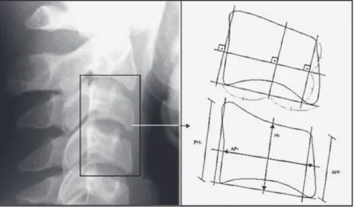
The following variables were obtained for both cervical vertebrae:
• (AH) anterior height of the vertebral body, • (AP) lateral anterior-posterior of the vertebral
body and
• (H) the height of the vertebral body, • (PH) posterior height of the vertebral body.
These variables were named AH3, AP3, H3, PH 3 when referring to the cervical vertebra C3, and AH4, AP4, H4, PH 4 when referring to cervical vertebra C4 (Figure 1).
The C3 and C4 vertebral segments were scratched manually and measured by a millimeter ruler and a digital caliper. Two repetitions of the scratch were made after at least a week and in a random manner. The intention was to obtain an average between the measurements made in each radiographic incidence. The segments were called vertebral age 1 and 2 for the Down syndrome individuals. The method pro-posed by Tanner and Whitehouse (2003), TW3, was used to obtain bone age using hand and wrist X-rays of the same individuals.10 All the measurements were
made by only one examiner, previously trained and author of this paper.
rESuLtS
In this study, the authors chose non-parametric statistical tests. These tests do not compare groups by the average but by the position of the data. Even though this method was not used to make the
com-Figure 1 - Body of cervical vertebrae C3 and C4 measured by means of lateral cephalometric X-rays: (AH) anterior height of the vertebral body, (AP) lateral anterior-posterior of the vertebral body and (H) height of the vertebral body, (PH) posterior height of the vertebral body.
Down
Syndrome Chronological Bone Vertebral 1
Female
Bone 0.308
Vertebral 0.287 0.114
Vertebral 0.073 0.039 0.046 table 2 - p-values for Down syndrome individuals, chro-nological, bone, vertebral, and vertebral 1 age for female individuals.
Non-Down
Syndrome Chronological Bone
Male Bone 0.367
Vertebral 0.042 < 0.001
Female Bone 0.788
Vertebral 0.048 0.037
table 1 - p-values for non-Down syndrome individuals, chronological, bone and vertebral age for male and fe-male.
parisons, descriptive statistics was used to allow us to understand the results, which can be seen in non-Down syndrome individuals. In this group, we ob-served that there were significant statistical differ-ences for both male and female individuals, among the different ages in overall manner. We used the Wil-coxon test to compare all the ages in pairs in order to discover precisely where the difference occurred (Table 1).
Regarding the p-values comparatively, a signifi-cant statistical difference was observed between ver-tebral bone age, bone age, and chronological age. In the sample studied, the vertebral bone age for both male and female individuals presented higher values than the other ages.
Below, we will show the results for the comparison made among all the ages for the group of Down syn-drome individuals. In this group, significant statistical differences could be noticed among the ages for the female individuals (Table 2).
According to the p-values observed, the differ-ences occurring between vertebral age 2 and vertebral
age 1 were revealed. Vertebral age 2 was that which had the highest values when compared to the other ages. Thus, in this sample of the female group of Down syndrome individuals, the formula designed by Caldas was not applied (Figures 2 and 3).
Significant statistical differences among the ages were not observed for the Down syndrome individuals of the male group, thus allowing the formula by Cal-das to be applied.
Based on these results, we can formulate a descrip-tion of the method for bone age in Down syndrome individuals.
• Female vertebral bone age = Óssea = -2.364 + 1.441 - H3/R2 = 84.2%
• Male vertebral bone age = Óssea = -1.004 + 0.759 x H3 + 0.580 x AH4/R2 = 84.1%
The results obtained made it possible to prove that the Caldas1 formula created for Brazilian boys and
girls cannot be applied to the female Down syndrome individuals of the studied sample. Thus, two new for-mulas were devised using the statistical method, one for women and another for men.
We determined that both models were significant and very explanatory. This means that they were well correlated with reference to the value of R2. We
deter-mined that the model is applicable, because there is no significant statistical difference between the bone age value (obtained by TW3) and the final bone age calculated through the model developed in this study.
In this context, it is worth mentioning that there is a difference between the final bone ages and the vertebral bone age only for the female group. There is no specific method for determining the bone age index of Down syndrome individuals.
DiSCuSSioN
After applying three methods to estimate the bone age of Down syndrome individuals, Santos (2007) noticed that there were statistical differences only using the Eklöf and Ringertz method, in referring to sex and chronological age.6 The TW31 and the
Greulich and Pyle methods were statistically the same. In relation to the Eklöf and Ringertz method in Down syndrome individuals, Sannomiya et al.(1998) made the same observation, but did not find significant sta-tistical differences among the sexes.8
Calles et al.(2004) observed that the Greulich and Pyle method is used for Down syndrome individuals, but it is not recommended to determine chronologi-cal ages between 10 and 13 years old for the female group and between 13 and 15 years old for the male group.2 They concluded that there was a significant
statistical difference in these age groups, when analyz-ing the chronological and bone ages. Sannomiya et
al. (2005) came to the same conclusion. This is why
the authors preferred to adopt the TW3 method in this study, and also because it is more updated.7
Mito et al. (2002) created a formula using lateral cephalometric X-rays of Japanese people with the mea-surements of vertebral bodies C3 and C4.. The for-mula obtains bone age according to the cervical ver-tebrae of these individuals.5 They concluded that this
result is reliable when compared to those obtained by hand and wrists X-rays using the TW2 method. In 2007, Caldas applied the Mito et al. (2002) formula in female and male Brazilians, and observed that it was only ap-plicable to Brazilian girls.1 With this in mind, he
cre-ated a formula to analyze the skeleton maturation of the cervical vertebrae in Brazilian boys and girls.
In this study, the authors applied the Caldas for-mula created for female and male Brazilians to non-Down syndrome individuals, and the result was differ-ent from the author’s, because the formula created was not statistically significant for the sample studied. Moreover, when applied to the Down syndrome indi-viduals sample, it was statistically significant only for the male but not for the female group.
CoNCLuDiNg rEmArKS
Based on the findings of this study, some impor-tant considerations can be made:
• The method proposed by Caldas, when applied to our samples of female and male non-Down syn-drome individuals, showed results with a statisti-cally significant difference between bone, verte-bral bone, and chronological age.
Figure 2 - Comparison of the chronological, vertebral bone and bone age for male and female non-Down syn-drome individuals. Male Female non-Down syndrome 11.5 10.5 11 10 9 9.5 10.36 9.92 11.28 10.18 10.26 10.92 Chronogical age Bone age Vertebral age
Figure 3 - Comparison of the chronological, vertebral bone 1 and bone 1 age in male and female Down syndro-me individuals. Down syndrome 12.86 Male Female 12.38 11.37 11.68 13.52 13.3 13 13.05 14 13.5 13 12 11 10 12.5 11.5 10.5 Chronogical age Bone age Vertebral age Vertebral age 1
• Thus, a similar method of evaluation was devel-oped for bone and vertebral bone age for both male and female Down syndrome individuals. rESumo
Avaliação e desenvolvimento de um método de avaliação de idade óssea em portadores de síndrome de Down
O objetivo deste trabalho foi avaliar a aplicabilida-de do método aplicabilida-desenvolvido por Caldas para medir a idade óssea vertebral em brasileiros, quando emprega-do em indivíduos portaemprega-dores da síndrome de Down. Foram estudados 57 prontuários de indivíduos com síndrome de Down, de ambos os sexos, com idades variando entre 5 e 18 anos. Esses prontuários conti-nham radiografias cefalométricas laterais e radiogra-fias de mão e punho, obtidas no mesmo dia, e também foram avaliados 48 prontuários de indivíduos não por-tadores de síndrome de Down. Para as radiografias de mão e punho, o método de Tanner e Whitehouse (TW3) foi usado para que pudéssemos obter a idade óssea. O método de Caldas foi empregado nas radio-grafias cefalométricas laterais, e assim obtivemos a ida-de óssea vertebral. A partir das informações sobre a idade óssea, idade óssea vertebral e idade cronológica, foi verificada uma diferença estatisticamente significa-tiva entre as três faixas etárias para ambos os sexos do grupo controle e grupo com síndrome de Down do sexo feminino. Portanto, este método foi aplicável ape-nas em indivíduos do sexo masculino portadores da síndrome de Down. Com base nos resultados, uma fórmula para obtenção da idade óssea de indivíduos com síndrome de Down foi desenvolvida.
DESCritorES
Síndrome de Down. Desenvolvimento ósseo. Vér-tebras cervicais. Crescimento. Radiologia.
§
rEFErENCES
1. Caldas MP. Avaliação da maturação esquelética na população brasileira por meio da análise das vértebras cervicais [tese]. Piracicaba: Faculdade de Odontologia de Piracicaba, Univer-sidade Estadual de Campinas; 2007.
2. Calles AC, Carinhena G. Avaliação da idade óssea em indivídu-os portadores da síndrome de Down por meio de radiografias da mão e punho. 7º Simpósio de informática na ortodontia e ortopedia funcional dos maxilares; 9-12 out 2004; São Paulo. [acesso em: mar.2008]. Disponível em: http: // www.cleber. com.br/orto2004/andreia.html
3. Coelho CRS, Loevy HT. Aspectos odontológicos da síndrome de Down. Ars Curandi Odontol.1982;8(3):9-16.
4. Greulich WW, Pyle SI. Radiographic atlas of skeletal develop-ment of the hand and wrist. Stanford: Stanford University Press; 1959.
5. Mito T, Sato K, Mitani H. Cervical vertebral bone age in girls. Am J Orthod Dentofac Orthop. 2002;122(4):380-85. 6. Santos LRA. Análise comparativa entre três métodos de
esti-mativa da idade óssea em indivíduos com síndrome de Down, por meio de radiografias de mão e punho [tese]. São José dos Campos: Faculdade de Odontologia de São José dos Campos, Universidade Estadual Paulista; 2007.
7. Sannomiya EK, Calles A. Comparação da idade óssea com a cronológica em indivíduos portadores da síndrome de Down pelo índice de Eklöf & Ringertz, por meio de radiografias de mão e punho. Cienc Odontol Bras. 2005;8(2):39-44.
8. Sannomiya EK, Médici-Filho E, Castilho JCM, Graziosi MAOC. Avaliação da idade óssea em indivíduos portadores da síndro-me de Down por síndro-meio de radiografias da mão e punho. Rev. Odontol. Unesp.1998;27(2):527-36.
9. Tanner JM, Whitehouse RH, Cameron N. Assessment of skel-etal maturity and prediction of adult height (TW2 Method). Institute of Education, University of London: Academic Press; 1983.
10. Tanner JM, Whitehouse RH, Cameron N, Healy MJR, Goldstein H. Assessment of skeletal maturity and prediction of adult height (TW3 Method). Austral Radiol. 2003;47:340-41
Recebido em 14/10/2010 Aceito em 17/12/2010