Characterization of Cortico-Striatal
Myelination in the Context of
Pathological Repetitive Behaviours
Eliana Lopes Rio da Silva Lousada
1,2,3Integrated Master in Bioengineering
Molecular Biotechnology
Supervisors: Eric Burguière, PhD3
Ana Paula Pêgo, PhD1,2,4,5
1- Faculdade de Engenharia, Universidade do Porto
2- Instituto de Ciências Biomédicas Abel Salazar, Universidade do Porto 3- Institut du Cerveau et de la Moelle Épinière, Sorbonnes-University, Paris 4- Instituto de Investigação e Inovação em Saúde, Universidade do Porto 5- Instituto de Engenharia Biomédica, Universidade do Porto
iii
v
A
BSTRACT
Several neuropsychiatric conditions are characterized by repetitive behaviours such as simple tics or ritualized sequences of complex behaviour. Although different types of repetitive behaviours are likely emerging from different aetiologies, a wide array of both clinical and preclinical evidence suggests that these behaviours are imprinted in dysfunctions within the cortico-striatal-thalamic-cortical (CSTC) circuitry. Numerous published studies have demonstrated white matter abnormalities in various brain regions comprising mainly frontal and prefrontal cortical projections (both of which crossing the corpus callosum into striatum) in patients with psychiatric disorders with repetitive behaviours, such as Obsessive-Compulsive Disorder. The conductive properties of myelin provide a fascinating tool for neural networks to adapt to environmental and experience-derived cues, considering the fact that minor alterations in myelin thickness or axonal coverage patterns can provide significant changes in conduction speed. Thus, we believe that aberrant myelination of cortico-striatal pathways may participate to the emergence of repetitive behaviours. Conversely, we hypothesized that the promotion of myelination in these pathways could rescue abnormal behaviour.
In order to address these questions, we assessed myelin-related differences between wild-type mice and a well-characterized model for repetitive behaviours, the Sapap3-KO mouse. We performed immunohistochemical analysis for the quantification of oligodendroglial cells, and complemented our results with the measurement of myelin thickness and density of myelinated axons. Additionally, aiming for an understanding of the topographical organization of the different cortico-striatal circuits, and their direct implication on the diversity of existing categories of repetitive behaviours, we performed a detailed analysis on the behavioural heterogeneity displayed by the Sapap3-KO mouse and correlated it with two different striatal regions: dorsomedial, and ventrolateral striatum. Our working hypothesis was that compulsions are imprinted within dysfunction of associative cortical areas that project to the dorsomedial striatum, whereas the pathological signatures of tic events are anatomically related to motor cortical areas who project to the ventrolateral part of the striatum.
We observed a slight, but non-significant, tendency towards a reduced number of mature oligodendrocytes in striatal regions, and, in opposition, a noteworthy increase in myelin thickness was demonstrated in the dorsomedial striatum. Contrary to what we expected, we did not find a correlation that corroborated our hypothesis. However, a noteworthy correlation between tic-like events and the number of mature oligodendrocytes in the dorsomedial striatum was observed.
Combination of these results seem to point towards a validation of the hypothesis that myelination abnormalities can be underlying pathological repetitive behaviours, but a further analysis and characterization will be required to prove such causal correlation.
Finally, targeting a possible behavioural rescue, we administered a drug that has been shown to promote remyelination - Clemastine, and quantified behavioural changes. However, under the tested conditions, no apparent effect of this drug was noted throughout the course of the experiment.
vii
R
ESUMO
Diversas patologias neuropsiquiátricas são caracterizadas por comportamentos repetitivos, que podem abranger desde simples e involuntários tiques, a sequências ritualizadas de comportamentos complexos. Apesar de se considerar que diferentes tipos de comportamentos repetitivos se encontrem associados a diferentes etiologias, um largo espectro de evidências clínicas e pré-clínicas sugerem que estes comportamentos estão correlacionados com disfunções nos circuitos cortico-estriato-tálamo-corticais. Estudos recentes têm vindo a demonstrar alterações na substânica branca em diversas regiões cerebrais, com princpal enfoque em projecções frontais e pré-frontais (projecções que atravessam o corpo caloso e o corpo estriado), em pacientes com perturbações psiquiátricas tais como transtorno obsessivo-compulsivo. As propriedades condutoras da mielina proporcionam uma ferramenta fascinante para as redes neuronais se modificarem perante sinalizações do microambiente e alterações que derivam de experiências adaptativas, uma vez que pequenas alterações de espessura ou dos padrões de cobertura axonal podem influenciar significativamente a velocidade de condução. Desta forma, colocamos como hipótese que uma mielinização aberrante de projecções cortico-estriatais pode participar no aparecimento e desenvolvimento de comportamentos repetitivos. Adicionalmente, propusemos testar se a promoção da mielinização destes feixes poderia atenuar as alterações comportamentais observadas.
De forma a endereçar estas questões, verificamos a existência de possíveis diferenças de mielinização entre murganhos wild-type e um modelo animal de comportamentos repetitivos, o murganho Sapap3-knockout (KO). Para tal, quantificamos células da oligodendroglia através de análise imunohistoquímica, e complementamos os nossos resultados medindo a espessura da mielina e a densidade de axónios mielinizados. Adicionalmente, com o intuito de inferir sobre a organização topográfica dos diferentes circuitos cortico-estriatais, e a sua implicação directa na diversidade de categorias comportamentais, avaliamos detalhadamente a heterogeneidade comportamental característica do modelo animal utilizado e correlacionamos com duas regiões estriatais: porções dorsomedial e ventrolateral do corpo estriado. A nossa hipótese prende-se à existência de correlação entre o surgimento de comportamentos compulsivos e disfunções entre áreas corticais associativas que projectam para a porção dorsomedial do estriado e, por outro lado, o aparecimento de tiques ter por base disfunções em áreas corticais motoras que projectam para a porção ventromedial do estriado.
Resultados preliminares indicam uma tendência, embora sem significância estatística, para um reduzido número de oligodendrócitos em regiões estriatais e, em oposição, um aumento da espessura na porção dorsomedial desta estrutura cerebral. Contrariamente ao que havíamos considerado, não foi encontrada uma correlação que corroborasse a nossa hipótese. No
entanto, uma correlação significativa foi observada entre a frequência de tiques e um reduzido número de oligodendrócitos na porção dorsomedial do corpo estriado.
A combinação dos resultados obtidos parece apontar para a validação da hipótese que alterações no processo de mielinização podem estar na base do surgimento de comportamentos repetitivos patológicos. Contudo, análise e caracterizações mais detalhadas deverão ser efectuadas para provar uma relação causal.
Por fim, com vista a uma possível atenuação de comportamento patológico, administramos um fármaco que tem vindo a ser descrito como promotor de remielinação – clemastina, e quantificamos alterações comportamentais. Contudo, nas condições testadas, nenhuma evidência do efeito deste fármaco foi denotado no decorrer desta experiência.
ix
A
CKNOWLEDGMENTS
I would like to start by thanking my grandmother, Conceição Ribeiro. You were the one who first introduced me to Science, the one who grabbed my hand through the discovery of what is today one of my greatest passions. It is with an indescribable honor that I graduate from the Institution where you worked for so many years.
I am entirely grateful to Eric Burguière, my supervisor, and Christiane Schreiweis. The enthusiasm and motivation that you bring to the lab every day is inspiring. You have the ability to provide more than just knowledge to your students. You work and stand everyday by our side, and you give us a feeling of home, even when home seems nowhere to be found. In addition, I would also like to acknowledge all the other members of our team. Each of you, in your own special manner, made my days feel lighter and brighter. And a special thank you to Lindsay Rondot for being constantly understanding and supportive, even in my frequent displays of coffee-machine-insulting-type-of-bad-mood!
To our collaborator, Brahim Nait-Oumesmar, and all the members of your group. You had your doors constantly open for my questions, my doubts, my ideas (and for the antibodies!), and you were capable of making me learn so much.
To Ana Paula Pêgo, in the quality of my co-supervisor, and in the quality of my Professor. You were always available (even if the e-mail reply came back one month later!) to listen to my bottomless bag of questions. Without realizing, the way you dedicate yourself to what you do had an impact on my motivation and commitment.
A very special gratitude goes out to my parents, and all the rest of my family. The amount of strength that I receive, and the amount of trust that I put on each one of you is reassuring. To my friends. Old ones, recent ones. Present ones, absent ones. Every single one of you contributed to define what I am today. A very special thank you for all the talks, all the laughs, and all the life that we shared. Cristiana Nogueira, Tiago Rodrigues, it is almost mandatory that I highlight the most bizarre and amazing set of coincidences that made us been so tightly together for so long. It is an enormous pleasure to close this chapter of my life with you. And finally, to Sara Marques. It would require another dissertation just to properly thank you for everything that you contributed for. Having in you a reason to go forward is the strongest motivation one can receive.
xi
“If I see anything vital around me, it is precisely that spirit of adventure, which seems indestructible and is akin to curiosity.”
xiii
C
ONTENTS
Abstract ... v Resumo ... vii Acknowledgments ... ix Contents ... xiii Figures List... xvAbbreviations and Symbols ... xvi
Chapter 1 – Introduction ... 19
1.1 Repetitive Behaviour ... 20
1.1.1 Cortico-striatal-thalamic-cortical pathways, the candidate circuitry of Repetitive Behaviour ... 20
1.1.2 Sapap3-KO mouse and the lack of inhibition ... 22
1.1.3 Potential implication of neuroplasticity ... 24
1.2 Myelination: The How’s and Why’s... 26
1.2.1 Oligodendroglial cell lineage ... 26
1.2.2 Dynamics of axon-OPC communication ... 27
1.2.3 Axonal ensheathment ... 27
1.3 Pathological expressions of white matter abnormalities ... 30
1.3.1 Myelination in motor skill learning ... 30
1.3.2 Social isolation ... 31
1.4 A pharmacological approach ... 32
Chapter 2 – Aim of the study ... 33
Chapter 3 – Methodology ... 35
3.2 Behavioural analysis ... 36
3.3 Immunohistochemistry ... 36
3.4 Electron Microscopy ... 37
3.5 Imaging analysis ... 38
3.5.1 Immunohistochemistry – Quantification of mature oligodendrocytes ... 38
3.5.2 Electron Microscopy – G-ratio measurement ... 39
3.5.3 Electron Microscopy – Quantification of myelinated axons ... 39
3.6 Pharmacological Treatment ... 39 3.6.1 Administration protocol ... 39 3.6.2 Behavioural analysis ... 40 3.7 Statistical analysis ... 40 Chapter 4 – Results ... 41 4.1 Behavioural analysis ... 42
4.2 Density of mature oligodendrocytes ... 42
4.3 Functional analysis... 45
4.4 Myelin thickness ... 47
4.5 Number of myelinated axons ... 47
4.6 Pharmacological Treatment ... 49
Chapter 5 – Discussion ... 51
Chapter 7 – Conclusion ... 57
xv
F
IGURES
L
IST
Figure 1: Hypothesized mechanisms underlying compulsive and tic symptoms. ... 21 Figure 2: Rodent sequential self-grooming behaviour... 22 Figure 3: Facial lesions on Sapap3-KO mouse due to excessive grooming. ... 23 Figure 4: Additional characteristic repetitive behaviours detected in the Sapap3-KO
mouse ... 24 Figure 7: Models of adaptive myelination in partially and fully myelinated axons. ... 29 Figure 8: Quantitative behavioural comparison between the KO and the
Sapap3-WT mice ... 43 Figure 9: Quantification of the density of mature oligodendrocytes ... 44 Figure 10: Correlation between behavioural quantifications and the number of
oligodendrocytes in DMS and VLS ... 46 Figure 11: Quantification of myelin thickness by g-ratio measurement ... 48 Figure 12: Quantification of the average density of myelinated axons ... 49 Figure 13: Quantitative behavioural comparison between Day 0 and Day 19 on
A
BBREVIATIONS AND
S
YMBOLS
∅ diameter
ACC anterior cingulate cortex
AMPA α-amino-3-hydroxy-5-methyl-4-isoxazolepropionic acid
BSA bovine serum albumin
CC1 adenomatous polyposis coli clone CC1 CNS central nervous system
CSTS cortico-striatal-thalamic-cortical
DMS dorsomedial striatum
DMSO dimethyl sulfoxide
DTI diffusion tensor imaging ECM extracellular matrix
fMRI functional magnetic resonance imaging
GA glutaraldehyde
GABA gamma-aminobutyric acid
I.P. intraperitoneally
KO knock-out
lOFC lateral orbitofrontal cortex
M1 motor cortical area M2 premotor cortical area
xvii MBP myelin basic protein
MSN medium-spiny neuron
NMDA N-methyl-D-aspartate
OCD Obsessive-Compulsive disorder OFC orbitofrontal cortex
OL oligodendrocyte
OPC oligodendrocyte progenitor cell
PB phosphate buffered
PBS phosphate buffered saline
PV parvalbumin
ROI region of interest
RT room-temperature
SMA supplementary motor area TEM transmission electron microscopy VLS ventrolateral striatum
1.1
R
EPETITIVEB
EHAVIOURRepetitive, ritualistic behaviour is a core feature of animal behaviour, including for humans, and can range from habitual thumb sucking to bedtime rituals as part of normal development in children. However, several neuropsychiatric disorders are characterized by a pathological expression of repetitive behaviours in the form of rumination, compulsions, tics, mannerisms, or stereotypies. Among these disorders are Autism Spectrum Disorders, Gilles de la Tourette Syndrome, and Obsessive-Compulsive Disorder (OCD) (Langen, Kas, Staal, Engeland, & Durston, 2011; Langen, Durston, Kas, Engeland, & Staal, 2011; Ridley, 1994). These symptoms can seriously impair daily functioning, with repercussions that go from impaired capacity to engage in relationships to the diminished ability to be occupationally functional, increasing stigma and diminishing the quality of life of the patients (Bokor & Anderson, 2014).
The term repetitive behaviour is broadly used, and sometimes refers to quite different classes of comportment, nevertheless linked by repetition, rigidity, and inappropriateness. Two clusters have been proposed: on the one hand, repetitive behaviours with predominantly motor features such as stereotypies or tics, and on the other hand repetitive behaviours of cognitive character such as compulsions or rituals (Turner, 1999), though it has been argued that grouping disparate behaviours into broad characteristics may oversimplify the subject. In fact, this distinction is not trivial. Different forms of repetitive behaviours are associated with different neuropsychiatric disorders and may derive from different neural mechanisms, although comorbidity exists (Worbe, 2015).
1.1.1 Cortico-striatal-thalamic-cortical pathways, the candidate circuitry of Repetitive Behaviour
The hypothesized mechanism mediating the expression of repetitive behaviour points to abnormalities in the cortico-striatal-thalamic-cortical (CSTC) pathways. Cortico-striatal loops are implied in processing limbic, cognitive and sensorimotor information, and learning, and are thought to be implicated in several neuropsychiatric conditions that exhibit movement disorders (such as repetitive behaviours). (Alexander, DeLong, & Strick, 1986)
Motor and non-motor information processing is thought to be mediated by functionally different, topographically organized CSTC circuits. The three proposed anatomo-functional subdivisions within this circuitry (sensorimotor, associative, and limbic) are therefore thought to be responsible for distinct classes of repetitive behaviour. The sensorimotor cortex projects to the dorsolateral part of the striatum (corresponding to the putamen in primates), and abnormalities in this loop might be responsible for stereotypical motor behaviour, where repetitive motor actions are continuously completed without the presence of a specific goal. (Tremblay, Worbe, Thobois, Sgambato-Faure, & Féger, 2015) Additionally, functional magnetic
21
resonance imaging (fMRI) studies have shown that supplementary motor area (SMA), a structure that highly projects into the ventrolateral striatum, has been described to be activated in the onset of a tic event in Tourette patients (Neuner, et al., 2014). In the light of this hypothesis, vocal and motor tics, which are a key characteristic in Gilles de la Tourette syndrome, might be due to changes in the circuits comprising both the dorso- and ventrolateral striatum (DLS and VLS) (Langen, Durston, Kas, Engeland, & Staal, 2011).
On the other hand, the associative cortex projects to the dorsomedial part of the striatum (DMS) (corresponding to part of the caudate nucleus in primates) and the limbic cortex to the more ventromedial part of the striatum (Tremblay, Worbe, Thobois, Sgambato-Faure, & Féger, 2015). These two loops are thought to be closely related to the pathological repetition of more complex sequences of actions, where the associative loop is considered to be associated with an exacerbated repetition of a goal, which can be related to its role in monitoring performance and correcting errors and may indicate a deficit in the interpretation of feedback and reward, or in the update of working memory (Groenewegen, Berendse, Cools, Voorn, & Mulder, 2009); and the limbic loop is associated with increased impulsivity and difficulty to suppress behaviour, regardless of the outcome, due to its implication in motivation as a component of behavioural control. (Langen, Durston, Kas, Engeland, & Staal, 2011)
Nonetheless, there is solid evidence that different cortico-striatal circuits are differently implied in different types of information processing, and, despite clinical and fundamental research in the field of repetitive behaviours and striatal sub-regions (Xu, Li, & Pittenger, 2016; Kalanithi, et al., 2005; Kataoka, et al., 2010), the hypothesis still remains to be causally tested whether dorsomedial striatum underlies repetitive behaviours of cognitive character and the ventro- and dorsolateral striatum repetitive behaviours of sensorimotor character.
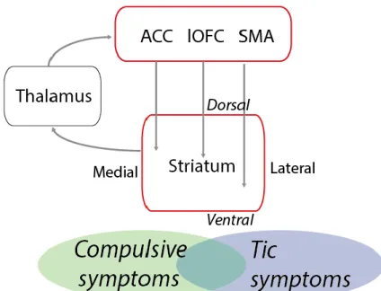
Figure 1: Hypothesized mechanisms underlying compulsive and tic symptoms.
In a simplified schematic manner, we hypothesize that compulsions are more closely related with dysfunctions within associative cortical areas, such as anterior cingulate cortex (ACC) and lateral orbitofrontal cortex (lOFC), who heavily project into the more dorsomedial part of the striatum, whereas
tics should be imprinted within more sensorimotor regions, namely supplementary motor area (SMA), that project into the lateral striatum.
1.1.2 Sapap3-KO mouse and the lack of inhibition
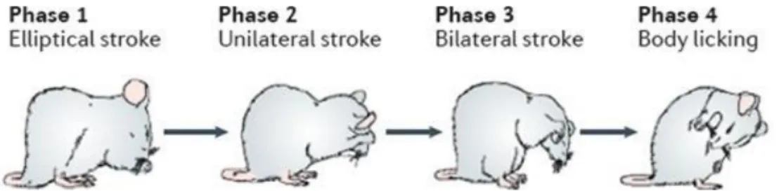
Assessing manifestations of neuropsychiatric disorders in animal models constitutes both a great challenge and a great opportunity to understand the neural mechanisms involved in the pathophysiology of these diseases. A powerful insight into repetitive behaviour can be achieved through the analysis of self-grooming in rodents (Figure 2), attending to the fact that it establishes a model of a complex, sequentially patterned, and repetitive behaviour. Self-grooming in rodents comprises a series of movements that form functional sequences that are crucial for hygiene maintenance, thermoregulation, and social communication, and its manifestation is known to be controlled by basal ganglia. Aberrant rodent self-grooming provides a great parallelism for symptomatic displays of several neuropsychiatric disorders (e.g. reduced grooming activity may be correlated with depression, or increased frequency and duration may be bridged with obsessive-compulsive disorder), thus numerous animal models have been proposed, which rely on distinct grooming phenotypes (Kalueff, et al., 2016).
Figure 2: Rodent sequential self-grooming behaviour.
Phase 1 comprises bilateral elliptical strokes nearby the nose, corresponding to the grooming of the nose and paws. Phase 2 corresponds to face grooming and consists of alternated unilateral strokes near the eyes. Phase 3 is again characterized by bilateral strokes, but now corresponding to head grooming, and starting behind the ears. Phase 4 comprises body and tail licking. Adapted by permission from Macmillan
Publishers Ltd: Nature Reviews Neuroscience (Kalueff, et al., 2016), copyright (2016)
One interesting mouse model that presents pathological repetitive behaviours, the Sapap3-KO mouse, has been developed and widely studied in this context, putatively presenting OCD-like behaviours (Welch, et al., 2007; Burguière, Monteiro, Feng, & Graybiel, 2013). SAPAP3 (SAP90/PDS-95-Associated Protein 3) belongs to the SAPAP family of proteins, which are postsynaptic density components of glutamatergic (excitatory) synapses, and it is the only one that is highly expressed in the striatum. Deletion of the Sapap3 gene leads to the development of self-inflicted, bilateral, facial lesions by the age of 4-6 months (Figure 3). These lesions are a consequence of disproportionate and injurious levels of self-grooming throughout all day, a behaviour that has been characterized as compulsive. Additionally, Sapap3-KO mice present increased anxiety-like behaviour when performing open-field, dark-light emergence, and elevated zero maze tests (Welch, et al., 2007).
23
Figure 3: Facial lesions on Sapap3-KO mouse due to excessive grooming.
Adapted by permission from Macmillan Publishers Ltd: Nature (Welch, et al., 2007), copyright (2007)
Studies in this mouse model have provided powerful insights into the neurocircuitry of repetitive behaviours. Chronic electrophysiological recordings have shown a significant increase in the baseline firing rates of medium spiny neurons (MSNs) in the DMS, but not in the lateral orbitofrontal cortex (lOFC), an associative cortex that has been shown to be differentially activated in patients with OCD and that projects to the DMS (Mar, Walker, Theobald, Eagle, & Robbins, 2011; Burguière, Monteiro, Feng, & Graybiel, 2013; Burguière, Monteiro, Mallet, Feng, & Graybiel, 2015). Furthermore, no thalamostriatal synaptic alterations were observed, indicating that the impaired circuitry may be restricted to cortico-striatal pathways (Wan, et al., 2014).
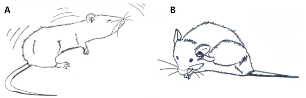
Remarkably, the Sapap3-mutant mice revealed an abnormally low number of parvalbumin (PV)-immunostained interneurons, which correspond to the fast-spiking interneurons (FSIs) within the striatum, known for their role in feed-forward inhibition of MSNs. Moreover, optogenetic stimulation of FSIs greatly increased the inhibition of MSNs and rescued compulsive behaviour (Burguière, Monteiro, Feng, & Graybiel, 2013). Altogether, these results indicate that a deficit in the inhibitory mechanism at the cortico-striatal level may be implicated in the development of repetitive behaviours in this mouse model. These mice were capable of learning a simple behavioural task as efficiently as their non-transgenic littermates, however, they displayed the inability to adapt to conditioned paradigms, showing they are bound to maladaptive compulsive response resulting from a defective inhibition (Burguière, Monteiro, Feng, & Graybiel, 2013). Surprisingly, on a detailed analysis and behavioural quantification in the Sapap3-KO mouse, our group detected, not only the well-established repeated syntactic self-grooming, but also brief and isolated myoclonic jerks (sudden and involuntary contraction of a muscle or group of muscles), which may correspond to tic-like behaviours (Figure 4A), indicating a more complex model than previously thought (unpublished data). Indeed, this phenotype is consistent with what can be observed in clinical practice, where patients often present comorbidity between compulsions and tic-like behaviours (Worbe, 2015). Moreover, an extremely evident behavioural aspect of this model is the increased number and duration of hind paw scratches (Figure 4B). In fact, this behaviour is usually the most obvious and severe phenotype of the
Sapap3-KO, and it remains poorly described. This behaviour might probably be paralleled with
Figure 4: Additional characteristic repetitive behaviours detected in the Sapap3-KO mouse
Apart from the already well-described disrupted self-grooming on the Sapap3-KO mouse, two other characteristic behaviours have been noted: A – myoclonic jerk: a simple and brief tic, mostly detected in the head; B – hind paw scratch: a repetitive, alternated unilateral movement.
Taken together all the aforementioned behavioural characteristics, Sapap3-KO mouse arises as a highly complex and interesting model of repetitive behaviours. Considering the multitude and diversity of the phenotype, it leaves space to raise questions on the topographical organization of the different types of repetitive behaviours observed.
1.1.3 Potential implication of neuroplasticity
Neuroplasticity within CSTC circuitry has been highlighted as a key player in compulsive, repetitive behaviour (Koob & Moal, 2005). An interesting approach that reiterates this thought was provided by the development of an optogenetic mouse model of obsessive-compulsive disorder. This model consists in the optogenetic stimulation of the OFC-DMS pathway in awake, behaving wild-type mice. Notably, whereas acute stimulation revealed no significant alterations on mice behaviour, chronic stimulation (15 minutes a day for 5 days) led to the development of abnormal levels of self-grooming, which persisted for 2 weeks after stimulation cessation (Ahmari, et al., 2013).
This optogenetically-introduced long-term change in the associative cortico-striatal pathway opens the window for new questions, such as the molecular signatures that underlie adult plasticity and their involvement in the development of abnormal behaviour.
Until a recent paradigm shift, neuroplasticity mechanisms were ultimately attributed to neuronal changes in neuron morphology, synaptic mechanisms and neurogenesis (Kulak & Sobaniec, 2004; Fuchs & Flügge, 2014). The fundamentals that describe these mechanisms are based on Hebbian plasticity (Hebb, 1949) and were elegantly made famous by Löuel with the sentence “neurons that fire together wire together” (Löwel & Singer, 1992). This principle determines that excitatory presynaptic neurons that fire milliseconds before the postsynaptic neuron fires will strengthen the connectivity between them and cause long-term potentiation, whereas if the presynaptic firing occurs milliseconds after the postsynaptic, the connectivity is weakened and cause long-term depression (Power & Schlaggar, 2016; Cohen, Quarta, Bravi,
25
Granato, & Minciacchi, 2017). In a broader level, morphological changes in dendrites have been reported as a stress-induced response, with observable regression of their length in the hippocampus (Watanabe, Gould, & McEwen, 1992) and prefrontal cortex (Cook & Wellman, 2004), and enhancement in the amygdala (Vyas, Mitra, Rao, & Chattarji, 2002). The length of the dendritic branches regulates the number of axons that are able to connect and therefore the number of synapses (Fuchs & Flügge, 2014). However, in the light of recent development in brain imaging, it was possible to detect fractional anisotropy in white matter during learning, which broke ground to understand the possible role for activity-dependent myelination in both motor and cognitive modifications resulting from functional experience (Fields, 2008).
One particularly interesting study in OCD patients measured white matter abnormalities using a diffusion tensor imaging (DTI) technique. DTI is a MRI technique that provides measurement for fractional anisotropy, mean and radial diffusivity, and provides information about white matter microstructure. Disperse white matter irregularities were found in OCD patients, with specific relevance to abnormalities in the internal capsule, which were positively correlated with the percentage of errors that were committed during a cognitive task (Magioncalda, Martino, Ely, Inglese, & Stern, 2016). Internal capsule in primates does not have a specific correspondent structure in rodents, but comprises cortico-striatal projections (Steiner & Tseng, 2010), and might therefore constitute an important parallel to what has been described in the Sapap3-KO mouse model for obsessive-compulsive disorder, in which optogenetic stimulation of the lOFC-DMS permitted behavioural rescue (Burguière, Monteiro, Feng, & Graybiel, 2013).
Combination of the abovementioned premises led us to question a potential role for myelin-related abnormalities in the development of cortico-striatal connectivity impairment, and its causal relationship with the emergence of pathological repetitive behaviours.
1.2 M
YELINATION:
T
HEH
OW’
S ANDW
HY’
SMyelination of neurons increases electrical conduction by working as an insulator, reducing transverse capacitance and increasing transverse resistance across the axonal membrane. Furthermore, conduction velocity is also potentiated by clustering voltage-gated sodium channels in gaps (Nodes of Ranvier) between myelin sheaths, which translates in a saltatory conduction of action potentials (Nave & Werner, 2014). Bearing in mind these aforementioned properties, it is not surprising that much attention has been attributed to myelination.
1.2.1 Oligodendroglial cell lineage
In the CNS, myelin sheaths are formed by one type of multipolar glial cells, the oligodendrocytes, which wrap around the axons by spreading their membrane and growing both radially and longitudinally. Oligodendrocytes are part of the oligodendroglial lineage, and derive from the differentiation of oligodendrocyte progenitor cells (OPCs). During embryonic development, undifferentiated progenitor cells migrate from ventral and dorsal domains. After birth, these cells start their differentiating process and go through different stages until the final differentiation into mature, myelinating oligodendrocytes (Figure 5) (Nishiyama, Komitova, Suzuki, & Zhu, 2009; Bradl & Lassmann, 2010). OPCs extend and multiply their ramified membrane until they reach their mature state. These protrusions are crucial for cell-cell communication with the axonal portion of the surrounding neurons, and will be determinant for their subsequent ensheathment.
Figure 5: Representation of the oligodendroglial lineage progression during differentiation
Oligodendrocyte progenitor cells (OPCs) expand and multiply their protrusions until reaching a star-shaped immature oligodendrocyte (Pre-OL). Further expansion and arborization of this protrusions leads to the full maturation of these cells (Mature OL), allowing the contact with the surrounding axons until reaching their myelinating state (Myelinating OL).
Adapted by permission from Macmillan Publishers Ltd: Nature Reviews Neuroscience (Nishiyama, et al., 2009), copyright (2009)
The biological cues that drive the OPCs into a differentiation process remains not fully understood. Several mechanical, electrical, and biochemical factors are believed to work together in providing the appropriate environment for these precursor cells to enter into a maturating state. OPCs are known to be mechanosensitive cells, who respond to variances in
27
the rigidity of the ECM. Their interaction with proteins of the cytoskeleton are thought to be essential for the extension and ramification of the protrusions (Lourenço, et al., 2016; Jagielska, Norman, Whyte, & Vliet, 2012; Domingues, Cruz, Chan, & Relvas, 2017). Other environmental cues from the extracellular milieu are held responsible for this process, as such the Notch signalling pathway, which is thought to be determinant in the timing of the differentiation (Shimizu, Osanai, & Ikenaka, 2017).
1.2.2 Dynamics of axon-OPC communication
Interestingly, from a biochemical point of view, OPCs have been shown to express receptors for glutamatergic (AMPA and NMDA receptors), GABAergic, and acetilcholinergic neurotransmitters (Barres, Koroshetz, Swartz, Chun, & Corey, 1990; Wyllie, Mathie, Symonds, & Cull-Candy, 1991; Bergles & Richardson, 2015). Complementary to this observation, axon-OPC synapses have been described both in grey and white matter, in all studied CNS regions (Zonouzi, et al., 2015; Bergles & Richardson, 2015; Chittajallu, Aguirre, & Gallo, 2004; Jabs, et al., 2005). These findings intuitively lead to the idea of an activity-dependent activation of a differentiation process in the OPCs, however, these synaptic inputs remain poorly understood. One curious aspect of the aforesaid findings is the fact that the activation of either glutamatergic or GABAergic synapses induces depolarization on the OPCs (Bergles, Roberts, Somogyi, & Jahr, 2000; Tanaka, et al., 2009), which presents as counter-intuitive, as one would expect a GABAergic synapse to induce hyperpolarization. This fact is related to the high intracellular concentration of Cl- in the OPCs,
and the GABAergic promotion of Cl- eflux.
Glutamatergic signaling through AMPA receptors enhances oligodendrocyte survival, but the impact of this cell-cell comunication has yet to be better elucidated and proven significant for the myelination process (Kougioumtzidou, et al., 2017). Nevertheless, recent studies have been gathering evidence that point to the idea that GABAergic axon-OPC communication may indeed have some impact on maintaining activity-dependent plasticity (Tanaka Y. , et al., 2009), and that the blockade of this synaptic transmission increases the proliferation of OPCs, but decreases the number of mature oligodendrocytes (Zonouzi, et al., 2015; Hamilton, et al., 2017).
The relevance of the concept of activity-dependent OPC differentiation is highly related to the idea of myelination as a form of adult plasticity. If proven correct, neuronal activity may be essential in potentiating and modulating de novo myelination and/or myelin replacement.
1.2.3 Axonal ensheathment
The most recently proposed model of myelin formation indicates that oligodendrocytes spread their membrane around the axon, wrapping it, and expanding their projections both in a radial and longitudinal manner. This growth takes place concomitantly and results in a greater number of wraps at the site of attachment, decreasing gradually towards the nodes (Figure 6) (Chang,
Redmond, & Chan, 2016). Afterwards, these lipid layers compact, extruding most of the cytoplasm and extracellular space, achieving, in this way, the insulating properties. Oligodendrocytes are capable of myelinating several axons simultaneously in response to certain attractive or inhibitory environmental cues (Chang, Redmond, & Chan, 2016; Fields, 2015). This characteristic particularity of oligodendrocytes might additionally contribute to their role in neuroplasticity.
Figure 6: Current model of myelination in the CNS.
On the left, a newly differentiated oligodendrocyte contacts the unmyelinated axon by extending one of its protrusions. Afterwards, it begins to wrap its membrane around the axon by spreading this lipidic bilayer in a radial and longitudinal manner. Finally, after approximately three layers of myelin membrane, intracellular compaction starts from the outermost layer towards the inner layers. Adapted by permission
from Macmillan Publishers Ltd: Nature Neuroscience (Chang, Redmond, & Chan, 2016), copyright (2016)
In the light of the hypothesis of an activity-dependent myelination as a form of neuroplasticity, it is interesting to understand how adult axonal remodelling may be determinant in fine-tuning conductive properties. Several factors such as sheaths’ thickness, length and axonal coverage patterns may constitute the adaptive changes that emerge as a response to learning and environmental fluctuations (Figure 7) (Bujalka & Emery, 2016; Tomassy, et al., 2014).
29
Figure 5: Models of adaptive myelination in partially and fully myelinated axons.
In partially myelinated axons (A), myelin remodeling may occur by the longitudinal extension of the already existing internodes or by de addition of de novo myelin sheats with different coverage patterns. In already fully myelinated axons (B), internodes may increase their thickness or demyelination may occur, with subsequent, differentially patterned, remyelination.
(Baraban, et al., 2016) This is an open access article under the terms of the Creative Commons Attribution
1.3 P
ATHOLOGICAL EXPRESSIONS OF WHITE MATTER ABNORMALITIESParallel to findings that adult myelination in humans can be triggered during learning, impaired oligodendroglial and myelin integrity have been detected in patients with psychiatric disorders, including depression or schizophrenia by in vivo neuroimaging as well as in postmortem studies (Roussos & Haroutunian, 2014; Edgar & Sibille, 2012). This dual approach leaves the thought that “myelin is not only a prerequisite, but also a consequence of normal activity” (Nave & Werner, 2014).
Several animal studies have been demonstrating an important role of myelination in different types of learning and other behaviours, thus reinforcing the idea of its involvement in neuroplasticity mechanisms.
1.3.1 Myelination in motor skill learning
Particular attention has been attributed to study the role of myelination in motor skill learning. In the first study of this kind, adult rats were assigned to three different experimental conditions, namely skilled reach for food pellets (SR), unskilled reach for food pellets (UR), and caged controls (CC). In all three groups, white matter differences were assessed by diffusion MRI fractional anisotropy, and significant difference in these values was observed between several white matter areas contralateral to the trained paw between the SR group and both control groups. Additionally, quantitative immunohistochemistry of myelin basic protein (MBP), a myelin marker, demonstrated a substantial increase in white matter subjacent to motor cortical area (M1) contralateral to the trained paw only in the SR group (Sampaio-Baptista, et al., 2013). Another interesting and detailed approach came from Gibson et al., 2014, where mice that were optogenetically stimulated in the premotor cortex (M2) have shown proliferative response of OPCs in ipsilateral premotor area and subcortical white matter projections into the corpus callosum. OPC differentiation was assessed by immunohistochemistry, showing increased expression of CC1 (marker for mature oligodendrocytes), and histone modifications that translate the epigenetic changes required for progression into differentiated states. Transmission electron microscopy (TEM) analysis demonstrated increased myelin thickness in layer VI of M2 axons and subcortical fibers entering corpus callosum. Complementing this finding, MBP expression levels also increased in these optogenetically stimulated mice in comparison to their wild-type littermates. Remarkably, chronically stimulated mice have shown enhanced speed in limb swing only in the stimulated limb when performing a CatWalk task, correlating better motor performance with increased myelin thickness (Gibson, et al., 2014). On the other hand, blocking differentiation of novel oligodendrocytes in adult mice prevented the animal from learning new motor tasks such as mastering a complex wheel (McKenzie, et al., 2014). Altogether, these results show that active myelination is required for adult motor skill learning.
31 1.3.2 Social isolation
Two different studies addressed the question on whether social isolation would impact myelination, one in juvenile and one in adult mice. The first study determined a critical period for oligodendrocyte maturation (P21 to P35), during which socially isolated mice developed impairment in neuregulin 1-ErbB3 signaling pathways in the oligodendrocytes of the medial prefrontal cortex. This results in morphological differences in these oligodendroglial cells (simpler, shorter processes, less branching and fewer internodes), whereas their density remains unaltered. This, in turn, translates into reduced MBP expression and myelin thickness, which culminated in compromised social interactions and working memory that were not reversible by the reintroduction into a social environment (Makinodan, Rosen, Ito, & Corfas, 2012). Conversely, eight weeks of social isolation in adult mice led to changes in chromatin organization and in ultrastructural changes in oligodendrocyte, resulting in myelin thinning. When put back into a social environment, these manifestations were rescued, along with the social behaviour of the mice with their littermates (Liu, et al., 2012).
1.4 A
PHARMACOLOGICAL APPROACHAs myelination has been resurfaced as a potential neuroplasticity mechanism involved in learning and behaviour, several studies aimed for a therapeutic approach by the enhancement of adult de novo myelination. A pharmacological therapy that has been in the limelight is the treatment of pathological behaviours with clemastine (Li, He, Fan, & Sun, 2015; Liu, et al., 2016). Clemastine is a Food and Drug Administration-approved drug that has been shown to promote remyelination. This small anti-muscarinic compound can easily pass the blood-brain barrier and promote OPC differentiation in the CNS (Li, He, Fan, & Sun, 2015) (Liu, et al., 2016). Mice that were submitted to cuprizone administration (cuprizone mouse model of demyelination) for six weeks developed schizophrenia-like behaviours. When treated with clemastine for three weeks after cuprizone withdrawal, they showed recovery of MBP intensity and an increase in mature oligodendrocytes, which promoted behavioural rescue and improvement of spatial working memory (Li, He, Fan, & Sun, 2015). Another approach that pronounces the pharmaceutical potential of clemastine was recently provided by Liu et al. These authors orally administrated this compound to two to eight week old, socially isolated mice, and demonstrated its ability to rescue social behaviour by increasing the levels of repressive histone methylation in oligodendroglial cells, a crucial step for OPC differentiation into oligodendrocytes (Liu, et al., 2016).
33
Considering recent findings on the role of myelination in learning, behaviour, and adult plasticity, we hypothesized that a deficit in myelination of cortico-striatal circuits may be underlying the emergence of repetitive behaviours in the Sapap3-KO mouse, as well as a topographical organization of these circuits being the substrate for the heterogeneity of these behaviours. Thus, we defined as aims of this work:
- Assess myelin-related differences between Sapap3-KO and Sapap3-WT mice in three regions of interest: lateral orbitofrontal cortex, dorsomedial and ventrolateral striatum; - Test causal correlation between compulsive-like behaviours and myelin deficits in
dorsomedial striatum, and tic-like behaviours with deficits in ventrolateral striatum; - Behavioural rescue by a pharmacological promotion of remyelination.
35
3.1 A
NIMALSAnimals used for immunohistochemistry and electron microscopy analysis were six- to eleven-month old male Sapap3-KO and non-transgenic littermates (Sapap3-WT), C57BL/6J background (Welch, et al., 2007). Animals were kept at the animal facilities of the “Institut du Cerveau et de la Moelle Épinière”, housed in Techniplast ventilated polycarbonate cages under positive pressure with hard-wood bedding and provided with food and fresh water, ad libidum. All animals were housed in environmentally controlled cages with 40 air changes per hour. The temperature was maintained at 21–23 °C and the relative humidity at 55 ± 10% with a 12-h light/dark cycle. Each study animal was assigned a unique number and identified by tattoos in the paws. All experiments were approved by the French ministry of research under the agreement number (APAFIS) #1418-2015120217347265.
3.2 B
EHAVIOURAL ANALYSISAll animals were filmed for 24hours in the automated behavioural boxes developed by the team, with food and water ad libidum, without being submitted to the performance of any behavioural task.
Videos were analyzed with Kinovea (version 0.8.15), an open-source software adapted to the study of movement and motor performance. 15 minute videos were selected for each animal between 8 and 9 a.m. when the mouse was awake, after an overnight period of adaptation to the new cage.
During the selected period, three types of behaviour were considered for quantification: myoclonic jerks, grooming events (with discrimination of the four different phases), and hind paw scratches. Frequency and duration of these behaviours were the parameters used for analysis.
3.3 I
MMUNOHISTOCHEMISTRYAnimals were divided in two different groups (Sapap3-KO and Sapap3-WT, N=7 per group). Animals from both groups were deeply anesthetized with Euthasol®, and transcardial perfused with 2% PFA (Sigma-Aldrich). The brain of each animal was collected and post-fixed overnight in 2% PFA. Samples were then rinsed in 1X PBS (Euromedex).
37
With the purpose of cryo-protecting the tissue for the upcoming freezing procedure, samples were immersed in a 15% sucrose solution for 24 hours, and subsequently immersed in 30% sucrose for 48 hours, until the brains sinks in the solution as sign it is completely impregnated. Samples were embedded in Tissue-Tek O.C.T. compound (Thermo Scientific) for 30 minutes, in order to fully impregnate the tissue and provide a stabilizing matrix, and frozen in dry ice. Brains were subsequently cut in a Microm HM 560 cryostat, in 12µm coronal slices, on a glass slide, and stored in -80ºC.
Slices were defrost at RT and rehydrated in 1X PBS.
With the purpose of increasing the intensity of the immunostaining, the slides were immersed in 10mM sodium citrate buffer (C6H5Na3O72H2O) and put in water-bath at 85ºC for 30 minutes.
The acidic environment provided by the citrate buffer disrupts the protein cross-linking caused by the PFA and enables a more efficient penetration of the antibodies. When taken off the water-bath, the samples were cooled-down to RT and rinsed in 1X PBS.
In order to decrease the non-specific binding of the antibodies, thus staining with less background noise, samples were saturated in 4% BSA/0.1% Triton X-100 for 1 hour. Triton X-100 (VWR Chemicals) is a detergent that lyses the lipids in the membranes, and therefore increases membrane permeability for the antibodies.
The antibodies anti-Olig2 rabbit (Millipore, #AB9610) and anti-CC1 mouse IgG2b (Calbiochem, #OP80) were diluted in 4% BSA/0.1% Triton X-100 with a concentration of 1/1000 and 1/100 respectively, and incubated overnight at 4ºC.
Samples were rinsed with 1X PBS and incubated with the secondary antibodies anti-Rabbit 488 Alexa and anti-Mouse IgG 568 Alexa (Invitrogen, #A21206 and #A21144 respectively) in a 1/2000 dilution, for 1 hour at RT, in dark. The slides were rinsed in dark, under agitation.
The slides were mounted with Mowiol® as a mounting medium, and left overnight to dry. Samples were imaged in a Zeiss Axio Scan.Z1, in fluorescence, objective Plan-Apochromat x20, Hamamatsu camera.
3.4 E
LECTRONM
ICROSCOPYAnimals (N=3 per group) were deeply anesthetized with Euthasol® and transcardial perfused with 5% glutaraldehyde (GA) (Electron Microscopy Sciences)/5mM CaCl2. The brain of each
animal was collected and post-fixed overnight in 5% GA. Samples were then rinsed in 0.12M phosphate buffered solution (PB) (pH=7.4).
Brains were cut in 200µm slices in the vibratome Leica VT1200 S, and the slices that comprised the target areas were selected. In order to be specific about our regions of interest, a 1mm ∅ biopsy punch was used to extract them. The following protocol was performed only in the collected punches.
Attending to the fact that myelin is a lipidic structure, another post-fixation step with 4% osmium tetroxide (Electron Microscopy Sciences) was added. After 1hour of incubation, samples were carefully rinsed.
As a first contrasting step, samples were immersed in 5% uranyl acetate (Electron Microscopy Sciences), a heavy metal who interacts with proteins and lipids. After 1hour of incubation, samples were carefully rinsed.
Afterwards, samples were dehydrated in increasing concentrations of ethanol and finally with 100% acetone. To prepare the samples for the embedding with epoxy resin, they were first let overnight, at 4ºC, immersed in a solution of 50:50 acetone and epoxy resin.
Samples were transferred into embedding molds and immersed in resin (Embed 812 Kit, Electron Microscopy Sciences). After 2hours the epoxy resin was replaced and left to polymerize at 60ºC for 48hours. Samples were then cut in an ultramicrotome Leica EM UC7 into 70nm slices and places in a TEM grid.
Finally, as a complementary contrasting agent, lead citrate (ChromaLys) was used under CO2 free
conditions.
Samples were imaged in the Transmission Electron Microscope HITACHI 120kV HT7700, camera AMT XR41-B. Magnification used for quantification of g-ratios was x26.0k, and x11.0k for the quantification of the density of myelinated axons.
3.5 I
MAGING ANALYSISAll images were analyzed with Fiji from ImageJ2 (Schindelin, et al., 2012; Schindelin, Rueden, Hiner, & Eliceiri, 2015).
3.5.1 Immunohistochemistry – Quantification of mature oligodendrocytes
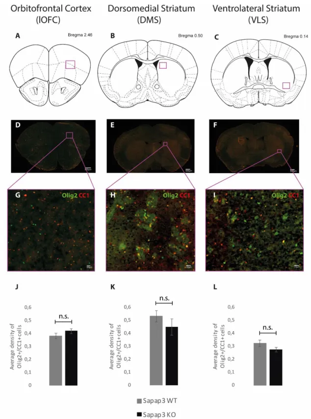
For the quantification of the density of oligodendrocytes, we first chose the slices that matched the Bregma levels of our regions of interest: lateral orbitofrontal cortex – Bregma +2.46; dorsomedial striatum – Bregma +0.50; ventrolateral striatum – Bregma +0.14. For each animal, four slices per area were chosen, only on the left hemisphere. Lateralization effects have been previously assessed, and no different values were found between the two hemispheres (data not shown).
We drew a square on the specific area of interest in each sample by using the ROI tool on Fiji, and counted all cells that were positively stained for Olig2 and CC1 (Olig2+/CC1+ cells). Olig2 is a nuclear marker for all cells of the oligodendroglial lineage, and CC1 is a cytosolic marker specific for mature oligodendrocytes. Combination of these two markers allowed us to be confident on the quantification of mature oligodendrocytes. To calculate the density, we divided the total number of cells by the area of the drawn ROI.
39
3.5.2 Electron Microscopy – G-ratio measurement
To assess myelin thickness, we analyzed the TEM images with x26.0k magnification that were previously collected from two regions of interest: dorsomedial and ventrolateral striatum. In order to be specific on the quantification of local striatal cells, myelinated axons within the fibers that cross the striatum (corresponding to internal capsule) were not considered. To obtain a satisfactory level of confidence in our results, we quantified ~150 myelinated axons per region, per animal.
The tool “Polygon selections” in Fiji was used to draw both the inner axonal area and the outer (myelinated) area of each axon. To calculate the g-ratio, we divided the inner area by the outer.
3.5.3 Electron Microscopy – Quantification of myelinated axons
For the quantification of the number of myelinated axons, we analyzed the TEM images with x11.0k magnification that were previously collected from two regions of interest: dorsomedial and ventrolateral striatum.
In order to be specific on the quantification of local striatal cells, myelinated axons within the fibers that cross the striatum (corresponding to internal capsule) were not considered.
For each animal, we analyzed ~20 images per region and counted the number of myelinated axons in each image.
3.6 P
HARMACOLOGICALT
REATMENT 3.6.1 Administration protocolThe animals used were seven- to fourteen-month old female Sapap3, C57BL/6J background, housed in the same conditions as previously described.
Sapap3-KO females were divided in two groups (N=6 per group): clemastine-treated (CT) and
the control, vehicle-treated (VT) groups.
Clemastine (Sigma-Aldrich) was dissolved in NaCl 0.3% DMSO (Sigma-Aldrich) and administrated daily to the CT group via I.P. injection at the concentration of 10mg/kg body weight (Apolloni, Fabbrizio, Parisi, Amadio, & Volonté, 2014) for 12 days. VT group was treated with equivalent volumes of NaCl 0.3% DMSO. Animals were weighted and health monitored daily.
3.6.2 Behavioural analysis
All animals were filmed for 24hours with food and water ad libidum at different time points: day 0, day 6, day 12, and day 7 after treatment cessation.
Videos were analyzed with Kinovea (version 0.8.15). 15 minute videos were selected for each animal between 1 and 2 a.m. when the mouse was awake, after a six hour period of adaptation to the new cage. Analyzed parameters were the same as previously described.
3.7 S
TATISTICAL ANALYSISAll data was visually inspected prior to statistical analysis, which was then performed in the programming environment R (a language and environment for statistic computing, R Development Core Team - R Foundation for Statistical Computing, Vienna, Austria, 2008). Due to small sample size, behavioural parameters (number of hind paw scratches, number of myoclonic jerks, and number of grooming events), as well as the number of myelinated axons in the two genotypes were assessed using the function “wilcox test” to perform non-parametric Mann Whitney U tests. Severity of individual behavioural readouts and the number of Olig2+/CC1+ cells in each of the three brain regions were correlated using the function “cor test”, the non-parametric Spearman correlation. Even though a small sample was used to measure the g-ratios, considering the apparent normal distribution, a Student’s T test was performed. All p-values > 0.05 were considered non-significant.
41
4.1 B
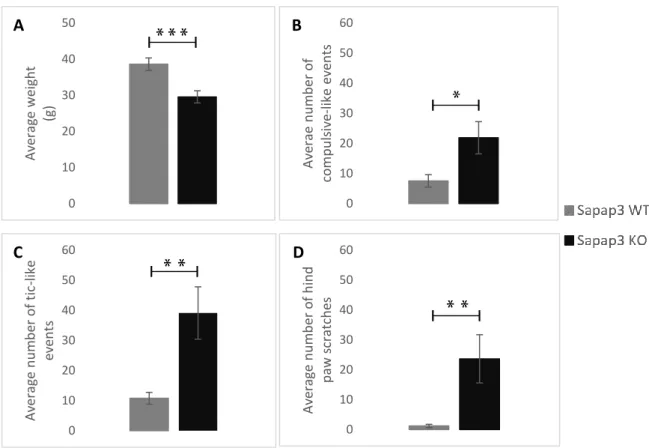
EHAVIOURAL ANALYSISAiming for a descriptive analysis on the behaviour of the Sapap3-KO mouse, we analyzed the categories that provide the most obvious phenotype. The first notable difference is on the average weight of the Sapap3-KO (p-value=0.00053) (Figure 8A), who are visibly smaller and lighter than their non-transgenic littermates (Sapap3-WT).
The subsequent analysis, targeting the validation of what has been described on the model, was to count the number of compulsive-like events (grooming events) on both groups. Results indicate a significant increase in the average frequency of grooming behaviour in the Sapap3-KO mice (p-value=0.0333) (Figure 8B), which goes in line with what has previously been stated (Welch, et al., 2007). Remarkably, the most significant displays of abnormal behaviour come from the phenotypes that have not yet been described in the literature. Both the average frequency of myoclonic jerks (tic-like events), and the hind paw scratches present a notorious increase in the Sapap3-KO mice (p-value=0.00695; p-value=0.00183, respectively) (Figure 8C-D). Altogether, these results seem to reinforce our working hypothesis of a complex model, who presents repetitive behaviours of mutually motor and cognitive character.
4.2 D
ENSITY OF MATURE OLIGODENDROCYTESThe first approach on assessing possible myelin-related deficits in the Sapap3-KO mouse relies on the evaluation of the cells that are actively myelinating the axons. Different values regarding the density of mature oligodendrocytes in our regions of interest (lateral orbitofrontal cortex, dorsomedial striatum, and ventrolateral striatum) (Figure 9A-C) may constitute the initial evidence of this matter. For that purpose, we double-immunostained our samples (N=7 per group) for Olig2: nuclear marker of all the cells that are part of oligodendroglial lineage, and CC1: specific cytosolic marker for mature oligodendrocytes, and counted all Olig2+/CC1+ cells (Figure
9D-I).
Our results indicate a slight, non-significant increase in the number of oligodendrocytes per µm2
in the lOFC (p-value=0.2285) of the Sapap3-KO mouse (Figure 9J). However, this tendency seems to be inverted both in the DMS (p-value = 0.3024) and VLS (p-value = 0.1332) (Figure 9K-L) where the number of oligodendrocytes counted by µm2 was reduced in the Sapap3-KO, in comparison
43
Figure 6: Quantitative behavioural comparison between the Sapap3-KO and the Sapap3-WT mice
A – Comparison between the average weights of the two groups of animals. Sapap3-KO animals are notoriously smaller and lighter than the Sapap3-WT group; B – Average number of compulsive-like events in the two groups validating the characteristic excessive grooming seen in the mutant mice; C – Average number of tic-like events (myoclonic jerks) and D – Average number of hind paw scratches, demonstrating the significant increase on the frequency of these pathological repetitive behaviour on the Sapap3-KO mouse. Bars represent average value and error bars show standard error. Mean and standard error were calculated from video analysis of 18 animals (N=9 per group). Represents * p-value < 0.05, ** p-value < 0.01, and *** p-value < 0.001. 0 10 20 30 40 50 Av era ge w eight (g) 0 10 20 30 40 50 60 Av era e n u m b er o f comp u ls iv e-like e ve n ts 0 10 20 30 40 50 60 Av era ge n u m b er o f t ic -like ev en ts 0 10 20 30 40 50 60 Av era ge n u m b er o f h in d p aw s crat ch es
A
B
C
D
Figure 7: Quantification of the density of mature oligodendrocytes
Figures A, B, and C represent a diagram of the Bregma levels we aimed in order to incorporate the defined ROIs (lOFC, DMS, and VLS, respectively). Figures D, E, and F correspond to the fluorescence imaging of the entire brain slice that match the Bregma levels we targeted, and a representation of where we outlined the abovementioned ROIs. Figures G, H, and I correspond to a zoom-in of the ROIs. Olig2+ cells are stained in green and CC1+ cells are stained in red. Figures J, K, L represent the quantification of the number of Olig2+/CC1+ positive cells per µm2 in the regions abovementioned. A slight decrease in the average
45
density of Olig2+/CC1+ cells in DMS (K) and VLS (L) was noticed in the Sapap3-KO group. In OFC (J), this tendency was inverted, with a slight increase in the average density of mature oligodendrocytes in the
Sapap3-KO group. Bars represent average value and error bars show standard error. Mean and standard
error were calculated from image analysis of 14 animals (N=7 per group), 4 images per animal. Represents n.s. = non-significant.
4.3 F
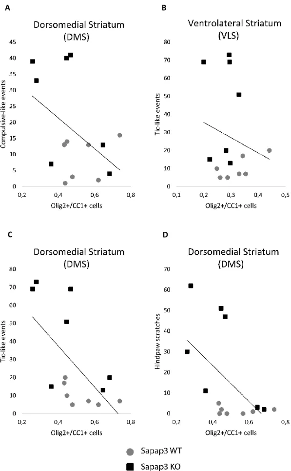
UNCTIONAL ANALYSISIn order to verify our hypothesis that compulsive-like behaviours are correlated with dysfunctions within associative circuits and their projections in the DMS, and tic-like behaviours within SMA-VLS circuitry, we tested a possible correlation between the behavioural quantifications and the density of Olig2+/CC1+ cells in these areas.
Contrary to what we expected, our results indicate that both the correlation between the compulsive-like events with the density of oligodendrocytes in the DMS, and the tic-like events with the density of oligodendrocytes in the VLS are not significant (value=0.3811, p-value=0.8986, respectively) (Figure 10A-B).
However, a surprising and noteworthy negative correlation was found between the frequency of myoclonic jerks and the density of Olig2+/CC1+ cells in the DMS (p-value=0.0231) (Figure
10C). This result demonstrates a clear indication that an increased number of tic-like events
relates to a decreased number of mature oligodendrocytes in this brain region. Additionally, a tendency towards an also negative correlation between the number of hind paw scratches and the density of oligodendrocytes was also denoted in the DMS (p-value=0.0585) (Figure 10D). All other tested correlations were non-significant (data not shown), indicating that there seems to be no apparent implication of myelin-related deficits in VLS nor lOFC. Additionally, myelination deficits that could be underlying compulsive-like behaviours do not appear to correlate with neither of the hypothesized areas.
Figure 8: Correlation between behavioural quantifications and the number of oligodendrocytes in DMS and VLS
We looked for a correlation between the frequency of compulsive-like events (grooming events) with a decreased number of oligodendrocytes in the DMS (A) and a correlation between the frequency of tic-like
A
B
47
behaviours (myoclonic jerks) with a decreased number of oligodendrocytes in the VLS (B). However, contrary to our hypothesis, these correlations were not verified. On the other hand, we detected a negative correlation between the number of tic-like events with the average density of mature oligodendrocytes (p-value=0.0231) (C), and a tendency towards an also negative correlation between the frequency of hind paw scratches and the average density of oligodendrocytes (p-value=0.0585) (D), both in DMS.
4.4 M
YELIN THICKNESSAiming for a more detailed characterization of possible myelin-related deficits in the Sapap3-KO mouse, we quantified myelin thickness by performing g-ratios (ratio between the diameter of the axon per se and the outer diameter of the myelinated axon) on electron microscopy images (N=3 per group) on our designated ROIs: DSM and VLS (Figure 11A-D).
Bearing in mind the first indication of a reduced number of oligodendrocytes in striatal regions, we would expect its translation into a reduction of myelin thickness. However, a decrease in the g-ratio (increased myelin thickness) was observed in DMS of the Sapap3-KO group (p-value=0.053) (Figure 11E). Considering the diversity of cells that can be found in striatal regions, we looked for a possible correlation between the g-ratio and the respective axonal diameter. This test would enable us to detect if there was a tendency that could be attributed to one specific type of striatal cells. However, no correlation was seen (data not shown), meaning that this increase in myelin thickness is consistent across the different striatal cells.
A difference in myelin thickness is a strong indication of myelination deficits on our mouse model, and thus, might be implicated on the connectivity deficits that have been shown to exist in the lOFC-DMS pathway.
In VLS, no difference in the g-ratios was detected (p-value=0.7616) (Figure 11F).
4.5 N
UMBER OF MYELINATED AXONSOur final approach on the characterization of myelination on the Sapap3-KO mouse was to seek for a reduced density of myelinated axons on the obtained TEM images, as a possible consequence of a reduced number of oligodendrocytes. No apparent difference in the average number of myelinated axons was detected in DMS (p-value=0.7000) (Figure 12A). However, our results demonstrate a slight, yet non-significant, tendency to an increase in the number of myelinated axons in VLS (p-value=0.2000) (Figure 12B).
Figure 9: Quantification of myelin thickness by g-ratio measurement
Figures A and B represent a diagram of the Bregma levels we aimed in order to incorporate the defined ROIs (DMS and VLS, respectively). Figures C and D correspond to the slices that match the target regions
49
(DMS and VLS, respectively) under the stereo microscope, with the regions where we removed the ROIs for analysis. Figures E and F represent a quantification of myelin thickness by the measurement of the g-ratio (N=3 per group). In the DMS (E) a significant increase in myelin thickness can be observed, while no difference is seen in VLS (F). Figure G corresponds to a TEM image in the DMS, with three visible myelinated axons. Figure H schematically demonstrates how a g-ratio measurement is performed, by diving the inner axonal diameter by the outer diameter of the myelinated axon. Bars represent average value and error bars show standard error. Mean and standard error were calculated from image analysis of 6 animals (N=3 per group), 150 g-ratios measured per animal. Represents n.s. = non-significant.
Figure 10: Quantification of the average density of myelinated axons
A tendency towards an increased number of myelinated axons can be seen in the DMS (A) and, more prominently, in VLS (B). Bars represent average value and error bars show standard error. Mean and standard error were calculated from image analysis of 6 animals (N=3 per group), 20 images per animal. Represents n.s. = non-significant.
4.6 P
HARMACOLOGICALT
REATMENTMotivated by the putatively reduced number of oligodendrocytes observed, we aimed for a behavioural rescue by the promotion of oligodendrocyte maturation. For that purpose, we used Clemastine, a drug that has been pointed out by its pro-remyelinating properties by inducing OPC differentiation. Animals were divided in two groups: clemastine-treated (CT) and vehicle treated (VT) (N=6 per group). Our administration protocol consisted on daily I.P. injections of 10mg/kg body weight for 12 days. To address behavioural changes, we quantified weight, compulsive-like and tic-like behaviours, and hind paw scratches on day 0 and on day 19 (seven days after treatment cessation).
0 5 10 15 20 25 Av era ge d en sity o f my elina ted ax o n s 0 5 10 15 20 25 Av era ge d en sity o f my elina ted ax o n s