Se x ho rm o ne m o dulatio n o f
se ro to nin-induce d co ro nary
vaso dilatio n in iso late d he art
1Departamento de Ciências Fisiológicas, Centro Biomédico,
Universidade Federal do Espírito Santo, Vitória, ES, Brasil
2Department of Pharmacology and Experimental Therapeutics,
Louisiana State University, Health Sciences Center, New O rleans, LA, USA M.R. Moysés1,
L.A. Barker2
and A.M. Cabral1
Abstract
The present study was designed to evaluate the differences in the coronary vasodilator actions of serotonin (5-HT) in isolated heart obtained from naive or castrated male and female rats that were treated with either estrogen or testosterone. Hearts from 12 groups of rats were used: male and female naive animals, castrated, castrated and treated with 17ß-estradiol (0.5 µg kg-1 day-1) for 7 or 30 days, and
castrated and treated with testosterone (0.5 mg kg-1 day-1) for 7 or 30
days. After treatment, the vascular reactivity of the coronary bed was evaluated. Baseline coronary perfusion pressure (CPP) was deter-mined and dose-response curves to 5-HT were generated. Baseline CPP differed between male (70 ± 6 mmHg, N = 10) and female (115 ± 6 mmHg, N = 12) naive rats. Maximal 5-HT-induced coronary vasodilation was higher (P<0.05) in naive female than in naive male rats. In both sexes, 5-HT produced endothelium-dependent coronary vasodilation. After castration, there was no significant difference in baseline CPP between hearts obtained from male and female rats (75 ± 7 mmHg, N = 8, and 83 ± 5 mmHg, N = 8, respectively). Castration reduced the 5-HT-induced maximal vasodilation in female and male rats (P<0.05). Estrogen treatment of castrated female rats restored (P<0.05) the vascular reactivity. In castrated male rats, 30 days of estrogen treatment increased (P<0.05) the responsiveness to 5-HT. The endothelium-dependent coronary vasodilator actions of 5-HT are greater in female rats and are modulated by estrogen. A knowledge of the mechanism of action of estrogen on coronary arteries could aid in the development of new therapeutic strategies and potentially de-crease the incidence of cardiovascular disease in both sexes. Co rre spo nde nce
M.R. Moysés
Programa de Pós-Graduação Departamento Ciências Fisiológicas CBM, UFES
Av. Marechal Campos, 1468 29050-090 Vitória, ES Brasil
Fax: + 55-27-335-7330
E-mail: mrmoyses@ npd.ufes.br
Received July 31, 2000 Accepted March 27, 2001
Ke y wo rds
·Coronary circulation
·Endothelial function
·Estradiol
·Serotonin
·Smooth muscle
Intro ductio n
Coronary artery disease is an important cause of morbidity and mortality. The dis-ease involves incrdis-eased vasoconstrictor re-sponses, enhanced interaction of circulating blood cells with blood vessel walls and pro-liferation and migration of vascular smooth
is known about the mechanisms underlying this phenomenon, epidemiological and ex-perimental evidence indicates that female sex hormones, in particular 17ß-estradiol, have protective effects against cardiovascu-lar diseases (5,6) because estrogen replace-ment therapy reduces the incidence of coro-nary artery disease (7,8).
A number of potential mechanisms for the protective effect of estrogens on coro-nary arteries have been proposed. Estrogen has a beneficial effect on plasma lipopro-teins, increasing HDL cholesterol and de-creasing LDL cholesterol (9). Although early studies suggested that 50 to 60% of the pro-tective effect of estrogens on coronary dis-ease was due to the favorable changes in plasma lipids, recent data suggest that this protection is closer to 25% (10,11). Estrogen also appears to inhibit cholesterol deposition on arterial walls (12). Moreover, estrogen has a number of other properties that may result in cardiovascular protection. For in-stance, a considerable body of data suggest that an important protective effect of estro-gen is to potentiate the actions of endotheli-um-derived relaxing factors (13-15). Additi-onally, estrogen can enhance the effects of calcium channel blocking agents (16). Stud-ies of the acute hemodynamic effects of estrogen have reported increases in cardiac output and peripheral blood flow in both humans and experimental animals (17). 17ß-Estradiol has been shown to induce relax-ation of precontracted coronary artery rings and to inhibit calcium influx in isolated car-diac myocytes (18). Only a few studies have addressed the actions of estrogen on the coronary vascular bed. Although there are clear indications that endothelial function is altered by estrogens, the effects of endoge-nous estrogens on the coronary vascular ac-tions of serotonin (5-HT) have not yet been elucidated for the entire coronary bed. The effects of 5-HT, an autacoid released during platelet aggregation, have been evaluated in some preparations of coronary arteries in
vitro and in vivo. 5-HT is a potent vasodila-tor of the coronary bed in isolated hearts and
in situ (19,20) and may have an important role in coronary tone in patients with healthy coronary arteries. 5-HT causes a dose-de-pendent increase in coronary blood flow, as well as an increase in coronary artery diam-eter that is mediated by endothelial relaxing factors (21). In contrast, in patients with coronary atherosclerotic lesions or impaired endothelium, 5-HT causes a dose-dependent vasoconstriction (20).
The present study was designed to evalu-ate the differences in vascular reactivity to 5-HT between males and females and to evalu-ate the effects of castration and treatment with sex hormones on the modulation of the vascular reactivity of the coronary vascular bed to 5-HT in isolated hearts.
Mate rial and Me tho ds
The investigation was conducted in com-pliance with the Guide for Biomedical Re-search, as stated by the Brazilian Societies of Experimental Biology, and with the guiding principles of other physiological societies for research involving animals. The experi-ments were performed on isolated perfused hearts from young (3 months old) male and female Wistar rats. Under ether anesthesia, half of the male and female rats used were castrated and left untreated for 30 days. Twelve groups of male and female animals were used in the present study: naive, cas-trated, castrated and treated with 17ß-estra-diol propionate (0.5 µg kg-1
day-1
; Sigma Chemical Co., St. Louis, MO, USA) for 7 or 30 days, and castrated and treated with tes-tosterone enanthate (0.5 mg kg-1
day-1
After the different treatment periods the rats were anesthetized with pentobarbital sodium (40 mg/kg, ip) and injected sc with heparin (100 units/kg). Fifteen minutes after heparin injection, the rats were killed and the hearts were excised immediately and per-fused at a constant flow. The studies on the responsiveness of the coronary vascular bed were performed on whole hearts using a Langendorff preparation for perfused iso-lated hearts. Briefly, using a Langendorff apparatus (Hugo Sachs Electronics, March-Hugstetten, Germany), the isolated hearts were perfused with modified Krebs solution containing 120 mM NaCl, 1.26 mM CaCl2.2 H2O, 5.4 mM KCl, 2.5 mM MgSO4.7 H2O, 2.0 mM NaH2PO4.H2O, 27.0 mM NaHCO3, 1.2 mM Na2SO4, 0.03 mM EDTA, and 11.0 mM glucose, equilibrated with 95% oxygen and 5% carbon dioxide at a controlled pres-sure of 100 mmHg to give a pH of 7.4, perfused at a rate of 10 ml/min with a peri-staltic pump (MS-Reglo 4 channels, Hugo Sachs Electronics), and kept at 36o
C. A fluid-filled balloon was introduced into the left ventricle through a steel cannula with a latex balloon and connected to a P23Db Statham Pressure Transducer for measuring the iso-volumetric cardiac force. The balloon was pressurized by a spindle syringe until it reached a preload of 10 mmHg.
Coronary perfusion pressure (CPP) was monitored with a P23Db Statham Trans-ducer connected to a sidearm of the aortic perfusion catheter. Once the preparation was stabilized, baseline CPP was measured after about one hour and dose-response curves to 5-HT (5-hydroxytryptamine creatine sulfate; Sigma) were constructed by single injec-tions administered at 10-min intervals. 5-HT (0.01 to 400 µg) was administered as a bolus injection in a volume of 0.1 ml saline. In the naive male and female groups, dose-response curves for 5-HT were constructed before and after endothelium denudation by infu-sion of sodium deoxycholate (deoxycholic acid, 2 mg/ml, for 10 min).
In all treatment groups, immediately prior to removing the hearts, blood samples were collected via puncture of the abdominal aorta to measure circulating levels of sex hor-mones. The blood samples were centrifuged immediately at 3000 rpm at 4o
C for 10 min to obtain plasma, which was kept at -20o
C for later measurement of testosterone and estra-diol concentration by radioimmunoassay (Diagnostic Products Corporation, Los An-geles, CA, USA).
At the end of each experiment the left and right ventricles were removed and wet weights were measured. In some experiments using naive male and female animals, after the completion of dose-response curves to 5-HT, the coronary vascular bed was perfused with 10% neutral buffered formalin at a flow rate of 8 ml/min for 1 h. After weighing, both ventricles were immersed in 10% neutral formalin. Serial tissue blocks were cut at approximately 3-mm intervals and perpen-dicular to the long axis of the heart. Histo-logical sections were stained with hema-toxylin/eosin.
Statistical analysis
Dose-response data were analyzed by nonlinear regression analysis for fit to the equation: E = Emax/(1 + (10X
/10B )n
), where E is the observed response, Emax is the maxi-mum response, X is the logarithm of the dose of HT, B is the logarithm of the dose of 5-HT producing one-half maximal response (log ED50), and n is a curve fitting parameter that defines the slope of the dose-responses curve. The Prism2 software (GraphPad Soft-ware, San Diego, CA, USA) was used.
sero-tonin-induced coronary vasodilation. The correlation between baseline CPP levels and ventricle weight was assessed by the Pearson partial correlation coefficient adjusted for the rat weight. We used the partial coeffi-cient of determination to measure the mar-ginal effects of baseline CPP levels and ven-tricle weight. The level of significance was set at 5%.
Re sults
Base line co ro nary pe rfusio n pre ssure
The data for baseline CPP in each treat-ment group are summarized in Table 1. Base-line CPP was significantly higher in female than in male naive rats. Castration had no effect on baseline CPP in hearts obtained from male rats. In female rats, castration caused a significant reduction (P<0.01) in baseline CPP relative to naive control fe-male rats.
In castrated female rats, the baseline CPP in hearts obtained after 7 or 30 days of estrogen was not significantly different from that of the naive female control group. Treat-ment of castrated female rats with testoster-one for 7 days resulted in a baseline CPP that was not significantly different from control, but treatment with testosterone for 30 days resulted in a significant (P<0.01) decrease in baseline CPP. In castrated male rats,
treat-ment with estrogen for 30, but not for 7, days resulted in a significant (P<0.01) increase in baseline CPP. Treatment with testosterone for 7 or 30 days did not significantly alter baseline CPP relative to control naive male rats.
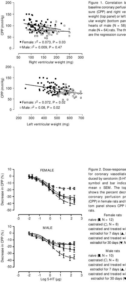
Figure 1 shows the results of the regres-sion analysis performed to determine if there was a correlation between right or left ven-tricular mass and baseline CPP that might explain the higher basal CPP observed in hearts obtained from female rats. No signifi-cant correlation was observed between base-line CPP and right ventricular weight. The slopes of the regression line of baseline CPP on left ventricular weight for both male and female rats were significantly different from zero. However, in each case the determina-tion coefficient (r2
) was small, 0.073 and 0.072, respectively, for the female and male groups. Thus, the difference in basal CPP (see Table 1, naive groups) in hearts from male and female rats was not explained by differences in ventricular weight. Addition-ally, it should be noted that there were no effects of castration and/or hormone replace-ment on ventricular weights.
Se ro to nin-induce d co ro nary re laxatio n
The dose-response curves for coronary vasodilation produced by 5-HT in isolated hearts obtained from male and female naive
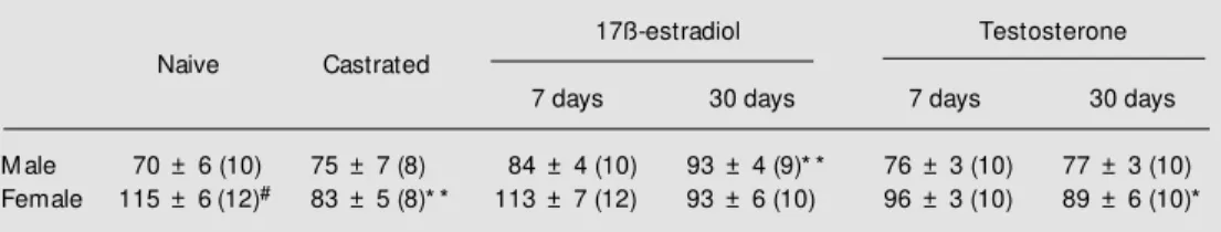
Table 1. Baseline values of coronary perfusion pressure (mmHg) in isolated hearts from the follow ing groups of male and female rats: naive, castrated, and castrated and long-term treated w ith 17ß-estradiol or testoster-one for 7 and 30 days.
17ß-estradiol Testosterone
Naive Castrated
7 days 30 days 7 days 30 days
M ale 70 ± 6 (10) 75 ± 7 (8) 84 ± 4 (10) 93 ± 4 (9)* * 76 ± 3 (10) 77 ± 3 (10)
Female 115 ± 6(12)# 83 ± 5 (8)* * 113 ± 7 (12) 93 ± 6 (10) 96 ± 3 (10) 89 ± 6 (10)*
Data are reported as means ± SEM . The number of experiments is given in parentheses.
* P<0.05 and * * P<0.01 compared to the respective naive control group (ANOVA follow ed by Dunnett’s test for comparison to control).
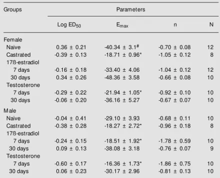
and castrated rats and rats castrated and treated with 17ß-estradiol for 7 or 30 days are shown in Figure 2. The dose-response parameters are shown in Table 2. The esti-mated maximal response for the 5-HT-in-duced decrease in CPP was greater in hearts obtained from naive female rats (Figure 2, upper panel) than that in hearts from naive male rats (Figure 2, lower panel; P<0.05). Although the Emax for 5-HT-induced coro-nary relaxation was higher in hearts from naive female rats, there were no significant differences in the potency of 5-HT between hearts obtained from naive male and female rats (Table 2).
In hearts obtained from castrated female rats, although the dose-response curve for 5-HT was slightly shifted to the left, the differ-ence was not significant. The maximum re-sponse was significantly (P<0.05) reduced compared to the curve for the naive female group (Figure 2, top panel). After 7 or 30 days of estrogen treatment of castrated fe-male rats, the Emax values and log ED50 val-ues for the actions of 5-HT were not signifi-cantly different (P>0.05) from those for the action of 5-HT on hearts obtained from na-ive female rats (Figure 2, top panel and Table 2).
In male rats, castration produced a sig-nificant decrease in the Emax for 5-HT when compared to the naive group (Figure 2, bot-tom panel). Seven days of treatment with estrogen did not alter the effects of castration on the responsiveness to 5-HT. However, after 30 days of estrogen treatment the re-sponsiveness to 5-HT was restored in cas-trated males (Figure 2, bottom panel). This treatment increased the Emax to a value simi-lar to that observed in naive female animals. None of the treatments produced significant changes in the potency of 5-HT (Table 2).
The effects of testosterone treatment for 7 or 30 days are shown in Figure 3. In both male and female rats, 30 but not 7 days of treatment with testosterone reversed the cas-tration-induced reduction in responsiveness
Figure 1. Correlation betw een baseline coronary perfusion pres-sure (CPP) and right ventricular w eight (top panel) or left ventric-ular w eight (bottom panel) from hearts of male (N = 58) and fe-male (N = 64) rats. The thick lines are the regression curves.
Figure 2. Dose-response curves for coronary vasodilation pro-duced by serotonin (5-HT). Each symbol and bar indicate the mean ± SEM . The top panel show s the percent decrease in coronary perfusion pressure (CPP) in female rats and the bot-tom panel show s CPP in male rats.
Female rats naive ( , N = 12) castrated ( , N = 8)
castrated and treated w ith 17ß-estradiol for 7 days ( , N = 12) castrated and treated w ith 17ß-estradiol for 30 days ( , N = 10)
M ale rats naive ( , N = 10) castrated ( , N = 8)
castrated and treated w ith 17ß-estradiol for 7 days ( , N = 10) castrated and treated w ith 17ß-estradiol for 30 days ( , N = 9)
C
P
P
(m
m
H
g)
200
300 150
100
50
0
200
150
100
50
0
C
P
P
(m
m
H
g)
50 100 150 200 250
Right ventricular w eight (mg)
700
200 300 400 500 600
Left ventricular w eight (mg) Female: r2 = 0.073, P = 0.03 M ale: r2 = 0.009, P = 0.47
Female: r2 = 0.072, P = 0.02 M ale: r2 = 0.08, P = 0.02
D
ec
re
as
e
in
C
P
P
(%
) 10
0
-20
-40 -50 -10
-30
2
-3 -2 -1 0 1 3
Log 5-HT (µg)
D
ec
re
as
e
in
C
P
P
(%
) 10
2 0
-20
-40 -50
-3 -2 -1 0 1
-10
-30
3 FEM ALE
(efficacy or maximal response) to 5-HT. No change was observed in the sensitivity (ED50) of the preparation to the different treatments (Table 2).
Endo the lium-de pe nde nt vaso dilatio n
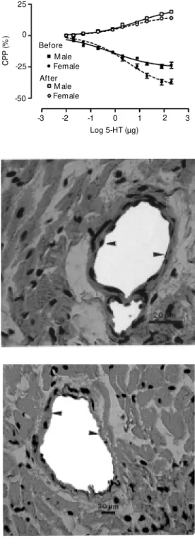
The results of studies in which the role of the endothelium in mediating 5-HT coro-nary vasodilation was examined are shown in Figures 4 and 5 and Table 3. The results presented in Figure 4 show that in the hearts obtained from naive male and female rats, 5-HT-induced vasodilation was abolished af-ter endothelial denudation. These experi-ments showed a significant difference in Emax (P<0.05) between the naive male and female groups before endothelial denuda-tion. The log ED50 values for 5-HT-induced vasodilation and vasoconstriction were not significantly different between female and male rats (Table 3). Figure 5 shows the histo-logical results of the removal of the coronary endothelium of female rats.
Ho rm o ne le ve ls
Castration resulted in significant reduc-tions in the levels of estradiol in female rats (from 44.0 ± 3.0 in naive rats to 29.6 ± 2.0 pg/ml in castrated rats). Similarly, castration resulted in a significant reduction in the levels of testosterone in male rats (from 0.67 ± 0.2 ng/ml in naive rats to undetectable values in castrated rats). In male rats, castra-tion did not alter the plasma levels of estra-diol. In castrated female rats, estradiol treat-ment for 7 or 30 days restored plasma levels to values not significantly different from those seen in naive female rats. In castrated male rats, treatment with testosterone for 7 days partially restored circulating levels of testosterone (0.37 ± 0.20 ng/ml) and treat-ment for 30 days resulted in plasma levels that were not significantly different from those of naive male rats. Interestingly, treat-ment with testosterone for 30 days of both
Table 2. Dose-response parameters for 5-HT-induced coronary vasodilation in hearts obtained from the follow ing groups of male and female rats: naive, castrated, and castrated and treated w ith either estrogen or testosterone.
Groups Parameters
Log ED50 Emax n N
Female
Naive 0.36 ± 0.21 -40.34 ± 3.1# -0.70 ± 0.08 12
Castrated -0.39 ± 0.13 -18.71 ± 0.96* -1.05 ± 0.12 8
17ß-estradiol
7 days -0.16 ± 0.18 -33.40 ± 4.06 -1.04 ± 0.12 12
30 days 0.34 ± 0.26 -48.36 ± 3.58 -0.66 ± 0.08 10
Testosterone
7 days -0.29 ± 0.22 -21.94 ± 1.05* -0.92 ± 0.10 10
30 days -0.06 ± 0.20 -36.16 ± 5.27 -0.67 ± 0.07 10
M ale
Naive -0.04 ± 0.41 -29.10 ± 3.93 -0.68 ± 0.11 10
Castrated -0.38 ± 0.28 -18.27 ± 2.72* -0.96 ± 0.18 8
17ß-estradiol
7 days -0.24 ± 0.15 -18.51 ± 1.92* -1.78 ± 0.59 10
30 days 0.09 ± 0.13 -38.08 ± 3.18 -0.76 ± 0.07 9
Testosterone
7 days -0.60 ± 0.17 -16.36 ± 1.73* -1.86 ± 0.75 10
30 days 0.06 ± 0.23 -30.17 ± 2.96 -0.81 ± 0.13 10
Emax is the maximum response, log ED50 is the logarithm of the dose of 5-HT (µg) producing one-half maximal response, and n is a curve-fitting parameter that defines the slope of the dose-response curve. Data are reported as means ± SEM .
* P<0.05 compared to the respective naive group (ANOVA). #P<0.05 compared to male naive controls (unpaired Student t-test).
D
ec
re
as
e
in
C
P
P
(%
)
10
2 0
-20
-40
-50
-3 -2 -1 0 1
-10
-30
3
D
ec
re
as
e
in
C
P
P
(%
)
10
0
-20
-40
-50 -10
-30
2
-3 -2 -1 0 1 3
Log 5-HT (µg) FEM ALE
M ALE Figure 3. Dose-response curves
for coronary vasodilation pro-duced by serotonin (5-HT). Each symbol and bar indicate the mean ± SEM . The top panel show s the percent decrease in coronary perfusion pressure (CPP) in female rats and the bot-tom panel show s CPP in male rats.
Female rats naive ( , N = 12) castrated ( , N = 8)
castrated and treated w ith tes-tosterone for 7 days ( , N = 10) castrated and treated w ith tes-tosterone for 30 days ( , N = 10)
M ale rats naive ( , N = 10) castrated ( , N = 8)
male and female rats was associated with an elevation in plasma 17ß-estradiol levels to 37.0 ± 3.0 pg/ml in females and 36.3 ± 4.0 pg/ml in males, values similar to those seen in naive female rats and castrated female rats treated with 17ß-estradiol.
D iscussio n
The present study was carried out on isolated hearts with intact whole coronary vascular beds in the absence of a pharmaco-logical elevation of vascular tone. Under these conditions, we found that basal CPP was significantly higher in hearts obtained from naive female rats than in hearts ob-tained from male rats. The influence of sex hormones in modulating this difference be-tween groups was demonstrated by castra-tion and subsequent hormone replacement. Castration of female rats reduced CPP to the same values as observed in naive male rats. In contrast, castration of male rats had no effect on basal CPP. Treatment of both cas-trated male and female rats with 17ß-estra-diol for 30 days increased basal CPP. In contrast, treatment of castrated male rats with testosterone had no effects on basal CPP. In castrated female rats, treatment with testosterone for 30 days resulted in an eleva-tion of basal CPP and also restored plasma 17ß-estradiol levels to those seen in naive female rats. Thus, the sex difference in basal CPP seems to be related to the presence of ovarian sex hormones, estrogen in particu-lar.
It may be argued that the augmented value of basal CPP in the female group was due to the smaller size of female hearts. However, the influence of left ventricular weight on basal CPP seems to be minor. The coefficients of determination were similar for both male (0.072) and female (0.073) hearts. Thus, we may conclude that ventric-ular mass is not responsible for the higher CPP in female rats.
The estrogen-related difference in basal
Figure 5. Histological sections perpendicular to the long axis of the heart. The tissues w ere stained w ith hematoxylin/eosin and serial blocks w ere cut at ap-proximately 3-mm intervals. Ar-row heads point to a myocardial vessel w ith the presence of typi-cal endothelial cells (top picture) and a denuded vessel follow ing treatment w ith deoxycholate (bottom picture).
Figure 4. Dose-response curves for coronary vasodilation pro-duced by serotonin (5-HT) in iso-lated hearts from naive male (N = 9) and female rats (N = 9) be-fore (closed symbols) and after (open symbols, N = 10) endothe-lial denudation by treatment w ith 2 mg/ml deoxycholic acid in the perfusate for 10 min. Each sym-bol and bar indicate the mean ± SEM .
C
P
P
(%
)
25
2 0
-50
-3 -2 -1 0 1
-25
3 Female
M ale Before
After
Female M ale
Log 5-HT (µg)
ap-pear to exert opposite effects. With respect to the high level of CPP observed in the female group, it is known that estrogen may have indirect effects on the cardiovascular system, e.g., through an interaction with the adrenergic nervous system (28). Many stud-ies have indicated a synergistic effect with sympathetic activity through inhibition of norepinephrine uptake (29), leading to an increase in vascular tonus. An increase in reactivity to norepinephrine has also been shown in oophorectomized (30) and prepu-beral rats (23) treated with estrogen. Like-wise, the renin-angiotensin system may also be responsible for the high CPP. This system has been associated with intramural renin-like enzyme and angiotensinogen levels in several organs, including the coronary circu-lation (31). Oelskers (32) demonstrated that 17ß-estradiol increased the synthesis of he-patic angiotensinogen, raising the content of angiotensin II and sodium, with a conse-quent increase in arterial pressure. Further experiments are necessary to elucidate the basis for the estrogen-dependent elevated CCP and whether this elevation has a role in the cardioprotective effect of estrogen ob-served in female rats.
5-HT acts on multiple receptors to pro-duce both vasodilator and vasoconstrictor
effects on coronary vascular smooth muscle (33). The relaxant effects mainly depend on the release of nitric oxide from the endothe-lium and the constrictor effects are due to direct actions on smooth muscle (34). The results of the present study confirm the en-dothelial dependence of 5-HT-induced cor-onary vasodilation. Our results are also con-sistent with the proposed role of estrogen in modulating endothelium-dependent vasodi-lation (13). The responsiveness to 5-HT, but not the potency of 5-HT, was higher in fe-male than in fe-male hearts. Thus, the influence of estrogen on 5-HT-induced coronary va-sodilation seems to involve an increase in the number of functional receptors since only the maximal response was altered. How-ever, this hypothesis needs to be further investigated. Following castration, the maxi-mal response produced by 5-HT was re-duced in both male and female rats and the sex difference in responsiveness was no longer seen. Estrogen treatment for either 7 or 30 days restored the responsiveness to 5-HT in the coronary beds of hearts from trated female rats. On the other hand, cas-trated male rats were relatively resistant to this action of 17ß-estradiol. In male rats, estrogen treatment for 30, but not 7, days restored the responsiveness to 5-HT to a level similar to that seen in hearts from naive female rats. These results demonstrate that 17ß-estradiol modulates the ability of 5-HT to produce an endothelium-dependent va-sodilation in the noncontracted coronary bed. In castrated female and male rats, testos-terone treatment for 7 days had no effect on the responsiveness to 5-HT. However, the responsiveness to 5-HT-induced coronary vasodilation in hearts from castrated male and female rats was restored after 30 days of treatment with testosterone. The increase in plasma 17ß-estradiol to levels similar to those seen in naive female rats following 30 days of treatment with testosterone in both male and female castrated rats suggests that the ability of testosterone to restore the
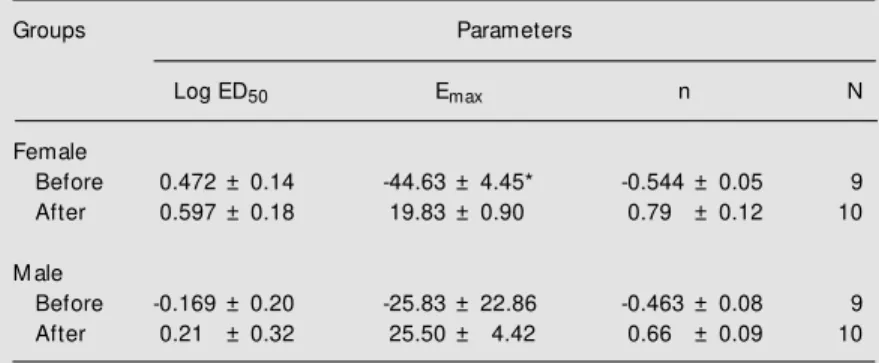
respon-Table 3. Dose-response parameters for 5-HT-induced coronary vasodilation in hearts obtained from naive male and female rats before and after endothelial denudation.
Groups Parameters
Log ED50 Emax n N
Female
Before 0.472 ± 0.14 -44.63 ± 4.45* -0.544 ± 0.05 9
After 0.597 ± 0.18 19.83 ± 0.90 0.79 ± 0.12 10
M ale
Before -0.169 ± 0.20 -25.83 ± 22.86 -0.463 ± 0.08 9
After 0.21 ± 0.32 25.50 ± 4.42 0.66 ± 0.09 10
Emax is the maximum response, log ED50 is the logarithm of the dose of 5-HT (µg) producing one-half maximal response, and n is a curve-fitting parameter that defines the slope of the dose-response curve. Data are reported as means ± SEM .
siveness to 5-HT might have been indirect and due to its conversion to 17ß-estradiol.
Taken together, our observations support the idea that estrogen modulates endotheli-um-dependent vasodilation. The ability of estrogen replacement to restore the endothe-lium-dependent vasodilator response in the intact coronary bed in castrated female rats is consistent with other studies that have shown this effect of estrogen replacement (13,15). Estrogen receptors are present in vascular endothelial cells and vascular smooth muscle cells (35,36) where they play a modulator role in the basal release of nitric oxide (NO) (37,38). Furthermore, 17ß-estra-diol has been recently reported to enhance expression of constitutive NO-synthase in cultured vascular endothelial cells and to induce both the neuronal and endothelial isoenzymes of NO-synthase in estrogen-treated animals (39,40). Corroborating these experimental studies, Rosselli et al. (26)
showed that serum nitrite and nitrate levels are higher in postmenopausal women sub-mitted to hormone replacement therapy, pro-viding indirect evidence of enhanced NO production induced by estrogen. Thus, es-trogen-induced enhancement of NO release may be an important underlying mechanism in the cardioprotective effect of estrogen.
Our results indicate that differences in coronary vascular reactivity to 5-HT in iso-lated hearts from male and female rats are probably modulated by 17ß-estradiol and may be associated with higher release of endothelium vasodilator autacoids. Knowl-edge of the mechanism of action of estrogen on coronary arteries could aid in the devel-opment of new therapeutic strategies in which the cardioprotective effects might be sepa-rated from other components of estrogen activity, potentially decreasing the incidence of cardiovascular disease in both sexes.
Re fe re nce s
1. Luscher TF & Vanhoutte PM (1990). The Endothelium: M odulator of Cardiovascu-lar Function. CRC Press, Boca Raton. 2. Crofton JT, Ratliff DL, Brooks DP & Share
L (1986). The metabolic clearance rate of and pressor responses to vasopressin in male and female rats. Endocrinology, 118: 1777-1781.
3. Freedman RR, Sabharw al SC & Desai N (1987). Sex differences in peripheral vas-cular adrenergic receptors. Circulation Research, 61: 581-585.
4. Zhang F, Ram JL, Standley PR & Sow ers JR (1994). 17ß-Estradiol attenuates volt-age-dependent Ca2+ currents in A7r5 vas-cular smooth muscle cell line. American Journal of Physiology, 266: C975-C980. 5. Kannel WB, Hjortland M C, M cNamara PM
& Gordon T (1976). M enopause and risk of cardiovascular disease: The Framing-ham Study. Annals of Internal M edicine, 85: 447-452.
6. Row land NE & Fregly M J (1992). Role of gonadal hormones in hypertension in the Dahl salt-sensitive rat. Clinical and Experi-mental Hypertension, 14: 367-375. 7. Stampfer M J, Colditz GA, Willett WC,
M anson JAE, Rosner B, Speizer FE &
Hennekens CH (1991). Postmenopausal estrogen therapy and cardiovascular dis-ease: Ten year follow -up from the Nurses’ Health Study. New England Journal of M edicine, 325: 756-762.
8. Grady D, Rubin SM & Petiti DB (1992). Hormone therapy to prevent disease and prolong life in postmenopausal w omen. Annals of Internal M edicine, 117: 1016-1037.
9. Walsh BW, Schiff I, Rosner B, Greenberg L, Ravnikar V & Sacks FM (1991). Effects of postmenopausal oestrogen replace-ment on the concentrations and metabo-lism of plasma lipoproteins. New England Journal of M edicine, 325: 1196-1204. 10. Bush TL, Barret-Connor E, Cow an LD,
Criqui M H, Wallace RB, Suchindran CM , Tyroler HA & Rifkind BM (1987). Cardio-vascular mortality and noncontraceptive use of estrogen in w omen: Results from the Lipid Research Clinics Program Fol-low Up Study. Circulation, 75: 1102-1109. 11. Barret-Connor E & Bush TL (1991). Estro-gen and coronary heart disease in w omen. Journal of the American M edical Associa-tion, 265: 1861-1867.
12. Adams M R, Clarkson TB, Kaplan JR &
Koritnik DR (1990). Ovarian secretions and atherosclerosis. In: Naftolin F, Gutmann JN, DeCherney AH & Sarrel PM (Editors), Ovarian Secretions and Cardiovascular and Neurological Function. Raven Press, New York, 151-159.
13. Gisclard V, M iller VM & Vanhoutte PM (1988). Effect of 17ß-estradiol on endo-thelium-dependent responses in the rab-bit. Journal of Pharmacology and Experi-mental Therapeutics, 244: 19-22. 14. Hayashi T, Fukuto JM , Ignarro LJ &
Chaudhuri G (1992). Basal release of nitric oxide from aortic rings is greater in fe-male rabbits than in fe-male rabbits: implica-tions for atherosclerosis. Proceedings of the National Academy of Sciences, USA, 89: 11259-11263.
15. Van-Buren GA, Yang D & Clark KE (1992). Estrogen-induced uterine vasodilatation is antagonized by L-nitroarginine methyl es-ter, an inhibitor of nitric oxide synthesis. American Journal of Obstetrics and Gyne-cology, 167: 828-833.
1264-1265.
17. M agness R & Rosenfeld CR (1989). Local and systemic estradiol-17ß: effects on uterine and systemic vasodilation. Ameri-can Journal of Physiology, 256: E536-E542.
18. Jiang C, Sarrel PM , Lindsay DC, Poole-Wilson PA & Collins P (1991). Endotheli-um-independent relaxation of rabbit coro-nary artery by 17ß-oestradiol in vitro. Brit-ish Journal of Pharmacology, 104: 1033-1037.
19. De Clerck F & Reneman RS (1985). Plate-let-derived serotonin and abnormal tissue perfusion. In: De Clerck F & Vanhoutte PM (Editors), Serotonin and the Cardio-vascular System. Raven Press, New York, 155-163.
20. Golino P, Piscione F, W illerson JT, Cappelli-Bigazzi M , Focaccio A, Villari B, Irdolfi C, Russolillo E, Condorelli M & Chiariello M (1991). Divergent effects of serotonin on coronary artery dimensions and blood flow in patients w ith coronary atherosclerosis and control patients. New England Journal of M edicine, 324: 641-648.
21. Riedel M , Oeltermann A, M ügge A, Creutzig A, Rafflenbeul W & Lichtlen P (1995). Vascular responses to 17ß-oestra-diol in postmenopausal w omen. European Journal of Clinical Investigation, 25: 44-47.
22. Farhat M Y, Abi-Younes S & Ramw ell PM (1996). Nongenomic effects of estrogen and the vessel w all. Biochemical Pharma-cology, 51: 571-576.
23. Cheng DY & Gruetter CA (1992). Chronic estrogen alters contractile responsive-ness to angiotensin II and norepinephrine in female rat aorta. European Journal of Pharmacology, 215: 171-176.
24. Gilligan DM , Badar DM , Panza JÁ, Quyyumi AA & Cannon RO (1994). Acute vascular effects of estrogen in postmeno-pausal w omen. Circulation, 90: 786-791. 25. Taddei S, Virdis A, Ghiadoni L, M attei P,
Sudano I, Bernini G, Pinto S & Salvetti A (1996). M enopause is associated w ith en-dothelial dysfunction in w omen. Hyper-tension, 28: 576-582.
26. Rosselli M , Imthurn B, Keller PJ, Jackson EK & Dubey RK (1995). Circulating nitric oxide (nitrite/nitrate) levels in postmeno-pausal w omen substituted w ith 17ß-es-tradiol and norethisterone acetate. Hyper-tension, 25: 848-853.
27. Rosano GM C, Sarrel PM , Poole-Wilson PA & Collins P (1993). Beneficial effect of oestrogen on exercise-induced myocar-dial ischaemia in w omen w ith coronary artery disease. Lancet, 342: 133-136. 28. Colucci WS, Gimbrone M A, M cLaughlin
M K, Halpern W & Alexander RW (1982). Increased vascular catecholamine sensi-tivity and alpha 1-adrenergic receptor af-finity in female and estrogen treated male rats. Circulation Research, 50: 805-811. 29. Hamlet M A, Rorie DK & Tyce GM (1980).
Effects of estradiol on release and dispo-sition on norepinephrine from nerve end-ings. American Journal of Physiology, 239: H450-H456.
30. Rorie DK & M uldoon SM (1979). In-creased reactivity of isolated rabbit saphe-nous vein after treatment w ith estrogen and progesterone. Blood Vessels, 16: 252-258.
31. Dzau VJ (1988). Circulating versus local renin-angiotensin system in cardiovascu-lar homeostasis. Circulation, 77 (Suppl I): 4-13.
32. Oelskers WKH (1996). Effects of estro-gens and progestoestro-gens on the
renin-aldo-sterone system and blood pressure. Ster-oids, 61: 166-171.
33. Houston DS & Vanhoutte PM (1988). Comparison of serotonergic receptor sub-types on the smooth muscle and endo-thelium of the canine coronary artery. Journal of Pharmacology and Experimen-tal Therapeutics, 244: 1-10.
34. Kelm M & Schrader J (1990). Control of coronary vascular tone by nitric oxide. Cir-culation Research, 66: 1561-1575. 35. Colburn P & Buonassisi V (1978).
Estro-gen binding sites in endothelial cell cul-tures. Science, 201: 817-819.
36. Horw itz KB & Horw itz CD (1982). Canine vascular tissue are targets for androgens, estrogens, progestins and glucocorti-coids. Journal of Clinical Investigation, 69: 750-758.
37. Losordo DW, Kearney M , Kim EA, Jeka-now ski J & Isner JM (1994). Variable ex-pression of the estrogen receptor in nor-mal and atherosclerotic coronary arteries in premenopausal w omen. Circulation, 89: 1501-1510.
38. Hayashi T, Yamada K, Esaki T, Kuzuya M , Satake S, Ishikaw a T, Hidaka H & Iguchi A (1995). Estrogen increases endothelial ni-tric oxide by a receptor mediated system. Biochemical and Biophysical Research Communications, 214: 847-855. 39. Schray-Utz B, Zeither AM & Busse R
(1993). Expression of constitutive NO syn-thase in cultured endothelial cells is en-hanced by 17ß-estradiol. Circulation, 88 (Suppl I): I-80 (Abstract).