Sao Paulo Med J. 2016; 134(5):457-60 457
CASE REPORTS
DOI: 10.1590/1516-3180.2016.0092220616A rare association of intussusception and celiac disease in a child
Associação rara entre intussuscepção e doença celíaca em criança
Vanessa Pacini Inaba Fernandes
I, Elizete Aparecida Lomazi
II, Maria Angela Bellomo-Brandão
IIUniversidade Estadual de Campinas (Unicamp), Campinas, SP, Brazil
ABSTRACT
CONTEXT: Intussusception is a common cause of acute intestinal obstruction in the pediatric population and it is normally idiopathic. Rare cases of chronic intussusception require investigation with greater attention.
CASE REPORT: We present a clinical case of a three-year-old boy with aqueous diarrhea, abdominal distension, vomiting and weight loss over a two-month period. During the investigation, abdominal ul-trasound showed imaging of intussusception. The intraoperative indings showed the intussusception had resolved spontaneously. In further investigation, it was found that the diarrhea was malabsorptive and, after the patient underwent upper gastrointestinal endoscopy, a diagnosis of celiac disease was made. After a gluten-free diet was introduced, the patient showed complete remission of symptoms and regained weight, and normal growth was reestablished.
CONCLUSION: If the clinical presentation of intussusception is unusual, etiological investigation should be undertaken. In this case report, celiac disease was the underlying cause.
RESUMO
CONTEXTO: Intussuscepção é uma causa frequente de obstrução intestinal aguda em pacientes pediátricos, e geralmente é idiopática. Raros casos de intussuscepção crônica devem ser investigados com maior atenção.
RELATO DE CASO: Apresentaremos um caso clínico de um menino de três anos com diarreia aquosa, distensão abdominal, vômito e perda de peso por dois meses, cuja investigação por ultrassonograia ab-dominal revelou imagem de intussuscepção. Os achados intraoperatórios mostraram que a intussuscep-ção havia se resolvido espontaneamente. Em investigaintussuscep-ção subsequente, foi identiicado que a diarreia era malabsortiva e, após a realização de endoscopia digestiva alta, foi feito diagnóstico de doença celíaca. Foi iniciada dieta com restrição de glúten e o paciente teve remissão completa dos sintomas, recuperou peso e o crescimento foi restabelecido.
CONCLUSÃO: Caso a apresentação clínica de intussuscepção não seja a habitual, deve-se prosseguir a investigação etiológica. No caso clínico apresentado, doença celíaca foi a causa subjacente.
IMD, MSc. Attending Physician, Pediatric Gastroenterology, Universidade Estadual de Campinas (Unicamp), Campinas, SP, Brazil.
IIMD, PhD. Professor of Pediatrics, Pediatric Gastroenterology, Universidade Estadual de Campinas (Unicamp), Campinas, SP, Brazil.
KEY WORDS: Child. Intussusception. Celiac disease. Diarrhea. Gluten-free diet.
PALAVRAS-CHAVE: Criança.
Intussuscepção. Doença celíaca. Diarreia.
CASE REPORTS | Fernandes VPI, Lomazi EA, Bellomo-Brandão MA
458 Sao Paulo Med J. 2016; 134(5):457-60 INTRODUCTION
Intussusception is a common cause of small bowel obstruc-tion in children under ive years of age, and its classical symp-toms include acute abdominal pain, red currant jelly stools and abdominal mass. However, these classical symptoms are not always present and it sometimes mimics acute viral gastroen-teritis, with diarrhea and vomiting. Invagination of the proxi-mal bowel into the distal bowel results in venous congestion and bowel wall edema.1 If not promptly diagnosed and treated,
this condition can lead to arterial obstruction, bowel necrosis and perforation. Almost 90% of the etiology of intussusception in children is ileocolic and idiopathic.1 he lead point of the
invagi-nation is lymphoid hyperplasia of the small bowel, but Meckel’s diverticulum, polyps and trauma can also lead to this problem. Intussusception is diagnosed by means of abdominal ultrasound and can be treated surgically or non-surgically.
his case report describes the presence of intussusception in a child with a chronic history of diarrhea, which is an unusual pre-sentation in children that should indicate the possibility of a dif-ferent diagnosis. Ater the case description, there is a brief discus-sion with a systematic search of data in the PubMed, Cochrane and LILACS databases (Table 1).
CASE REPORT
A three-year-old boy with a history of liquid diarrhea for a cou-ple of days, several times a day, with intermittent vomiting and without blood on stools or fever, was irst seen by a primary care physician. At physical examination, the child was found to be hydrated, without abdominal pain or distention, and was able to take oral luids and medication. He was diagnosed as hav-ing acute gastroenteritis and was advised to take oral hydrate solution and to return to the primary care center if the symp-toms worsened. Fourteen days later, the child returned and the mother complained that he had been sufering from intermit-tent diarrhea, vomiting and abdominal disintermit-tention. At examina-tion, he did not show any weight loss, was hydrated and had an overall good clinical condition. At this point, the diagnosis was persistent diarrhea and he was advised to start a lactose-free
diet, take albendazole for ive days and collect parasitological stool samples.
here was no short-term follow-up on this case by the primary care team because the family requested a second opinion from a diferent general pediatrician. he second physician made the interpretation that the child possibly had lactose intolerance and ordered a lactose intolerance test with blood analysis. When the patient returned to the original primary care center, two months ater the initial onset, he was still presenting aqueous diarrhea three to six times a day, abdominal distension, intermittent vomiting and now weight loss. Parasitological stool samples were negative and blood analysis suggested an iron deicient anemia. he lactose intolerance test was not available at that moment. he diagnosis at this point was chronic diarrhea with probable malabsorption. he child was referred to a tertiary gastroenterology center for fur-ther investigation, with upper gastrointestinal endoscopy, serum xylose tests and stool analysis for fat and pH.
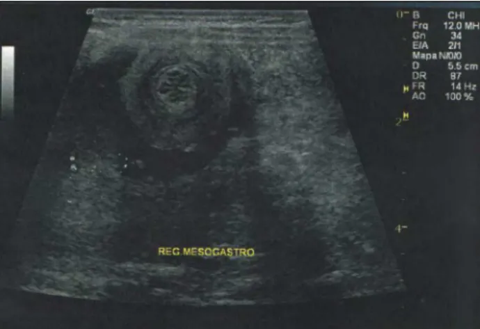
Abdominal ultrasound was performed and identiied an onionskin image of part of the small bowel (Figure 1), sug-gestive of intussusception. On the day of the ultrasound, the patient presented a distended but sot palpable abdomen with-out abdominal pain and was referred for hospital admission. Ater hospital admission, laparoscopic intervention demon-strated dilation of the proximal small bowel, without any mass or enlarged lymph nodes.
he intraoperative indings showed that the intussusception had resolved spontaneously. During the hospital stay, further investigations on chronic diarrhea and malabsorption were conducted, and the most relevant results were the following: low albumin of 3.2 g/dl; increased international normalized ratio (INR) of 1.95, which resolved ater vitamin K administration; low serum sodium (Na) of 133 mEq/l; high presence of fat in stools (12 g of fat in 161.7 g of stool ater 72 hours) and normal stool pH of 6.0; low serum xylose of 6.08 mg/dl, suggestive
Table 1. Database search results for the relationship between intussusception and celiac disease and child. Search performed on February 22, 2016
Database Search strategy (descriptors) Articles found Articles included
MEDLINE (PubMed)
(Intussusception) AND
(Celiac disease) AND (child) 24 4 (Intussusception) AND (child) 2633 1 Cochrane
Library
(Intussusception) AND
(Celiac disease) 0 0 LILACS (Intussusception) AND
onionskin-A rare association of intussusception and celiac disease in a child | CASE REPORTS
Sao Paulo Med J. 2016; 134(5):457-60 459 of malabsorption; negative HIV serological test; normal
immunoglobulin A (IgA) antibody level of 247 mg/dl; and normal sweat test (Na of 44.01 mEq/l and Cl of 35.6 mEq/l). All of these laboratory and reference values are shown in Table 2. To investigate malabsorption, upper gastrointestinal endoscopy was performed, and macroscopic evaluation showed a mosaic pattern of mucosa suggestive of celiac disease. On microscopic evaluation, inlammation of the duodenum mucosa was observed along with atrophic intestinal villi and crypt hypertrophy, compatible with Marsh III celiac disease, which was further conirmed by elevated tissue transglutaminase antibody concentration of 200 U/ml.
he child was then administered a gluten-free diet and his weight, stool consistency and stool frequency recovered com-pletely, thus resolving the abdominal distention. He was discharged ater a month in hospital, with body weight of 16.1 kg. One month later, at an outpatient visit, he was weighing 17.4 kg. One year ater admission, his weight had increased to 19.8 kg.
DISCUSSION
Idiopathic intussusception is a common cause of small bowel obstruction in children between the ages of 3 months and 5 years and has been recently correlated with underlying celiac disease.1-4
It is the most common cause of intestinal obstruction at pediat-ric ages and needs prompt intervention, which may be surgical or nonsurgical.
Non-operative methods for treating intussusception include barium enema and hydrostatic or pneumatic reduction. While non-operative methods are used more commonly than surgical inter-vention, the latter may still be needed when there are complications (peritonitis, perforation or profound shock) or when non-operative intervention is unsuccessful, or in situations of lack of a trained pro-fessional for non-invasive approaches.1
Most cases of intestinal intussusception at pediatric ages are idiopathic, and the lead point is generally lymphoid hyperplasia of the small bowel. he classical symptoms of intussusception include acute abdominal pain, red currant jelly stools and abdominal mass in a child with normal nutritional status. If the child fails to thrive and has chronic diarrhea, pain or blood stools, further investiga-tion should be conducted ater treating the intussuscepinvestiga-tion, as was done in our patient. he diferential diagnoses include Meckel’s diverticulum, polyps, trauma, celiac disease and enteropathy-asso-ciated T-cell lymphoma, among others.1
Celiac disease is a chronic inlammatory intestinal disease that occurs in about 1 to 2% of the general population. Untreated celiac disease has therefore been correlated with intussuscep-tion, and this association has been documented by a number of case reports.2-4 he proposed cause of intussusception in cases
of celiac disease is difuse inlammation and thickening of the intestinal wall, which lead to hyperperistalsis and increased dila-tion of the small bowel. his, in turn, could be the lead point for intussusception, which can develop singly or multiply, chroni-cally and with self-resolution or the need for surgical interven-tion. Less frequently, but with worse prognosis, this lead point could also be associated with a focal lead point in lymphomas.4
he majority of studies on this subject are case descriptions or series of cases, and there is a lack of case-control or random-ized clinical studies. In a recent retrospective study on patients undergoing imaging for abdominal pain, intussusception was found more frequently in patients with untreated celiac disease (less than nine months before celiac disease was diagnosed) than in the general population (1.2% versus 0.07% respectively).2
In a large case-control study evaluating the risk of late celiac dis-ease in patients with intussusception, no signiicant association was found.3 Nonetheless, using a prospective cohort approach,
a post-hoc analysis found that 12 out of 29,060 individuals with celiac disease were given a diagnosis of intussusception ater the onset of celiac disease, with a modest but signiicant increase risk of intussusception ater celiac disease had been diagnosed (odds ratio, OR = 1.95; 95% conidence interval, CI = 1.01–3.77; P = 0.046). In that study, patients with celiac disease without symptoms were not serially investigated with abdominal imag-ing, which may have reduced the absolute numbers of patients with subclinical intussusception who did not seek acute care and were unaccounted for.
Because intussusception in celiac patients may be chronic and painless, it is probably underdiagnosed. Patients with celiac disease may undergo imaging for chronic abdominal pain and intussusception may be identiied by chance, as it is usually not the irst diferential diagnosis in these cases. While in adults most cases of intussusception are investigated because it is always a pathological condition, in children this is only done when an
Patient’s values Reference values
Albumin (g/dl) 3.2 3.8-5.4 International normalized
ratio (INR) 1.95 1-1.2 Sodium (Na) (mEq/l) 133 136-145 Stool weight (g/day) 161.7 < 160 (< 1% of
body weight) Fat in stool (g/day) 12 < 2 pH in stool 6.0 5.0-7.0 Serum xylose (mg/dl) 6.08 > 20 IgA antibody (mg/dl) 247 33-308 Sweat test Na (mEq/l) 44.01 < 50 Sweat test Cl (mEq/l) 35.6 < 60 Tissue transglutaminase
antibody (U/ml) 200 < 10
Table 2. Patient’s laboratory result values and reference values for normality
CASE REPORTS | Fernandes VPI, Lomazi EA, Bellomo-Brandão MA
460 Sao Paulo Med J. 2016; 134(5):457-60
abnormal clinical presentation or physical examination ensues, since it is considered to be a common cause of idiopathic intes-tinal obstruction.
What makes our case description original is the chronic pre-sentation of intussusception with spontaneous resolution, with-out the need for surgical intervention. his inding has only infre-quently been described. he majority of case descriptions found in the literature describe an emergency situation of intussuscep-tion, in a child who failed to thrive.4,5 It will only be determined
whether spontaneous resolution in these situations is more fre-quent when routine abdominal ultrasound becomes part of the celiac disease protocol workup. A large serial study on pediat-ric populations with celiac disease with routine abdominal ultra-sound might help in resolving this question.
CONCLUSION
Intussusception is a common cause of idiopathic acute intes-tinal obstruction in children. If the clinical presentation is unusual, this may prompt further investigation. In this case, celiac disease was the pathological condition behind the ind-ings, with clinical presentation of chronic painless intussuscep-tion, without any need for surgical intervention and with signs and symptoms of malabsorption.
REFERENCES
1. Wong CW, Chan IH, Chung PH, et al. Childhood intussusception:
17-year experience at a tertiary referral centre in Hong Kong. Hong Kong
Med J. 2015;21(6):518-23.
2. Reilly NR, Aguilar KM, Green PH. Should intussusception in children
prompt screening for celiac disease? J Pediatr Gastroenterol Nutr.
2013;56(1):56-9.
3. Ludvigsson JF, Nordenskjöld A, Murray JA, Olén O. A large nationwide
population-based case-control study of the association between
intussusception and later celiac disease. BMC Gastroenterol.
2013;13:89.
4. Fishman DS, Chumpitazi BP, Ngo PD, Kim HB, Lightdale JR. Small bowel
intussusception in celiac disease: revisiting a classic association. J
Pediatr Gastroenterol Nutr. 2010;50(3):237.
5. Gheibi S. Association between Celiac Disease and Intussusceptions
in Children: Two Case Reports and Literature Review. Pediatr
Gastroenterol Hepatol Nutr. 2013;16(4):269-72.
Sources of funding: None
Conlicts of interest: None
Date of irst submission: May 9, 2016
Last received: June 13, 2016
Accepted: June 22, 2016
Address for correspondence:
Vanessa Pacini Inaba Fernandes
Universidade Estadual de Campinas
Rua Antonio Lapa, 1.032
São Paulo — Campinas (SP) — Brasil
CEP 13025-242
Tel/Fax. (+55 19) 3252-2903