The efect of sleep apnea severity on cardiac autonomic
activity during night time in obstructive sleep apnea patients
O efeito da gravidade da apneia do sono sobre atividade cardíaca autonômica
durante o período noturno em pacientes com apneia obstrutiva do sono
Gulay Ozkececi
I,
Sevinc Sarinc Ulasli
II, Onder Akci
I, Alaettin Avsar
III, Mehmet Unlu
IV, Ersel Onrat
IIIAfyon Kocatepe University, Afyonkarahisar, Turkey
ABSTRACT
CONTEXT AND OBJECTIVE: Impaired autonomic cardiac function is an important consequence of obstructive sleep apnea (OSA). This impairment is mainly due to intermittent hypoxia episodes following apneas. However, the impact of apnea severity on autonomic cardiac function remains unclear. The aim of this study was to evaluate the relationship between the severity of sleep apnea and heart rate turbulence (HRT) and heart rate variability (HRV) in OSA.
DESIGN AND SETTING: Observational cross-sectional study conducted in the Departments of Cardiology and Pulmonary Diseases, Afyon Kocatepe University, Turkey.
METHODS:106 patients with OSA and 27 healthy volunteers were enrolled. Based on apnea hypopnea index (AHI) values, obstructive sleep apnea severity was classiied as follows: mild OSA (AHI ≥ 5 and < 15), moderate OSA (AHI ≥ 15 and ≤ 30) and severe OSA (AHI > 30). HRV and HRT parameters were assessed via 24-hour digital Holter electrocardiogram recordings for all subjects.
RESULTS: HRV and HRT results were signiicantly lower among OSA patients than among control subjects (P < 0.05). However, there were no signiicant diferences in HRT and HRV between the three patient subgroups. Correlations did emerge between AHI and the NN-interval parameter RMSSD and between oxygen desaturation and turbulence slope (respectively: r = -0.22, P = 0.037; and r = -0.28, P = 0.025).
CONCLUSION: HRT and HRV results deteriorate in OSA. Correlations between apnea severity and these parameters seem to be present.
RESUMO
CONTEXTO E OBJETIVO: Função autonômica cardíaca prejudicada é consequência importante da apneia obstrutiva do sono (AOS). Este prejuízo deve-se principalmente a episódios de hipóxia intermitente após apneias. No entanto, o impacto da gravidade da apneia na função cardíaca autonômica permanece obs-curo. O objetivo deste estudo foi avaliar a relação entre gravidade da apneia do sono com turbulência da frequência cardíaca (TFC) e variabilidade da frequência cardíaca (VFC) em pacientes com AOS.
DESENHO E LOCAL: Estudo observacional transversal conduzido nos Departamentos de Cardiologia e Doenças Pulmonares, Afyon Kocatepe University, Turkey.
MÉTODOS: 106 pacientes com AOS e 27 voluntários saudáveis foram recrutados. Com base nos valores do índice de apneia-hypopneia (IAH), a gravidade da apneia obstrutiva do sono foi classiicada assim: AOS leve (IAH ≥ 5 e < 15), AOS moderada (IAH ≥ 15 e ≤ 30) e AOS grave (IAH > 30). Parâmetros da VFC e TFC foram avaliados por meio de gravações de eletrocardiograma digital Holter de 24 horas para todos os sujeitos.
RESULTADOS: Os resultados da VFC e TFC foram signiicativamente menores nos pacientes com OSA, em comparação com indivíduos controle (P < 0,05). No entanto, não houve diferenças signiicativas em VFC e TFC, entre os três subgrupos de pacientes. Correlações surgiram entre IAH e o parâmetro do intervalo-NN, RMSSD, e entre dessaturação de oxigênio e declive da turbulência (respectivamente; r = -0,22, P = 0,037; e r = -0,28, P = 0,025).
CONCLUSÃO:Os resultados da VFC e TFC deterioram em AOS. Parece haver relação entre a gravidade da apneia e tais parâmetros.
IMD. Assistant Professor, Department of
Cardiology, Faculty of Medicine, Afyon Kocatepe University, Afyonkarahisar, Turkey.
IIMD. Associate Professor, Department of
Pulmonary Diseases, Faculty of Medicine, Hacettepe University, Ankara, Turkey.
IIIMD. Professor, Department of Cardiology,
Faculty of Medicine, Afyon Kocatepe University, Afyonkarahisar, Turkey.
IVMD. Professor, Department of Pulmonary
Diseases, Faculty of Medicine, Afyon Kocatepe University, Afyonkarahisar, Turkey.
KEY WORDS: Heart rate. Arrhythmias, cardiac. Death, sudden, cardiac. Sleep apnea syndromes. Sleep apnea, obstructive.
INTRODUCTION
Obstructive sleep apnea (OSA) is characterized by recurrent total apnea or partial hypopnea due to narrowed upper air-ways during sleep.1 he current estimates for the prevalence of
moderate-to-severe sleep-disordered breathing (apnea-hypop-nea index, AHI, measured as events/hour, ≥ 15) are 10% among 30-49 year-old men; 17% among 50-70 year-old men; 3% among 30-49 year-old women; and 9% among 50-70 year-old women, according to the study by Peppard et al.2 he prevalence
of obstructive sleep apnea syndrome among Brazilian railroad workers has been found to be 35%.3
OSA is associated with cardiovascular diseases, includ-ing cardiac arrhythmias.4 myocardial infarction,5 chronic
heart failure6 and pulmonary hypertension.7 The
mecha-nism underlying cardiovascular diseases is complex and not fully understood in relation to OSA. Arrhythmias are con-sidered to arise from changes in the cardiac autonomic bal-ance due to hypoxia during apnea.8
here is a signiicant correlation between autonomic dys-function and cardiovascular mortality.9 Baroreceptor relex
sen-sitivity, heart rate turbulence (HRT) and heart rate variability (HRV) are parameters relecting cardiac autonomic functions. HRV and baroreceptor relex sensitivity are believed to evaluate diferent aspects of autonomic control. While a moderate rela-tionship has been determined between HRV and baroreceptor relex sensitivity, there is a strong relationship between HRT and baroreceptor relex sensitivity. herefore, it has been suggested that HRT should be used as an evaluation parameter instead of baroreceptor relex sensitivity.10,11
HRV12,13 and HRT14 provide important information
regard-ing autonomic cardiac function. HRV measures the oscillation in successive cardiac cycles as well as the oscillations between instantaneous heart rates.13 Previous studies have determined
that a reduction in HRV is predictive of increased cardiac mor-tality.15 HRT is deined as the sinus rhythm cycle-length
luctua-tion ater isolated premature ventricular beats.16 HRT evaluation
has been deemed appropriate for risk estimation ater acute myo-cardial infarction17 and as a prognostic evaluator for heart
fail-ure18 and other pathological conditions.16
Autonomic cardiac functions in OSA patients have been the focus of attention for researchers. Previous studies revealed dete-riorations in HRT and a relationship between HRT and the sever-ity of the apnea.19-21 However, the results relating to HRV in these
patients have been divergent; reductions and no changes in HRV have been reported in diferent studies.20-22
OBJECTIVE
herefore, the aims of the present study were to investigate whether both HRT and HRV parameters are impaired in patients
with OSA and to make correlations between these parameters and disease severity.
METHODS
Study design, ethics and setting
his study was designed as an observational cross-sectional study with a convenience sample. Interim power analyses were per-formed and detected a 90% statistical power.
he study was conducted in our Departments of Cardiology and Pulmonary Diseases between March 1, 2014, and November 1, 2014. he Ethics Committee of the Afyon Kocatepe University School of Medicine approved this study. All patients and control subjects gave their informed consent prior to inclusion.
Participants
he study sample included subjects consulted at a sleep labora-tory for clinically suspected OSA. hese were all the consecutive patients examined over an eight-month period between March 2014 and November 2014. Ater polysomnographic examination, sub-jects with ive or more obstructive apnea, mixed apnea or hypopnea events per hour were diagnosed as having OSA in accordance with the criteria of the American Academy of Sleep Medicine. Subjects presenting simple snoring, with apnea-hypopnea index less than 5 and no systemic diseases were enrolled in the control group. Subjects with diabetes mellitus, hypertension, ischemic heart disease, heart failure, renal disease, chronic inlammatory diseases, disorders of the autonomic nervous system or endocrine system, histories of drug use afecting the autonomic nervous system, pulmonary diseases or a smoking habit were excluded from the study.
Systolic and diastolic blood pressure of all patients and controls were measured from the right arm using a mercury manometer, ater they had rested in a seated position for at least ive minutes. Blood pressure was measured using a mechani-cal sphygmomanometer before polysomnographic evaluation. Patients whose oice systolic blood pressure was ≥ 140 mmHg and/or diastolic blood pressure was ≥ 90 mmHg were considered to present arterial hypertension. Body weight and height were also assessed. Body mass index (BMI) was calculated as weight in kilograms divided by the square of height in meters (kg/m2).
HRT and HRV analysis
the analyses. TO relects the initial heart rate acceleration ater a premature ventricular beat, whereas TS represents the subsequent heart rate deceleration following a premature ventricular beat. TO was determined as the diference between the mean of the two RR intervals ater premature ventricular beats and the mean of the two RR intervals before premature ventricular beats divided by the mean value of two RR intervals before premature ventricular beats. TS was deined as the maximum positive value of the slope of a regression line computed over any sequence of ive subsequent sinus RR intervals within the irst 15 sinus intervals ater premature ventricular beats. TO values determined for all suitable premature ventricular beats were averaged to obtain a inal TO value. TO < 0% and TS > 2.5 ms/RR are considered to be normal values.22
he HRV parameters used in the present study were selected in accordance with guidelines from the European Society of Cardiology and the North American Society of Pacing and Electrophysiology.13 he following time domain HRV
param-eters were computed: standard deviation of all normal-to-nor-mal (NN) intervals (SDNN), standard deviation of NN interval averages during all ive-minute segments across all recordings (SDANN), mean of the standard deviation of all NN intervals for all ive-minute segments across all recordings (SDNN index), integral of the density distribution of NN intervals divided by the maximum of the density distribution (HRV triangular index) and root mean square of the sum of squares for diferences between adjacent NN intervals (RMSSD).
Polysomnography
Full-night polysomnography (PSG) was recorded for all subjects using a digital PSG system (E series, Compumedics, Abbotsford, Victoria 3067, Australia) within the Department of Chest Diseases, Afyon Kocatepe University. Respiratory and physical changes were recorded during sleep. For all patients, electroen-cephalography, electrooculogram and submental electromyogra-phy data were obtained. Oronasal airlow was measured using a nasal cannula placed in the nose. horacoabdominal movements were evaluated by means of sensors placed on the chest and abdo-men to determine respiratory patterns. A pulse oximeter and electrocardiography electrodes were used to measure oxygen sat-uration and heart rate, respectively. Leg movements were exam-ined using electromyography sensors positioned on the anterior tibialis muscle. Sleep stages were scored in accordance with stan-dard criteria from the American Academia of Sleep Medicine.23
Apnea is deined as a drop in the peak signal excursion by ≥ 90% of the pre-event baseline for at least 10 seconds. Hypopnea is an event in which there is a ≥ 30% reduction in nasal pres-sure signal excursions from baseline and an associated ≥ 4% desaturation from the pre-event baseline for at least 10 seconds. Obstructive events were deined as ongoing thoracoabdominal
efort in cases of partial or complete airlow cessation. he apnea-hypopnea index (AHI) was the number of apnea or apnea-hypopnea events recorded during the study per hour of sleep. he severity of OSA was deined as mild (AHI ≥ 5 and < 15), moderate (AHI ≥ 15 and ≤ 30) or severe (AHI > 30). he oxygen desaturation index (ODI) was also calculated, as the total number of oxygen desatu-ration events divided by total dudesatu-ration of sleep.
All polysomnographic data was interpreted by a sleep special-ist who was blind to the subjects’ HRT and HRV analysis results.
Statistical analysis
Statistical analyses were performed using SPSS version 20.0 (IBM Co., Armonk, NY, USA). Data were expressed as the mean ± SD, medians (with interquartile range), or number (and %). Assumptions of normal distribution were tested using the Kolmogorov-Smirnov test. Categorical variables between groups were compared using a chi-square test. Diferences between the patient group and control subjects were tested using Student’s t test for parametric variables and the Mann-Whitney Utest for nonparametric variables. Comparisons between more than two groups (patients with mild, moderate or severe OSA and control subjects) in relation to variables with homogenous distributions were also analyzed using one-way analysis of variance (ANOVA) and Tukey’s test for post-hoc analyses. Comparisons between more than two groups in which the variables were not normally distributed were made using the Kruskal-Wallis test. In addition, analysis of covariance (ANCOVA) was performed to compare HRT and HRV parameters among the groups, controlling for BMI. Correlations between AHI, TO with AHI, and total oxy-gen desaturation were analyzed using Spearman’s rho correlation coeicients. An alpha level of P < 0.05 was accepted as signiicant.
RESULTS
Demographic characteristics
he study subjects consisted of 106 newly diagnosed patients with OSA (mean age 49.2 ± 11.2 years) and 27 healthy controls (mean age 47.2 ± 7.9 years). he patients included 30 with mild OSA (mean age 49.1 ± 10.4 years), 34 with moderate OSA (mean age 51.2 ± 6.8 years) and 42 with severe OSA (mean age 54.4 ± 8.3 years) OSA. here were no diferences between the groups in terms of age or gender. However, there was a signiicant difer-ence between the groups in terms of BMI. he subjects’ demo-graphics are depicted in Table 1 and Table 2.
HRV and HRT parameters
Table 1. Demographics, polysomnographic results and HRV and HRT parameters among control subjects and patients with OSA
Subjects without OSA n = 27
Patients with OSA
n = 106 P-value
Age (years) 47.2 ± 7.9 49.2 ± 11.2 0.1*
Gender (F/M) 11/16 40/66 0.84†
BMI (kg/m2) 27.7 ± 5.6 33.7 ± 6.3 0.001‡
AHI 1.75 ± 1.5 30 ± 23.5 < 0.001‡
Total duration of sleep 394 (304-396) 325 (188-519) 0.06*
Sleep eicacy 86.5 ± 6.8 77.9 ± 11.1 0.125‡
Total oxygen desaturation 3 (2-13) 101 (34-622) < 0.001*
ODI 2.2 (0.3-3.2) 27.1 (3.2-96.8) < 0.001*
TO (%) -3.19 ± 1.6 -2.2 ± 1.4 0.004‡
TS (ms/RR) 8.3 (3.8-8.6) 6.2 (2-17.3) 0.89*
SDNN (ms) 143 ± 37 90 ± 25 < 0.001‡
SDNNI (ms) 63 ± 27 45 ± 13 < 0.001‡
SDANN (ms) 116 (88-157) 74 (29-142) < 0.001*
RMSSD (ms) 42 ± 36 25 ± 10 0.003‡
TI (ms) 25 (23-41) 22 (8-50) < 0.001*
SBP (mmHg) 108.7 ± 33.2 123.1 ± 10.8 0.049‡
DBP (mmHg) 72.2 ± 14.6 73 ± 8.1 0.51‡
Mean HR (bpm) 70 (59-71) 74 (56-97) 0.14*
Data were shown as mean ± SD or median (with interquartile range). Categorical variables are deined as percentages. *Mann-Whitney Utest; †chi-square test; ‡Student’s ttest.
OSA =obstructive sleep apnea; F = female; M = male; BMI = body mass index; AHI = apnea-hypopnea index; ODI = oxygen desaturation index; TO = turbulence onset; TS = turbulence slope; SDNN = standard deviations of all (normal-to-normal) NN intervals; SDNNI = mean of the standard deviation of all NN intervals for all 5-min segments of the entire recording; SDANN = standard deviation of averages of NN intervals in all 5-min segments of the entire recording;TI = triangular index; RMSSD = the square root of the mean of the sum of the squares of diferences between adjacent NN intervals; SBP = systolic blood pressure; DBP = diastolic blood pressure; HR = heart rate; BPM = beats per minute.
Table 2. Polysomnographic results and HRT and HRV parameters compared between subgroups
Parameters Subjects without OSA n = 27
Patients with mild OSA n = 30
Patients with moderate OSA n = 34
Patients with severe OSA
n = 42 P-value
Age (years) 47.2 ± 7.9 49.1 ± 10.4 51.2 ± 6.8 54.4 ± 8.3 0.1
Gender (F/M) 11/16 17/13 9/25 14/28 0.07
BMI (kg/m2) 27.7 ± 5.7 31.8 ± 6.1* 32.4 ± 5.3* 37.1 ± 6.4* 0.002
AHI 1.75 ± 1.5 10.7 ± 2.6 21.7 ± 3.6 58.7 ± 19 < 0.001
Total duration of sleep 372 ± 45.3 322 ± 26.9 338.2 ± 77.7 305 ± 69.3 0.249
Sleep eicacy 88 (78-92) 81 (67-92) 80 (61-98) 75 (52-97) 0.269
Total oxygen desaturation 5.2 ± 5.2 92 ± 90 130 ± 72.5 330 ± 132 < 0.001
ODI 2.2 (0.3-3.2) 11.8 (7-67.7) 24.8 (6.4-51) 56.6 (3.2-97) < 0.001
TO (%) -3.19 ± 1.6 -2.06 ± 1.2* -2.06 ± 0.8* -1.9 ± 1.2* 0.005
TS (ms/RR) 8.3 (3.8-8.6) 5.5 (3.2-17.2) 8.3 (2.2-14) 6.4 (2-13.8) 0.98 SDNN (ms) 143 ± 37 96 ± 29* 95 ± 2* 79 ± 20* < 0.001
SDNNI (ms) 63 ± 27 45 ± 13 * 47 ± 13* 42 ± 14* < 0.001
SDANN (ms) 116 (88-157) 69 (29-142) * 77 (58-132)* 73 (38-140) * < 0.001
RMSSD (ms) 30 (20-81) 23 (10-56) * 23 (10-37) * 33 (8-60) 0.007
TI (ms) 25 (23-41) 20 (8-44)* 21.5 (13-50)* 22 (10-42) * < 0.001
SBP (mmHg) 108.7 ± 33.2 121.6 ± 11.9* 120 ± 7.7* 119 ± 6* 0.03
DBP (mmHg) 72.2 ± 14.6 69.7 ± 7.3 74.7 ± 8.9 75.1 ± 7.4 0.1
Mean HR (bpm) 66 ± 10 74 ± 9 71 ± 10 75 ± 11 0.10
Data are shown as mean ± SD or median (with interquartile range). Categorical variables are deined as percentages.
Comparisons between patients with mild, moderate or severe OSA and control subjects were analyzed using the Kruskal-Wallis test or ANOVA and Tukey’s test for post-hoc analyses and signiicant p values were presented bold
*P < 0.05: between subjects without OSA and patients with mild, moderate and severe OSA
not be calculated due to a lack of ventricular premature beats during Holter recording.
No signiicant diferences were found between OSA patients and control subjects in terms of total sleep time, sleep eicacy, age, TS, blood pressure, and heart rate. he TO, SDNN, SDNNI, SDANN, RMSSD, and TI values were signiicantly lower among OSA patients than among control subjects (Table 1). ANCOVA was performed to compare HRT and HRV parameters between the groups, controlling for BMI. However, signiicant diferences between groups continued ater eliminating the inluence of BMI.
he subjects were divided into four groups according to their AHI values. he subjects without OSA (n = 27) (AHI < 5), patients with mild OSA (AHI 5-15) (n = 30), patients with mod-erate OSA (n = 34) and patients with severe OSA (AHI > 30) (n = 42) were compared with regard to HRV, HRT and polysom-nographic parameters (Table 2). Post-hoc analyses were per-formed. No signiicant diferences between mild OSA patients, moderate OSA patients and severe OSA patients in terms of HRT and HRV parameters emerged. However, there were sig-niicant diferences between mild OSA patients and control sub-jects, between moderate OSA patients and control subjects and between severe OSA patients and control subjects in terms of TO, SDNN, SDNNI, SDANN, RMSSD and TI values (Table 2).
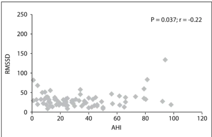
When the correlation between AHI (which relects disease severity) and TO was investigated, no statistically signiicant cor-relation was observed (P = 0.356; r = -0.11). here was no statis-tically signiicant correlation between total oxygen desaturation and TO (P = 0.631; r = -0.06). here was also no statistically sig-niicant correlation between total oxygen desaturation and TO (P = 0.631; r = -0.06). However, RMSSD and AHI were negatively correlated (P = 0.037; r = -0.22; Figure 1).
A signiicant correlation between total oxygen desaturation and TS (P = 0.025; r = -0.28) was observed (Figure 2). In addition, there were negative correlations between ODI and SDNN, SDANN
and TI (respectively: P = 0.002, r = -0.28; P < 0.001, r = -0.34; and P = 0.01, r = -0.23).
DISCUSSION
he indings from the present study revealed that the patients with OSA had lower values for HRV and HRT than those of the control subjects. Such reductions were observed even in patients with mild OSA. Although there was no signiicant diference between patient groups in terms of HRV and HRT parameters, signiicant correlations between TS and total oxygen desatura-tion, between RMSSD and AHI, and between ODI and SDNN, SDANN and TI were found.
Previous studies revealed that HRV and HRT worsen in OSA.19-21,24-27 he authors of these studies thought that this
deterio-ration might be due to hypoxia during periods of apnea. Ari et al.19
reported that HRT worsened in OSA while HRV did not change and there was a relationship between HRT and AHI. Aytemir et al.20 observed increased myocardial vulnerability and autonomic
nervous system imbalance in OSA cases. hey found that HRT, HRV and QT dynamicity parameters were signiicantly worse among patients with OSA. Furthermore, a correlation was revealed between AHI and HRT. However, they did not ind any relation-ship between AHI and HRV. hese authors20 also showed the
pres-ence of autonomic balance changes in favor of the sympathetic nervous system at night and hypoxemia relating to apnea. Erdem et al.25 investigated HRT parameters among patients with pure
OSA. hey found that the OSA group had a signiicantly higher mean TO than the control group, and that the AHI of the OSA group was positively correlated with TO. Our study supports the indings of previous studies. TO was signiicantly higher in patients with OSA than in the control group and, although there were no correlations between AHI and HRT parameters, there was a corre-lation between total oxygen desaturation and TS in our study.
Figure 1. Relationship between apnea hypopnea index (AHI) and root mean square of the successive diferences (RMSSD).
P = 0.037; r = -0.22
0 0 50 100 150 200 250
20 40 60 80 100 120
AHI
RMSSD
Figure 2. Relationship between total oxygen desaturation and turbulence slope (TS).
P = 0.025; r = -0.28
0 0 50 100 150 200 250
20 40 60 80 100 120
TS
Yang et al.21 also showed that HRV did not change while
HRT worsened during sleep among patients with OSA, and that alterations in nighttime HRT correlated with sleep-disordered breathing severity. his indicates the existence of abnormalities in autonomic cardiac activity within moderate-to-severe OSA, even in the absence of evident cardiac disease. In contrast, we did not determine any signiicant diferences between the patient groups (mild, moderate and severe OSA) in terms of HRT and HRT parameters. hese diferences in results between the pres-ent study and the study by Yang et al.21 might be attributable to
diferences in study group selection. Yang et al.21 grouped their
patients as mild and moderate to severe, while we grouped our patients as mild, moderate and severe. In addition, merely one overnight polysomnographic assessment might not provide enough information regarding the severity of OSA. he duration of OSA might also be a factor afecting the deterioration of auto-nomic cardiac function.
D’Addio et al.27 investigated the efects of
pathologi-cal respiratory patterns on HRT among patients with severe OSA. hey found that TS increased during apnea but observed decreases during normal intervals following an apnea event (here, OSA patients showed a higher sympathetic tone). his sup-ports the idea that autonomic cardiac function is impaired due to hypoxia during apnea.
Unlike Yang21 and Ari et al.,19 Lado et al.28 found that all HRV
parameters decreased during sleep among patients with mod-erate and severe OSA, compared with a normal, healthy group. In addition, among patients with severe low-frequency band and high-frequency band OSA indices, the total HRV power was lower during intervals labeled as apneas than those labeled as normal. In the present study, although the lowest values for HRV parameters in OSA patients compared with controls were determined; this decrease did not reach statistically signii-cance level among the patient groups. However, we found cor-relations between some HRV parameters and the values of ODI and AHI. herefore, these correlations constitute the strength of our research.
he relatively small sample size was a limitation for the present study. Moreover, we only evaluated night-time measurements on HRV and HRT. hus, further research evaluating both day and night time values is needed in order to contribute important and new data to the literature, given that these parameters are afected by the breathing pattern of OSA patients during sleep. he arrhythmia mechanism in OSA is closely related to apnea and hyperventilation events, which depend on the sympathovagal balance.29 here is dominance
of parasympathetic tone and decreased sympathetic activity during sleep among healthy subjects.30 However, this situation
can change in OSA. Parasympathetic activity, with slowing
of the heart rate, is dominant during periods of apnea. When subsequent apnea termination and temporary arousal from sleep occur, sympathetic activity predominates with resultant heart rate acceleration.31 herefore, increased sympathetic tone and
barorelex dysfunction can cause cardiac arrhythmia and sudden death. Sympathetic tone can be evaluated through provocative testing and spectral HRV analysis. he lack of power of the spectral analysis of heart rate and absence of provocative testing are other limitations of the present study. Lastly, HRT parameters could not be calculated in 21% of the patients, who did not present a premature ventricular beat.
CONCLUSION
HRT and HRV parameters were diferent in OSA patients than in control subjects. Correlations could be made between the sever-ity of apnea that could be determined through AHI, ODI and total oxygen desaturation, and such parameters seemed to be present. hese subjects should be followed up in order to moni-tor any further adverse outcomes.
REFERENCES
1. American Academy of Sleep Medicine. International Classiication of
Sleep Disorders (ICSD-3). 3rd ed. Darien: American Academy of Sleep
Medicine; 2014.
2. Peppard PE, Young T, Barnet JH, et al. Increased prevalence of
sleep-disordered breathing in adults. Am J Epidemiol. 2013;177(9):1006-14.
3. Koyama RG, Esteves AM, Oliveira e Silva L, et al. Prevalence of and
risk factors for obstructive sleep apnea syndrome in Brazilian railroad
workers. Sleep Med. 2012;13(8):1028-32.
4. Harbison J, O’Reilly P, McNicholas WT. Cardiac rhythm disturbances
in the obstructive sleep apnea syndrome: efects of nasal continuous
positive airway pressure therapy. Chest. 2000;118(3):591-5.
5. Hung J, Whitford EG, Parsons RW, Hillman DR. Association
of sleep apnoea with myocardial infarction in men. Lancet.
1990;336(8710):261-4.
6. Malone S, Liu PP, Holloway R, et al. Obstructive sleep apnoea in
patients with dilated cardiomyopathy: efects of continuous positive
airway pressure. Lancet. 1991;338(8781):1480-4.
7. Sajkov D, Cowie RJ, Thornton AT, Espinoza HA, McEvoy RD. Pulmonary
hypertension and hypoxemia in obstructive sleep apnea syndrome.
Am J Respir Crit Care Med. 1994;149(2 Pt 1):416-22.
8. Parish JM, Somers VK. Obstructive sleep apnea and cardiovascular
disease. Mayo Clin Proc. 2004;79(8):1036-46.
9. Lown B, Verrier RL. Neural activity and ventricular ibrillation. N Engl J
Med. 1976;294(21):1165-70.
10. La Rovere MT, Pinna GD, Hohnloser SH. Barorelex sensitivity and
heart rate variability in the identiication of patients at risk for
life-threatening arrhythmias: implications for clinical trials. Circulation.
11. La Rovere MT, Maestri R, Pinna GD, Sleight P, Febo O. Clinical and
haemodynamic correlates of heart rate turbulence as a non-invasive
index of barorelex sensitivity in chronic heart failure. Clin Sci (Lond).
2011;121(6):279-84.
12. Kleiger RE, Stein PK, Bigger JT Jr. Heart rate variability: measurement
and clinical utility. Ann Noninvasive Electrocardiol. 2005;10(1):88-101.
13. Heart rate variability: standards of measurement, physiological
interpretation and clinical use. Task Force of the European Society
of Cardiology and the North American Society of Pacing and
Electrophysiology. Circulation. 1996;93(5):1043-65.
14. Schmidt G, Malik M, Barthel P, et al. Heart-rate turbulence after
ventricular premature beats as a predictor of mortality after
myocardial infarction. Lancet. 1999;353(9162):1390-6.
15. Tsuji H, Larson MG, Venditti FJ Jr, et al. Impact of reduced heart rate
variability on risk for cardiac events. The Framingham Heart Study.
Circulation. 1996;94(11):2850-5.
16. Bauer A, Malik M, Schmidt G, et al. Heart rate turbulence: standards
of measurement, physiological interpretation, and clinical use:
International Society for Holter and Noninvasive Electrophysiology
Consensus. J Am Coll Cardiol. 2008;52(17):1353-65.
17. Bauer A, Malik M, Barthel P, et al. Turbulence dynamics: an
independent predictor of late mortality after acute myocardial
infarction. Int J Cardiol. 2006;107(1):42-7.
18. Koyama J, Watanabe J, Yamada A, et al. Evaluation of heart-rate
turbulence as a new prognostic marker in patients with chronic heart
failure. Circ J. 2002;66(10):902-7.
19. Arı H, Arı S, Yazıcı F, Koca V, Bozat T. [Cardiac autonomic function and
cardiac arrhythmias in patients with obstructive sleep apnea]. Turk
Kardiyol Dern Ars. 2011;39(4):292-9.
20. Aytemir K, Deniz A, Yavuz B, et al. Increased myocardial vulnerability
and autonomic nervous system imbalance in obstructive sleep
apnea syndrome. Respir Med. 2007;101(6):1277-82.
21. Yang A, Schäfer H, Manka R, et al. Inluence of obstructive sleep apnea
on heart rate turbulence. Basic Res Cardiol. 2005;100(5):439-45.
22. Cygankiewicz I. Heart rate turbulence. Prog Cardiovasc Dis.
2013;56(2):160-71.
23. Berry RB, Budhiraja R, Gottlieb DJ, et al. Rules for scoring respiratory
events in sleep: update of the 2007 AASM Manual for the Scoring
of Sleep and Associated Events. Deliberations of the Sleep Apnea
Deinitions Task Force of the American Academy of Sleep Medicine. J
Clin Sleep Med. 2012;8(5):597-619.
24. Song MK, Ha JH, Ryu SH, Yu J, Park DH. The efect of aging and severity
of sleep apnea on heart rate variability indices in obstructive sleep
apnea syndrome. Psychiatry Investig. 2012;9(1):65-72.
25. Erdem A, Dogan OT, Yontar OC, et al. The pure efects of obstructive
sleep apnea syndrome on cardiac autonomic functions: heart rate
turbulence analysis. Eur Rev Med Pharmacol Sci. 2013;17(20):2778-83.
26. Szymanowska K, Piatkowska A, Nowicka A, Cofta S, Wierzchowiecki
M. Heart rate turbulence in patients with obstructive sleep apnea
syndrome. Cardiol J. 2008;15(5):441-5.
27. D’Addio G, De Felice A, Insalaco G, Romano M, Cesarelli M. Efects
of pathological respiratory pattern on heart rate turbulence in sleep
apnea. Stud Health Technol Inform. 2014;205:506-10.
28. Lado MJ, Méndez AJ, Rodriguez-Liñares L, Otero A, Vila XA. Nocturnal
evolution of heart rate variability indices in sleep apnea. Combut Biol
Med. 2012;42(12):1179-85.
29. Schäfer H, Koehler U, Hasper I, Ewig S, Lüderitz B. [Sleep apnea and
cardiovascular risk]. Z Kardiol. 1995;84(11):871-84.
30. Verrier RL, Muller JE, Hobson JA. Sleep, dreams, and sudden death:
the case for sleep as an autonomic stress test for the heart. Cardiovasc
Res. 1996;31(2):181-211.
31. Adlakha A, Shepard JW Jr. Cardiac arrhythmias during normal
sleep and in obstructive sleep apnea syndrome. Sleep Med Rev.
1998;2(1):45-60.
This study was presented as a poster at the 31st National Cardiology
Congress of Turkey, in October 2015
Conlicts of interest: None
Sources of funding: None
Date of irst submission: December 17, 2015
Last received: January 5, 2016
Accepted: January 7, 2016
Address for correspondence:
Gulay Ozkececi
School of Medicine
Department of Cardiology
Afyon Kocatepe University
03000 Afyonkarahisar, Turkey
Fax: +90 272 2463300