Mimic the Crowded Cytoplasm
Ronald Hancock*, Yasmina Hadj-Sahraoui
Laval University Cancer Research Centre, Hoˆtel-Dieu Hospital, Que´bec, Que´bec, Canada
Abstract
Cell nuclei are commonly isolated and studied in media which include millimolar concentrations of cations, which conserve the nuclear volume by screening the negative charges on chromatin and maintaining its compaction. However, two factors question if these ionic conditions correctly reproduce the environment of nuclei in vivo: the small-scale motion and conformation of chromatin in vivo are not reproduced in isolated nuclei, and experiments and theory suggest that small ions in the cytoplasm are not free in the soluble phase but are predominantly bound to macromolecules. We studied the possible role in maintaining the structure and functions of nuclei in vivo of a further but frequently overlooked property of the cytoplasm, the crowding or osmotic effects caused by diffusible macromolecules whose concentration, measured in several studies, is in the range of 130 mg/ml. Nuclei which conserved their volume in the cell and their ultrastructure seen by electron microscopy were released from K562 cells in media containing the inert polymer 70 kDa Ficoll (50% w/v) or 70 kDa dextran (35% w/v) to replace the diffusible cytoplasmic molecules which were dispersed on cell lysis with digitonin, with 100mM K-Hepes buffer as the only source of ions. Immunofluorescence labelling and experiments using cells expressing GFP-fusion proteins showed that internal compartments (nucleoli, PML and coiled bodies, foci of RNA polymerase II) were conserved in these nuclei, and nascent RNA transcripts could be elongated. Our observations are consistent with the hypothesis that crowding by diffusible cytoplasmic macromolecules is a crucial but overlooked factor which supports the nucleus in vivo by equilibrating the opposing osmotic pressure cause by the high concentration of macromolecules in the nucleus, and suggest that crowded media provide more physiological conditions to study nuclear structure and functions. They may also help to resolve the long-standing paradox that the small-scale motion and irregular conformation of chromatin seen in vivo are not reproduced in nuclei isolated in conventional ionic media.
Citation:Hancock R, Hadj-Sahraoui Y (2009) Isolation of Cell Nuclei Using Inert Macromolecules to Mimic the Crowded Cytoplasm. PLoS ONE 4(10): e7560. doi:10.1371/journal.pone.0007560
Editor:Peter Sommer, Institut Pasteur Korea, Republic of Korea
ReceivedMay 12, 2009;AcceptedAugust 8, 2009;PublishedOctober 23, 2009
Copyright:ß2009 Hancock, Hadj-Sahraoui. This is an open-access article distributed under the terms of the Creative Commons Attribution License, which permits unrestricted use, distribution, and reproduction in any medium, provided the original author and source are credited.
Funding:The authors have no support or funding to report.
Competing Interests:The authors have declared that no competing interests exist.
* E-mail: [email protected]
Introduction
Cell nuclei are commonly isolated and studied in media which have evolved from empirical observations of the conditions which maintain their ultrastructural features and functions such as transcription [1–6], or are based on the estimated concentrations of ions in the cell [5]. In these media, the volume of nuclei is conserved by including cations which screen the negative charges on chromatin and thus maintain its compaction [7,8]; without these cations, nuclei swell and may lyse [1,6,8,9] due to the osmotic pressure exerted by their high internal concentration of macromolecules [10].
It is tacitly believed that the stability and functioning of nuclei in these media reflects the fact that they reproduce the ionic environment in the cytoplasm which surrounds the nucleus in vivo, but this idea is not consistent with all the available experimental evidence. Firstly, persuasive biophysical considerations suggest that the cytoplasm in vivo contains nofreeions ([11]. Both K+[12–14]
and Na+ ions [12,15] in the cell are believed to be bound
predominantly to macromolecules, and Mg2+ ions to ATP and
probably also to negatively-charged macromolecules [11] or are sequestered by mitochondria and the sarcoplasmic reticulum [16–18]. Secondly, chromatin isolated from nuclei prepared in conventional ionic media, or reconstituted in similar conditions, does not reproduce completely the properties of chromatin within the cell
which shows constrained motion on a 100 nm-scale [19] and an irregular conformation [20–22], whereas in isolated nuclei the small-scale motion of chromatin is supressed [19] and chromatin isolated from them has a regular and symmetrical conformation termed the 30 nm fibre [23]. The binding of significant amounts of Mg2+
and Ca2+ions to nuclei, and possibly to chromatin, during their isolation
in ionic media [24] may contribute to this discrepancy.
nuclei which conserve their structure, internal compartmentalisation, and transcriptional activity can be isolated in media which contain an inert, volume-occupying polymer to replace the cytoplasmic macro-molecules which are dispersed upon cell breakage, with no ionic components other than 100mM K-Hepes buffer. These observations
are consistent with the idea that crowding forces due to cytoplasmic macromolecules are an important but overlooked parameter which supports the structure and functions of the nucleus in vivo.
Results
Isolation and structure of nuclei in media containing an inert polymer
For isolating nuclei we used media which contained uncharged, inert polymers which have been used widely to study effects of crowding on macromolecular systems [37,49–53]. The polymers were Ficoll (average Mr 70 kDa), an approximately spherical
branched and crosslinked polymer of sucrose [54], and dextran (average Mr69.9 kDa), a flexible linear random coil polymer of
glucose [55]. Macromolecules of this size cannot traverse the nuclear envelope [47,48,56] and therefore exert a crowding or osmotic effect on the exterior of nuclei. The only ionic component of the polymer solutions was 100mM K-Hepes buffer, pH 7.4. To maintain the volume of isolated nuclei at the level in vivo, polymer solutions which were close to saturation were required and at their density and viscosity cells could not be washed by centrifugation; therefore, after pelleting growing cells the medium was carefully removed and the cells were resuspended directly in polymer solution supplemented with digitonin, which permeabilises the cytoplasmic membrane while conserving the nuclear membrane [47]. After mixing on a vortexer at maximum speed to disperse cytoplasmic material, nuclei were released and were centrifuged onto slides for optical microscopy or pelleted for electron microscopy.
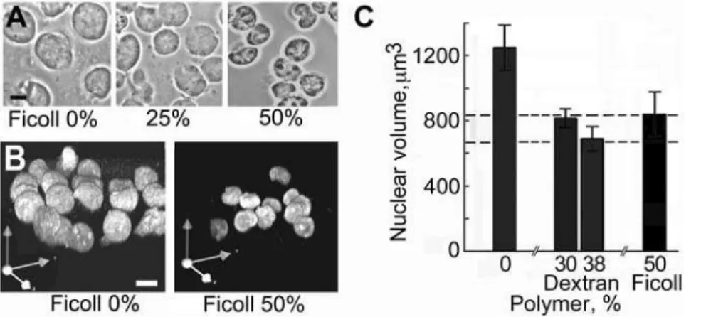
Nuclei which were structurally well conserved and free of cytoplasmic material as seen by phase-contrast and electron micro-scopy were released in solution containing 50% Ficoll or 35% dextran and 100mM K-Hepes buffer (Figure 1). In these nuclei the nucleoli,
which are prominent in phase-contrast images of intact K562 cells (Figure 1A), were conserved (Figure 1C, D) and were seen as more electron-dense structures by electron microscopy (Figure 1F, G). The rather homogeneous distribution of chromatin in isolated nuclei (Figure 1F, G) resembled that in the nuclei of intact cells processed in the same conditions (Figure 1B). The main features of the nuclear envelope and pores were conserved in isolated nuclei (Figure 1H). Nuclei of comparable purity and ultrastructure were released from Raji cells in the same conditions (unpublished results). We confirmed that polymers of this size are excluded from nuclei in the conditions used here, as observed in other studies [47,56]: nuclei isolated in dextran and deposited on slides were incubated in the same polymer solution containing fluorescein-labelled dextran of the same size, and
linescans of the fluorescence signal were recorded across approxi-mately mid-plane confocal sections of nuclei (Figure 1E). The mean fluorescence intensity in the nuclear interior was 3.260.4% (SEM, n = 10) of that in the external medium after incubation for 30 min, and 1266% after 1 h.
Nuclear volume responds to the concentration of polymer
The mean volume of nuclei in 50% Ficoll or 35% dextran was 8406220mm3 or 6806210mm3, respectively (SEM, n = 30),
compared with their volume of 7506210mm3 (SEM, n = 30) measured in living cells. The nuclear volume responded to the concentration of polymer in the medium (Figure 2), and these changes of volume were reversible (unpublished results).
The internal compartmentalisation of nuclei is conserved in polymer solutions
Within the nucleus, many macromolecules form regions of high local concentration which are termed compartments and include
Figure 1. Nuclei of K562 cells released in polymer solution containing 100mM K-Hepes.Cells suspended in 70 kDa Ficoll (50% w/ v) or 70 kDa dextran (35% w/v) containing digitonin were vortexed to disperse cytoplasmic material, and nuclei were centrifuged and fixed in the same polymer solution. (A) intact cells in growth medium, phase contrast. (B) intact cells, electron microscope section. (C, D) nuclei isolated in Ficoll or dextran respectively, phase contrast. (E) nuclei incubated as in D in dextran with addition of fluorescein-labelled dextran of the same size, fluorescence image of an approximately midplane 0.5mm confocal
section after 1 h; lower panel shows a scan of fluorescence intensity along the white line. (F, G) nuclei isolated in Ficoll and processed as in B, electron microscope images of sections. (H) nuclear pores (arrowheads) in nuclei isolated in Ficoll seen in transverse (left) and tangential (right) electron microscope sections. Bars 10mm (A, C, D, F); 1mm (B, G); 0.2mm (H).
nucleoli, PML and Cajal bodies, speckles, transcription facto-ries, and other types [57,58]. As an indication of the global conservation of nuclear architecture in nuclei isolated in polymer solution, we examined these compartments by immunofluores-cence labelling of their characteristic protein components. In nuclei isolated in 50% Ficoll (Figure 3) and in 35% dextran (unpublished results), all the compartments examined (nucleoli, PML and Cajal bodies, speckles, and transcription factories) were conserved. Mean numbers of 4.9 nucleoli, 4.7 PML bodies, and 1.5 Cajal bodies were counted in nuclei of cells isolated in 50% Ficoll (n = 100), whereas the corresponding numbers were 4.2, 4.5, and 1.4 respectively in nuclei isolated from the same cells in a conventional ionic medium [5].
Experiments with cells expressing a GFP-fusion of the protein PML isoform IV or of coilin (Figure 3B) confirmed that PML and Cajal bodies were conserved in nuclei isolated in polymer solution; these cells were washed with polymer solution and permeabilised and extracted in situ because after detachment by trypsin, digitonin lysis, and deposition on slides their nuclei were masked by cosedimenting extracellular matrix material. The mean number of PML bodies in nuclei isolated from cells expressing GFP-PML isoform IV was,20 (n = 100), close to the value of,22 reported in living cells [59], while in nuclei isolated from cells expressing GFP-coilin the mean number of Cajal bodies was,12, compared with 4–5 observed in living cells by Platini et al [60].
The conservation of Cajal bodies and nucleoli in nuclei in polymer solutions which contained only 100mM K-Hepes buffer (Figure 3) was unexpected, because after their isolation Cajal bodies require Mg2+
at 0.5 mM for their stability [61] and nucleoli require Mg2+, Ca2+ and/or polyamines at concentrations of
0.2–3 mM [62–64]. This discrepancy could be understood if these compartments were stabilised by free cations or polyamines in pools which were conserved within nuclei isolated using digitonin. We therefore examined the effect of replacing digitonin for cell lysis by Triton X-100, an ionic surfactant which removes the membrane components of the nuclear envelope [65] and therefore should allow any free ions to escape. Nucleoli, PML and Cajal bodies, and RNA pol II foci were still present in nuclei isolated by Triton lysis in 50% Ficoll (Figure 4) and in 35% dextran (unpublished results).
Nuclei isolated in polymer solution conserve transcriptional activity
In nuclei isolated in polymer solution, RNA pol II was still concentrated in foci (Figure 3) which could represent transcription factories where RNA pol II-mediated transcription occurs [58], and we tested if these nuclei could incorporate [a-32P]UTP into elongating RNA chains. For these experiments, the K+
concen-tration in the transcription medium had to be increased to ,1 mM to neutralise the nucleotide triphosphate RNA precur-sors. Nuclei isolated and incubated in 50% Ficoll incorporated [a-32P]UTP at an initial rate of ,0.25 pmoles UTP/min/106 nuclei (Figure 5A), and a similar rate was seen in nuclei isolated in
Figure 2. Influence of the concentration of polymer in the medium on nuclear volume.Nuclei were released as described in Figure 1, centrifuged onto slides, incubated for 30 min in a solution of the same polymer at the concentration shown, and fixed in the same solution. (A) nuclei in Ficoll, phase-contrast. (B) nuclei in Ficoll stained with YOYO, 3D volumes rendered from serial confocal sections to confirm that the changes of diameter in (A) represent changes of volume. (C) mean nuclear volume as a function of polymer concentration. Error bars show 95% confidence limits (n = 30) and horizontal dashed lines 95% confidence limits for the volume of nuclei in living cells labelled with DRAQ5. All differences of mean volume from that in 0% polymer were significant at p#0.01, calculated by the Welch t-test. Bars 10mm.
doi:10.1371/journal.pone.0007560.g002
Figure 3. Internal compartments in nuclei isolated in 50% Ficoll, 100mM K-Hepes.(A) distribution of nucleolin (nucleoli), PML (PML bodies), coilin (Cajal bodies), RNA polymerase II (pol II) (transcription factories) and SC35 (speckles). Nuclei of K562 cells were isolated in polymer solution, deposited on slides, and fixed as described in Figure 1. In the upper right panel, approximate nuclear outlines were traced from an overexposed image. (B) distribution of GFP-coilin or GFP-PML isoform IV fusion proteins in nuclei of HeLa or U2OS cells, respectively. Cells were washed with polymer solution and permeabi-lised and extracted in situ. Images are maximum intensity projections from deconvoluted confocal series. Bar 5mm.
35% dextran (unpublished results). For comparison, the rate of incoporation was ,0.5 pmoles UTP/min/106 in nuclei isolated and incubated in an ionic medium [5] with the same RNA precursors (unpublished results) and ,1 pmole/min/106 cells after permeabilisation by Triton X-100 [66,67]. Run-on tran-scription was essentially polymer-dependent; in the absence of polymer the rate was,5% of that when a polymer was present (Figure 5). Inhibition of RNA pol II by a-amanitin reduced the incorporation at 30 min to 63626% (SEM, n = 5) of the control value (Figure 5A).
Isolation of nuclei in medium containing a polymer which permeates the nuclear envelope
Handling and pipetting Ficoll and dextran solutions at concentrations which maintained the volume of isolated nuclei was inconvenient due to their viscosity and density, and we therefore explored if these could be replaced by a less viscous polymer; this implied, of course, that the polymer should be of smaller size and consequently would permeate through the nuclear envelope and exert crowding effects on macromolecules within the nucleus. Nuclei could be isolated using 12% 8 kDa poly(ethylene glycol) (PEG) in 100mM K-Hepes, pH 7.4; their appearance in
phase-contrast and electron microscope images was similar to that of nuclei isolated in a non-permeating polymer (Figure 6A, B) and the internal compartments examined were conserved (Figure 6D). Their volume was 7606180mm3 (SEM, n = 60) compared with
7506210mm3(SEM, n = 30) in living cells.
Nascent RNA transcripts were elongated in nuclei isolated in 12% PEG at an initial rate,two- fold greater than that in nuclei isolated in 50% Ficoll (Figure 5B). The nuclear volume varied over an,four-fold range with the concentration of PEG (Figure 6E), offering an interesting experimental approach to manipulate nuclear volume; at the highest concentration of PEG tested (40%) nuclei contracted and their periphery became crenellated (Figure 6C). Similar properties were seen in nuclei isolated in 10.5 kDa dextran (12%) (unpublished results).
Discussion
In this work, we show that cell nuclei which conserve their ultrastructure, internal compartmentalisation, and transcriptional activity can be stabilised by osmotic effects in solutions where an inert, volume-occupying polymer at an appropriate concentration replaces the diffusible cytoplasmic macromolecules which are dispersed upon cell lysis, and that they do not require the mM concentrations of cations which are commonly used in isolation media. The conservation of the structure of nuclei in these conditions is entirely consistent with the well-documented effects of macromolecular crowding in favouring the formation and stability of assemblies of macromolecules [35–37,49–53]. Further, the ability of a crowded environment to replace ions for conserving the structure and functions of nuclei is quite typical of a growing number of observations on macromolecular assemblies, although the nucleus is probably the largest assembly for which this phenomenon has been observed. Crowding agents can replace the requirement for a high ionic strength to polymerise the capsid protein CA of HIV-1 [53] and attenuate the need for Mg2+
ions for the association between 30S and 50S ribosomal subunits [49] and of K+ions for productive folding of the chaperonin GroEL
[51], and isolated chromosomes ofEscherichia coli[50,52] and of eukaryotic cells (unpublished results) are stabilised by inert polymers which reproduce their crowded environment in vivo, without the divalent cations and/or polyamines which were used earlier. The dependence of transcription on the addition of an inert polymer in nuclei isolated in 100mM K-Hepes observed here is consistent with a crowding-mediated assembly and functioning of the transcriptional machinery, a model which was first proposed to understand the requirement for polyvinylpyrrolidone or Ficoll to prepare nuclear extracts which initiate RNA pol II transcription series. Bar 5mm.
doi:10.1371/journal.pone.0007560.g004
Figure 5. Run-on incorporation of [a-32P]UTP into nascent RNA in nuclei isolated and incubated in polymer solution in 100mM K-Hepes.K+
was added to the medium to,1 mM to neutralise the nucleotide triphosphate RNA precursors. (A) nuclei isolated and incubated in 50% Ficoll; data points and error bars show means
6SEM (n = 5) for 30 min, and means and ranges (n = 2) for other points. (B) nuclei isolated and incubated in 12% 8 kDa PEG; data points and error bars show means6SEM (n = 9 for 30 min, n = 3 for other points). Full lines show incorporation in the complete system, dashed lines with addition ofa-amanitin (200mg/ml) to inhibit RNA pol II, and dotted
correctly [68]; transcription in permeabilised cells in an ionic buffer is also stimulated by PEG and BSA [69] or Ficoll [70], although the underlying mechanism was not discussed.
Does the conservation of the structure and functions of the nucleus by crowding or osmotic forces observed here play a significant role in the cell? A considerable body of experimental evidence supports this hypothesis. Firstly, crowding effects do indeed occur in the cytoplasm in vivo, as deduced from the anomalous diffusion of tracer macromolecules [30,38], and the volume of the nucleus in vivo is modulated by the concentration of macromolecules in the cytoplasm; nuclei of amphibian oocytes contract when BSA or polyvinylpyrrolidone are injected into the cytoplasm [71] and the effects of solute molecules on nuclear size in permeabilised macrophages led to the conclusion that ‘‘high concentrations of macromolecules such as those found inside cells can influence the size of the nucleus by directly affecting nuclear structure’’ [72]. Secondly, consideration of quantitative data shows that the polymer solutions which were found here to maintain the volume of isolated nuclei close to that in vivo reproduce quite closely the osmotic pressure which diffusible cytoplasmic macro-molecules are predicted to exert on the nucleus in vivo; 50% 70 kDa Ficoll and 35% 70 kDa dextran solutions have an osmotic pressure of,210 and,270 kPa, respectively [73,74] whereas a value of,200 kPa was measured directly in the cytoplasm of frog muscle [27]. This latter value is equivalent to that of a solution of BSA at,170 mg/ml [75], whereas the concentration of diffusible macromolecules is ,130 mg/ml in the fluid phase of the cytoplasm of rabbit muscle cells [26] and that of the fifteen most abundant diffusible proteins alone is 82 mg/ml in muscle fibres [29]. These high values are not unique to muscle cells, and in
fibroblasts the diffusion of Ficoll in the cytoplasm resembles that in a solution containing 124 mg/ml of ‘‘background’’ macromole-cules in the fluid phase together with 37 mg/ml of filamentous macromolecules [28].
Thirdly, these arguments have to be considered in the light of the doubts, cited in the Introduction, if media currently used to conserve the stability and functions of nuclei reproduce the ionic properties of the cytoplasm in vivo. Levels of cytoplasmic ions measured by biophysical methods cannot be interpreted as concentrations without knowing the extent to which they are bound to charged macromolecules, which itself can cause uncertainty in the calibration of analytical methods [12,16,18]. Indeed, a strong case has been made that the cytoplasm does not contain free small ions at significant levels and that ‘‘there is no cytoplasmic ‘bulk’ concentration of ions and metabolites (as is often assumed in biophysical models and in the design and interpretation of in vitro experiments)’’ [11]. However, nuclei isolated in conventional ionic media avidly bind extra Mg2+and
Ca2+
from buffers to levels many-fold the endogenous levels [24], an effect which could provide clues to resolving the paradox cited in the Introduction that chromatin from nuclei isolated in ionic media, or reconstituted in similar conditions, does not reproduce completely the properties of chromatin within the nucleus in vivo. That nuclei can be stabilised by osmotic effects has been observed, in fact, since sucrose was first used in isolation media [76] and inert macromolecules or BSA have been used previously in media for isolating nuclei [77–81]. However, in general Arthur Kornberg’s 7th commandment ‘‘Correct for extract dilution with molecular crowding’’ [82] has been overlooked when isolating and studying nuclei and their components. The specific features of the
Figure 6. Nuclei isolated in solution containing the permeant polymer 8 kDa PEG and 100mM K-Hepes.Cells were washed in 12% PEG, and nuclei were released by homogenisation in the the same solution containing digitonin and centrifuged onto slides or processed for electron microscopy. (A) nuclei in 12% PEG, phase-contrast. (B) sectioned nuclei in 12% PEG, electron microscopy. (C) nuclei deposited on slides and incubated for 30 min with PEG at the concentration shown in 100mM K-Hepes, pH 7.4, phase-contrast. (D) proteins characteristic of compartments in nuclei
isolated and incubated in 12% PEG, visualised by immunofluorescence as described in Figure 3. Images are maximum intensity projections from deconvoluted confocal series. (E) mean nuclear volume (n = 30) as a function of concentration of PEG; error bars are as defined in Figure 2, and dashed lines show 95% confidence limits for the volume of nuclei in living cells. All differences of nuclear volume from that in 0% PEG were significant at p#0.005. Bars 10mm.
monolayers with transfer to tetracycline-free medium for 4–6 h to allow GFP-coilin expression. Media contained 10% FCS (Invitro-gen), 100 units/ml penicillin, and 100mg/ml streptomycin. Cells were harvested when cultures reached ,half the maximum cell density.
Polymer solutions
Ficoll (average Mr 70 kDa, Fluka), dextran (average Mr
69.9 kDa, Sigma-Aldrich), and PEG (average Mr 8 kDa, Fluka
molecular biology grade) were dissolved in bidistilled H2O at 60–
70uC and the solutions were deionised by shaking with the mixed-bed resin AG 501-X8(D) (Bio-Rad) for 6–8 h. K-Hepes (Sigma-Aldrich) buffer, pH 7.4 was added to a final concentration of 100mM and solutions were passed through an 0.45mm filter and stored at220uC; their pH was verified before each experiment. Polymer Mrvalues are from the suppliers and concentrations are
expressed as w/v.
Isolation of nuclei
All steps were at room temperature (,20uC) and cells and nuclei were handled using cut-off micropipette tips. Using 50% Ficoll or 35% dextran solutions, cells could not be washed by centrifugation because of their high density and viscosity and they were centrifuged (60g, 6 min), the growth medium was removed carefully by micropipette and then with an absorbent paper wick, and the cells were resuspended at,1–56106cells/ml in polymer solution supplemented with 100mg/ml digitonin (Boehringer, high purity) diluted from a stock solution of 10 mg/ml in H2O
prepared fresh every month. Using PEG solution, cells were washed in 12% PEG by centrifugation (600g, 10 min) and resuspended in PEG solution supplemented with digitonin. In one series of experiments, digitonin was replaced by 0.5% v/v Triton X-100 (Sigma-Aldrich). After 10 min, cells in Ficoll or dextran were mixed on a vortexer at maximum speed for 2 to 3 min; cells in PEG could be broken in the same way or by,50 gentle hand strokes in a 2 ml glass homogeniser with a teflon piston (Wheaton), until .95% of the cells had released their nucleus as seen by phase-contrast microscopy. Two volumes of the same polymer solution without digitonin were added, and aliquots of the suspension containing ,106 nuclei were centrifuged onto polylysine-coated slides in a cytological centrifuge (Cytospin 2, Shandon) (4000g, 40 min in Ficoll or dextran or 1000g, 10 min in PEG). These methods could not be used for adherent HeLa and U2OS cells because after detachment by trypsin, digitonin lysis, and deposition on slides their nuclei were masked by cosediment-ing extracellular matrix material; instead, they were grown on cover glasses, growth medium was removed carefully with absorbent paper, and the cells were washed with polymer solution and then permeabilised and extracted in situ in 500ml of the same
solution containing 50mg/ml digitonin or 0.5% v/v Triton X-100
10 min. Five ml of a transcription mix prepared in the same polymer solution were added to give final concentrations of 100mM ATP, CTP, GTP, and UTP, 10 units/ml RNAguard (GE
Healthcare), and 20mCi [a-32P]UTP (6000 Ci/mmol; Perkin-Elmer), and samples were mixed and incubated at 35uC. To inhibit RNA pol II,a-amanitin (200mg/ml; Sigma-Aldrich) was
added to the suspension of nuclei 10 min before adding the transcription mix. Incorporation was arrested by adding 10% TCA to duplicate samples, and after 30 min on ice the precipitates were collected on GF/B filters, washed exhaustively with 10% TCA containing disodium pyrophosphate (10 g/l) followed by 70% ethanol, dried, and subjected to liquid scintillation counting.
Immunofluorescence
Nuclei deposited on slides were fixed by overlaying them for 10 min with the same polymer solution as the preceding step containing 2% paraformaldehyde, permeabilised with 0.5% Triton X-100 in PBS for 10 min, and incubated overnight at 4uC in casein blocking reagent (Roche). All subsequent incuba-tions were at room temperature in a humidified box, and washes and antibody dilutions were in PBS. Slides were incubated for 1 h with primary antibodies recognising nucleolin (Research Diagnos-tics, mAb; dilution 1/100 or 1/200), RNA pol II (M. Vincent, mAb CC-3; 1/100), SC35 (Sigma-Aldrich, mAb S-4045; 1/100), PML (Santa Cruz, mAb sc-966; 1/1000); coilin (E.K.L. Chan, rabbit R288; 1/100), or Daxx (Santa Cruz, rabbit sc-7152; 1/100). After washing, slides were incubated for 1 h with an appropriate secondary antibody conjugated with Alexa 488, 568, or 594 (Molecular Probes) diluted 1/100, and washed again. GFP fusion proteins were visualised after fixing nuclei as described above. DNA was stained with DAPI and preparations were mounted in SlowFade (Molecular Probes).
Imaging
Serial 0.5mm or 2mm confocal sections were acquired on an
MRC1024 (BioRad) with a 60x NA 1.4 oil-immersion objective or an Ultraview ERS (Perkin-Elmer) with a 60x NA 1.4 Plan Apo objective, and phase-contrast images on a Nikon E800 with a 100x NA 1.3 oil-immersion objective and a CoolSNAP camera (Roper Scientific). To measure nuclear volumes, living cells were labelled for 10 min with 1mM DRAQ5 (Biostatus, UK) and fixed cells with 10 nM YOYO-1 (Molecular Probes) or 0.5mg/ml propidium
iodide in PBS. Stacks of 0.5mm confocal sections were acquired from 30 randomly-selected nuclei and their areas in successive sections were summed. Differences between mean nuclear volumes were assessed by the Welch t-test to calculate 95% confidence intervals and p-values. Deconvolution, maximum intensity projections, area measurements, and linescans across 0.5mm confocal sections through nuclei incubated with
Imaging), 3D volumes were rendered using Volocity 4 (Improvi-sion), and Photoshop 7.0 was used to pseudocolour and merge images and for assembly of Figures. When shown, approximate nuclear outlines were traced from an overexposed image. For examination by electron microscopy, nuclei isolated in polymer solution were pelleted (5000g, 30 min in Ficoll and dextran or 700g, 10 min in PEG) and suspended in the same polymer solution; nuclei isolated in ‘‘physiological buffer’’ [5] were pelleted (200g,10 min) and resuspended in the same buffer. After fixation by addition of paraformaldehyde (2%) and glutaraldehyde (0.1%) for 1 h on ice, the nuclei were centrifuged and embedded in 1.5% low melting point agarose (Sigma-Aldrich) in PBS. Fragments were cut from the agarose, dehydrated, embedded in Poly/Bed 812 (Polysciences), and 90 to100 nm-thick sections were placed on nickel or copper grids and stained with uranyl acetate and lead
citrate. Digital images were acquired on a Jeol 1200 electron microscope.
Acknowledgments
We thank Edward K. L. Chan and Michel Vincent for gifts of antibodies, Jason Swedlow for HeLa cells expressing GFP-coilin, David Bazett-Jones for U2OS cells expressing GFP-PML isoform IV (originally from J. Taylor, Medical College of Wisconsin), and Anne Loranger, Carl St-Pierre, and Alain Goulet for expert support of microscopy. We are also gratefull to Steven Zimmerman for comments which improved the manuscript.
Author Contributions
Conceived and designed the experiments: RH. Performed the experiments: RH YHS. Analyzed the data: RH YHS. Wrote the paper: RH.
References
1. Anderson NG, Wilbur KM (1952) Studies on isolated cell components IV. The effect of various solutions on the isolated rat liver nucleus. J Gen Physiol 35: 781–796.
2. Allfrey VG, Littau VC, Mirsky AE (1964) Methods for the purification of thymus nuclei and their application to studies of nuclear protein synthesis. J Cell Biol 21: 213–231.
3. Busch H, Daskal Y (1977) Methods for isolation of nuclei and nucleoli. Meth Cell Biol 16: 1–44.
4. Olins AL, Olins DE (1972) Physical studies of isolated eucaryotic nuclei. J Cell Biol 53: 715–736.
5. Jackson DA, Yuan J, Cook PR (1988) A gentle method for preparing cyto- and nucleo-skeletons and associated chromatin. J Cell Sci 90: 365–378. 6. Hancock R (2004) A role for macromolecular crowding effects in the assembly
and function of compartments in the nucleus. J Struct Biol 146: 281–290. 7. Aaronson RP, Woo E (1981) Organization in the cell nucleus: divalent cations
modulate the distribution of condensed and diffuse chromatin. J Cell Biol 90: 181–186.
8. Engelhardt M (2004) Condensation of chromatin in situ by cation-dependent charge shielding and aggregation. Biochem Biophys Res Comm 324: 1210– 1214.
9. Dahl KN, Engler AJ, Pajerowski JD, Discher DE (2005) Power-Law Rheology of Isolated Nuclei with Deformation Mapping of Nuclear Substructures. Biophys J 89: 2855–2864.
10. Hancock R (2007) Packing of the polynucleosome chain in interphase chromosomes: Evidence for a contribution of crowding and entropic forces. Sem Cell Dev Biol 18: 668–675.
11. Spitzer J, Poolman B (2005) Electrochemical structure of the crowded cytoplasm. Trends Biochem Sci 30: 536–541.
12. Edelmann L (1989) The physical state of potassium in frog skeletal muscle studied by ion-sensitive microelectrodes and by electron microscopy. Scanning Microsc 3: 1219–1230.
13. Kellermayer M, Ludany A, Jobst K, Szucs G, Trombitas K, et al. (1986) Cocompartmentation of proteins and K+within the living cell. Proc Natl Acad Sci U S A 83: 1011–1015.
14. Ling GN (1990) The physical state of potassium ion in the living cell. Scanning Microsc 4: 737–750.
15. Negendank M, Shaller C (2005) Multiple fractions of sodium exchange in human lymphocytes. J Cell Physiol 104: 443–459.
16. Lu¨thi D, Gu¨nzel D, McGuigan JAS (1999) Mg-ATP binding: its modification by spermine, the relevance to cytosolic Mg2+
buffering, changes in the intracellular ionized Mg2+
concentration and the estimation of Mg2+
by 31P-NMR. Exp Physiol 84: 231–252.
17. Hudder A, Nathanson L, Deutscher MP (2003) Organization of Mammalian Cytoplasm. Mol Cell Biol 23: 9318–9326.
18. Gu¨nther T (2006) Concentration, compartmentation and metabolic function of intracellular free Mg2+
. Magnes Res 19: 225–236.
19. Davis SK, Bardeen CJ (2004) The Connection between Chromatin Motion on the 100 nm Length Scale and Core Histone Dynamics in Live XTC-2 Cells and Isolated Nuclei. Biophys J 86: 555–564.
20. Bednar J, Horowitz RA, Dubochet J, Woodcock CL (1995) Chromatin conformation and salt-induced compaction: three-dimensional structural information from cryoelectron microscopy. J Cell Biol 131: 1365–1376. 21. van Holde K, Zlatanova J (2007) Chromatin fiber structure: Where is the
problem now? Sem Cell Dev Biol 18: 651–658.
22. Dekker J (2008) Mapping in Vivo Chromatin Interactions in Yeast Suggests an Extended Chromatin Fiber with Regional Variation in Compaction. J Biol Chem 283: 34532–34540.
23. Robinson PJJ, Fairall L, Huynh Van AT, Rhodes D (2006) EM measurements define the dimensions of the ‘‘30-nm’’ chromatin fiber: Evidence for a compact, interdigitated structure. Proc Natl Acad Sci U S A 103: 6506–6511.
24. Naora H, Naora H, Mirsky AE, Allfrey VG (1961) Magnesium and Calcium in Isolated Cell Nuclei. J Gen Physiol 44: 713–742.
25. Fulton AB (1982) How crowded is the cytoplasm? Cell 30: 345–347. 26. Arrio-Dupont M, Cribier S, Foucault G, Devaux PF, d’Albis A (1996) Diffusion
of Fluorescently Labeled Macromolecules in Cultured Muscle Cells. Biophys J 70: 2327–2332.
27. Maughan DW, Godt RE (2001) Protein osmotic pressure and the state of water in frog myoplasm. Biophys J 80: 435–442.
28. Hou L, Lanni F, Luby-Phelps K (1990) Tracer diffusion in F-actin and Ficoll mixtures. Towards a model for cytoplasm. Biophys J 58: 31–43.
29. Maughan DW, Henkin JA, Vigoreaux JO (2005) Concentrations of Glycolytic Enzymes and Other Cytosolic Proteins in the Diffusible Fraction of a Vertebrate Muscle Proteome. .
30. Weiss M, Elsner M, Kartberg F, Nilsson T (2004) Anomalous Subdiffusion Is a Measure for Cytoplasmic Crowding in Living Cells. Biophys J 87: 3518–3524. 31. Pielak GJ (2005) A model of intracellular organization. Proc Natl Acad Sci U S A
102: 5901–5902.
32. Sear RP (2005) The cytoplasm of living cells: a functional mixture of thousands of components. J Phys: Condens Matter 17: S3587–S3595.
33. Luby-Phelps K, Taylor DL, Lanni F (1986) Probing the structure of cytoplasm. J Cell Biol 102: 2015–2022.
34. Rivas G, Ferrone F, Herzfeld J (2004) Life in a crowded world. EMBO Rep 5: 23–27. Mol Cell Proteomics 4: 1541–1549.
35. Ellis RJ (2001) Macromolecular crowding: obvious but underappreciated. Trends Biochem Sci 26: 597–604.
36. Chebotareva NA, Kurganov BI, Livanova NB (2004) Biochemical effects of molecular crowding. Biochemistry (Mosc) 69: 1239–1251.
37. Zhou HX, Rivas G, Minton AP (2008) Macromolecular crowding and confinement: biochemical, biophysical, and potential physiological consequenc-es. Annu Rev Biophys 37: 375–397.
38. Guigas G, Kalla C, Weiss M (2007) Probing the Nanoscale Viscoelasticity of Intracellular Fluids in Living Cells. Biophys J 93: 316–323.
39. Cuneo P, Magri E, Verzola A, Grazi E (1992) ‘Macromolecular crowding’ is a primary factor in the organization of the cytoskeleton. Biochem J 281: 507–512. 40. Herzfeld J (2004) Crowding-induced organization in cells: spontaneous alignment and sorting of filaments with physiological control points. J Mol Recognit 17: 376–381.
41. Birbeck MSC, Reid E (1956) Development of an improved medium for the isolation of liver mitochondria. J Biophys Biochem Cytol 2: 609–624. 42. Wicker U, Bu¨cheler K, Gellerich FN, Wagner M, Kapischke M, et al. (1993)
Effect of macromolecules on the structure of the mitochondrial inter-membrane space and the regulation of hexokinase. Biochim Biophys Acta 1142: 228–239. 43. Laterveer FD, Gellerich FN, Nicolay K (2005) Macromolecules Increase the Channeling of ADP from Externally Associated Hexokinase to the Matrix of Mitochondria. Eur J Biochem 232: 569–577.
44. Antonenkov VD, Sormunen RT, Hiltunen JK (2004) The behavior of peroxisomes in vitro: mammalian peroxisomes are osmotically sensitive particles. Am J Physiol Cell Physiol 287: C1623–C1635.
45. Parsegian VA, Rand RP, Rau DC (1995) Macromolecules and water: probing with osmotic stress. Meth Enzymol 259: 43–94.
46. Singh-Zocchi M, Andreasen A, Zocchi G (1999) Osmotic pressure contribution of albumin to colloidal interactions. Proc Natl Acad Sci U S A 96: 6711–6715. 47. Cassany A, Gerace L (2009) Reconstitution of nuclear import in permeabilized
cells. Methods Mol Biol 464: 181–205.
48. Hicks GR, Raikhel NV (1995) Protein import into the nucleus: an integrated view. Annu Rev Cell Dev Biol 11: 155–188.
49. Zimmerman SB, Trach SO (1988) Effects of macromolecular crowding on the association ofE. coliribosomal particles. Nucleic Acids Res 16: 6309–6326. 50. Cunha S, Woldringh CL, Odijk T (2001) Polymer-mediated compaction and
59. Dellaire G, Ching RW, Dehghani H, Ren Y, Bazett-Jones DP (2006) The number of PML nuclear bodies increases in early S phase by a fission mechanism. J Cell Sci 119: 1026–1033.
60. Platani M, Goldberg I, Swedlow JR, Lamond AI (2000) In Vivo Analysis of Cajal Body Movement and Joining in Live Human Cells. J Cell Biol 151: 1561– 1574.
61. Lam YW, Lyon CE, Lamond AI (2002) Large-Scale Isolation of Cajal Bodies from HeLa Cells. Mol Cell Biol 13: 2461–2473.
62. Muramatsu M, Smetana K, Busch H (1963) Quantitative aspects of isolation of nucleoli of the Walker carcinosarcoma and liver of the rat. Cancer Res 25: 693– 697.
63. Vandelaer M, Thiry M, Goessens G (1996) Isolation of Nucleoli from ELT Cells: A Quick New Method That Preserves Morphological Integrity and High Transcriptional Activity. Exp Cell Res 228: 125–131.
64. Andersen JS, Lyon CE, Fox AH, Leung AKL, Lam YW, et al. (2002) Directed Proteomic Analysis of the Human Nucleolus. Curr Biol 12: 1–11.
65. Aaronson RP, Blobel G (1974) On the attachment of the nuclear pore complex. J Cell Biol 62: 746–754.
66. Wansink DG, Schul W, van der Kraan I, van Steensel B, van Driel R, et al. (1993) Fluorescent Labeling of Nascent RNA Reveals Transcription by RNA Polymerase II in Domains Scattered Throughout the Nucleus. J Cell Biol 122: 283–293.
Hydrodynamic resistance of concentration polarization boundary layers in ultrafiltration. J Membr Sci 22: 117–135.
75. Vilker VL, Colton CK, Smith KA (1981) The osmotic pressure of concentrated protein solutions: Effect of concentration and pH in saline solutions of bovine serum albumin. J Coll Interface Sci 78: 548–566.
76. Hogeboom GH, Schneider WC, Palade GE (1948) Cytochemical Studies of Mammalian Tissues. I. Isolation of Intact Mitochondria from Rat Liver; Some Biochemical Properties of Mitochondria and Submicroscopic Particulate Material. J Biol Chem 172: 619–635.
77. Callan HG (1949) Some physical properties of the nuclear membrane. Exp Cell Res suppl. 1: 48.
78. Battin WT (1959) The osmotic properties of nuclei isolated from amphibian oocytes. Exp Cell Res 17: 59–75.
79. Macgregor HC (1962) The behavior of isolated nuclei. Exp Cell Res 26: 520– 525.
80. Takahashi Y, Asao T (1974) Study of the nuclei isolated from newt embryos by the use of Ficoll. Dev Growth Differ 16: 281–294.
81. Walmsley ME, Davies HG (1975) Ultrastructural and biochemical observations on interphase nuclei isolated from chicken erythrocytes. J Cell Sci 17: 113–139. 82. Kornberg A (2000) Ten Commandments: Lessons from the Enzymology of
![Figure 5. Run-on incorporation of [ a - 32 P]UTP into nascent RNA in nuclei isolated and incubated in polymer solution in 100 mM K-Hepes](https://thumb-eu.123doks.com/thumbv2/123dok_br/18308380.348476/4.918.115.828.93.246/figure-incorporation-nascent-nuclei-isolated-incubated-polymer-solution.webp)
