CASE REPORT
Journal of Evolution of Medical and Dental Sciences/ Volume 2/ Issue 49/ December 09, 2013 Page 9528
MALIGNANT SMALL ROUND CELL TUMOUR (PNET) OF PALATE: A RARE
CYTOLOGICAL DIAGNOSIS
Sarita Nibhoria1, Kanwardeep Kaur Tiwana2, Varinder Nibhoria3, Navtej Singh Bamra4
HOW TO CITE THIS ARTICLE:
Sarita Nibhoria, Kanwardeep Kaur Tiwana, Varinder Nibhoria, Navtej Singh Bamra. Malignant small round cell tumour PNET of palate: a rare cytological diagnosis . Journal of Evolution of Medical and Dental Sciences 2013; Vol2, Issue 49, December 09; Page: 9528-9531.
ABSTRACT: Primitive neuroectodermal tumour is a malignant small round cell tumour affecting mainly the older children and adults. Palate is a very rare site for this tumour. We here report a case of 3 year male child who presented with a firm nodule of 2 x 1 cm size in the palate. Fine needle aspiration of the lump was performed and a cytodiagnosis of malignant small round cell tumour of palate was suggested. Subsequent histopathological examination supported it while Immunohistochemistry i.e. CD 99 positivity confirmed the diagnosis of PNET.
KEY WORDS: Malignant small round cell tumour, Primitive neuroectodermal tumour (PNET), Fine needle aspiration cytology.
INTRODUCTION: Primitive neuroectodermal tumour is a malignant small round cell malignancy of presumably primitive, neuroectodermal tissue or pluripotential, migratory neural crest cells arising from the soft tissue or bone, predominantly in older children and adults.1 The cell of origin of this tumour is uncertain. The overall incidence rate of PNET is approximately 0.62 per million population in the U.S.A.2 Palate is a very rare site for this tumour. The vast majority (> 90%) of tumours and tumour like lesions in the oral cavity are benign.3,6 Cancer of the oral cavity is extremely rare in children and adolescents. According to the SEER data, the age adjusted incidence of oral cancers for this population was 0.24 per million. 7, 8
Here we present a cytodiagnosis of malignant small round cell tumour of palate which was confirmed on IHC as PNET.
CASE REPORT: A 3 year old male child presented to us with a firm nodule in palate of about 2 x 1 cm size. Cervical lymphadenopathy was absent. Hematological findings were normal except for the slightly raised total leucocyte count. The Peripheral blood film showed mild neutrophilia and no immature cells were found.
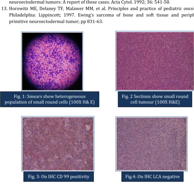
Fine needle aspiration was performed. The cytologic findings revealed cell rich smears comprising of dyscohesive sheets of monomorphic population of small and large round cells with scant pale-blue cytoplasm. The cells showed a high nucleo-cytoplasmic ratio. A cytodiagnosis of malignant small round cell tumour was suggested (Fig.1).
CASE REPORT
Journal of Evolution of Medical and Dental Sciences/ Volume 2/ Issue 49/ December 09, 2013 Page 9529 On Immunohistochemistry cells were positive for CD-99 and non-immunoreactive for LCA (Leucocyte common antigen) (Fig 3).
DISCUSSION: Malignant small round cell tumours are characterized by small, round, relatively undifferentiated cells. They generally include Ewing’s sarcoma, peripheral neuroectodermal tumour, rhabdomyosarcoma, synovial sarcoma, non-(odgkin’s lymphoma, retinoblastoma, neuroblastoma, hepatoblastoma and nephroblastoma. Differential diagnosis of small round cell tumours is particularly difficult due to their undifferentiated or primitive character.
Fine needle aspiration cytology is successful diagnostic tool when used by a skilled cytopathologist for documenting primary and recurrent MSRCT’s in pediatric patients.9 The exact categorization is not possible at the light microscopic level in the case of poorly differentiated tumours which poses a considerable challenge to the diagnostic skill of the cytopathologist.10,11
Primitive neuroectodermal tumour (PNET) is a malignant small round cell tumour arising from the soft tissue or bone, predominantly affecting the older children and adults. The term PNET includes malignant small round cell tumours of the thoracopulmonary region Askin’s tumour ,
Extra skeletal Ewing’s sarcoma, peripheral neuroblastoma, and peripheral epithelioma.12 The cell of origin of this tumour is uncertain. Originally, it was thought that EWS / PNET might arise from the neuroectoderm, but recent data have suggested that this tumour is more likely to originate from primitive stem cells and that the degree of malignancy appears to depend upon the stage of stem cell arrest during differentiation.13 The overall incidence rate of PNET is approximately 0.62 per million population in the U.S.A. Palate is a very rare site for this tumour. Cancer of the oral cavity is extremely rare in children and adolescents with age adjusted incidence for this population was 0.24 per million.
The present case highlights the palate as a rare primary site for malignant small round cell tumour (PNET) as well as importance of Fine needle aspiration cytology as an important diagnostic tool which equals to that of cumbersome histopathological examination.
REFERENCES:
1. Rajawanshi A, Srinivas R, Upasna G. Malignant small round cell tumors. J Cytol. 2009 Jan- Mar; 26 (1): 1 -10.
2. Smoll NR, Drummond KJ. The incidence of medulloblastomas and primitive neuroectodermal tumours in adults and children. J. Clin. Neurosci. 2012 ; 19 ( 11) : 1541 -4.
3. Das S, Das AK: A review of pediatric oral biopsies from a surgical pathology service in a dental school. Pediatr Dent. 1993 May-June; 15(3): 208-11.
4. Ulmansky M, Lustman J, Balkin N. Tumors and tumor like lesions of the oral cavity and related structures in Israeli children. Int J Oral Maxillofac Surg. 1999; 28 (4): 291-4.
5. Trobs RB, Mader E, Friedrich T, et al. Oral Tumors and tumor like lesions in infants and children. Pediatr Surg Int. 2003; 19 (9-10) : 639-45.
6. Tanaka N, Murata A, Yamaguchi A, et al. Clinical features and management of oral and maxillofacial tumors in children. Oral Surg Oral Med Oral Pathol Oral Radiol Endod. 1999; 88(1): 11-5.
CASE REPORT
Journal of Evolution of Medical and Dental Sciences/ Volume 2/ Issue 49/ December 09, 2013 Page 9530 8. Berstein L, Gurney JG. Carcinomas and other malignant epithelial neoplasms. In: Ries LA, Smith
MA, Gurney JG, et al; eds. Cancer incidence and survival among children and adolescents : United States SEER Program 1975-1995. Bathesda, Md: National Cancer Institute, SEER Program, 1999; NIH Pub. No. 99-4649; Chapter 11, pp 139-148.
9. Cohen MC, Pollano D, Tomarchio SA, Drut R. Cytologic characteristics of peripheral neuroectodermal tumors in fine needle aspiration smear: A retrospective study of three pediatric cases. Diagn Cytopathol 1997; 16: 513-7.
10.Akhtar M, Ali MA, Sabbah R, et al. Fine needle aspiration biopsy diagnosis of round cell malignant tumors of childhood: A combined light and electron microscopic approach. Cancer. 1985; 55: 1805-17.
11.Das DK, Bhambhani S, Chachra KL, et al. Small round cell tumors of the abdomen and thorax: Role of fine needle aspiration cytologic feature in the diagnosis and differential diagnosis. Acta Cytol. 1997; 41: 1035-47.
12.Silverman JF, Berns LA, Holbrook CT, et al. Fine needle aspiration cytology of primitive neuroectodermal tumors: A report of these cases. Acta Cytol. 1992; 36: 541-50.
13.Horowitz ME, Delaney TF, Malawer MM, et al. Principles and practice of pediatric oncology. Philadelphia: Lippincott; 1997. Ewing’s sarcoma of bone and soft tissue and peripheral primitive neuroectodermal tumor; pp 831-63.
Fig. 1: Smears show heterogeneous population of small round cells (100X H& E)
Fig. 2 Sections show small round cell tumour (100X H&E)
CASE REPORT
Journal of Evolution of Medical and Dental Sciences/ Volume 2/ Issue 49/ December 09, 2013 Page 9531
AUTHORS:
1. Sarita Nibhoria
2. Kanwardeep Kaur Tiwana 3. Varinder Nibhoria
4. Navtej Singh Bamra
PARTICULARS OF CONTRIBUTORS:
1. Associate Professor, Department of Pathology, Guru Gobind Singh Medical College, Faridkot. 2. Associate Professor, Department of Pathology,
Guru Gobind Singh Medical College, Faridkot. 3. Medical Officer, Civil Hospital, Jaitu, Faridkot. 4. Professor, Department of Pathology, Guru
Gobind Singh Medical College, Faridkot.
NAME ADRRESS EMAIL ID OF THE CORRESPONDING AUTHOR:
Dr. Kanwardeep Kaur Tiwana, H.No. 75, Medical College Campus,
Sadiq Road, Guru Gobind Singh Medical College, Faridkot, Punjab, PIN – 151203.
Email – [email protected]