TOXICOPHORES AND QUANTITATIVE STRUCTURE -TOXICITY
RELATIONSHIPS FOR SOME ENVIRONMENTAL POLLUTANTS
N. N. Gorinchoy
a, I. Ya. Ogurtsov
a, A. Tihonovschi
a, I. Balan
a, I. B. Bersuker
a,b,
A. Marenich
band J.Boggs
ba Institute of Chemistry, Academy of Sciences of Moldova, Academiei str. 3, MD 2028 Kishinev, Republic of Moldova bDepartment of Chemistry & Biochemistry, University of Texas at Austin, USA
*E-mail: [email protected]; Phone 373 22 739649
Abstract: The electron-conformational (EC) method is employed to reveal the toxicophore and to predict aquatic toxicity quantitatively using as a training set a series of 51 compounds that have aquatic toxicity to fi sh. By performing conformational analysis (optimization of geometries of the low-energy conformers by the PM3 method) and electronic structure calculations (by ab initio method corrected within the SM54/PM3 solvatation model), the Electron-Conformational Matrix of Congruity (ECMC) was constructed for each conformation of these compounds. The toxicophore defi ned as the EC sub-matrix of activity (ECSA), a sub-matrix with matrix elements common to all the active compounds under consideration within minimal tolerances, is determined by an iterative procedure of comparison of their ECMC’s, gradually minimizing the tolerances. Starting with only the four most toxic compounds, their ECSA (toxicophore) was found to consists of a 4x4 matrix (four sites with certain electronic and topologic characteristics) which was shown to be present in 17 most active compounds. A structure-toxicity correlation between three toxicophore parameters and the activities of these 17 compounds with R2=0.94 was found. It is shown that the same toxicophore with larger tolerances satisfi es the compounds with les activity, thus explicitly demonstrating how the activity is controlled by the tolerances quantitatively and which atoms (sites) are most fl exible in this respect. This allows for getting slightly different toxicophores for different levels of activity. For some active compounds that have no toxicophore a bimolecular mechanism of activity is suggested. Distinguished from other QSAR methods, no arbitrary descriptors and no statistics are involved in this EC structure-activity investigation.
Keywords: aquatic toxicity, electron-conformational method, QSAR
Abbreviations: Tph –toxicophore
EC – electron-conformational
ECMC – electron-conformational matrix of congruity ECSA – electron-conformational sub-matrix of activity QSAR – quantitative structure-activity relationships
Introduction
Many chemicals (organic environmental pollutants) and drugs, in addition to their useful properties, possess different types of toxicity to environmental living organisms. The QSAR methodology is usually used to reveal relationships between the chemical structure of the compound and the toxicity under consideration in order to predict the latter in new chemicals. The main problem in this approach is to choose the molecular features (descriptors) that properly represent the possible interaction of the toxicant with the bioreceptor to produce the toxicity, and to correlate the descriptors with the toxicity by means of some regression relationships. There are many monographs, review articles and original works devoted to this problem (see e.g. [1-3] and references therein). One of the latest versions of the QSAR approach to this problem is the so-called support vector machine (SVM) model [4, 5] in which the hypersurface in multi- dimensional space of the descriptors separates two classes of chemicals, toxic and not toxic. A rule to classify
chemicals into four classes was proposed by Verhaar et al. [6].
In all these approaches to QSAR problems there is a common shortcoming: the choice of descriptors is not
directly based on fi rst principles, meaning it is arbitrary, and their weight is evaluated by means of statistical comparison
with the activities. Since such a set of descriptors is necessarily incomplete and they are non-orthogonal to each other with unknown overlap, the comparison with activities may lead to chance correlations, some (or all) descriptors being thus artifacts with no physical meaning implied in their initial choice (see also [7]).
In the present paper we explore some problems of chemical toxicity, more precisely aquatic toxicity to fi sh, using
the electron-conformational (EC) method of pharmacophore identifi cation and quantitative bioactivity predictions which
is free from the above shortcomings (see the reviews of this method in [8], and also in [1], p. 455; an earlier less accurate qualitative version of this method was reviewed in [9]). Distinguished from the traditional QSAR approaches, the EC method does not employ arbitrary descriptors and statistics in its evaluation. Instead one (a unique) descriptor based on
fi rst principles is used, the electronic structure and topology of the molecule, calculated by means of quantum-chemical
activities allows us to reveal a group of matrix elements that are common to the active compounds under consideration and represent the numerical picture of pharmacophore, the necessary condition of activity (no statistics is involved in this process). This approach has been applied successfully to study several types of biological activities (see, e.g., [10-14]).
There are at least two recent publications where some approaches for prediction of aquatic toxicity were proposed.
A rule-based system to classify chemicals into four classes was proposed by Verhaar ei al [6]. Using this approach it is
possible to calculate the toxicity of chemicals belonging to one of those four classes based on the octanol-water partition
coeffi cient (P) of the compound. The other approach applies CODESSA descriptors to correlate with toxicity through
the use of regression relationships [15]. The latter publication is based on a data set comprising 293 diverse chemicals
with toxicity to Poecilia reticulata (guppy). A signifi cant contribution of HYBOT descriptors in modeling polar and
non-polar narcosis was reported earlier [16]. These publications represent important progress in the fi eld of construction
of structure-ecotoxicity models. Nevertheless the creation of stable predictive models of ecotoxicological properties of organic chemicals is still a long way from realization.
In this paper we intend to construct an alternative (to [15, 16]) approach for the toxicity to Poecilia reticulata
(guppy) based on the EC method (ECM). We apply the ECM only to one of four groups (Class 3) discussed in [15, 16], which consist of 51 compounds (Table 1, numbers 212-262 in [15, 16]).
Table 1
Name and experimental toxicity -log(LC50exp) of the compounds in the data set
No Name -logLC50exp No Name -log LC
50 exp
1 4-Dinitrobenzylbromide 0.30 27 1,3-Butadienediepoxide -1.49
2 Hexachlorobutadiene 0.20 28 Benzaldehyde -1.57
3 1-Chloro-2,4-dinitrobenzene 0.19 29 1,2,7,8-Diepoxyoctane -1.67
4 1,4-Dichloro-2-butene 0.16 30 Styrene oxide -1.77
5 α,α’-Dichloro-m-xylene 0.16 31 Octanal -1.79
6 2,4,α-Trichlorotoluene -0.08 32 l-Chloro-2-butene -1.82
7 2-sec-Butyl-4,6-dinitrophenol -0.17 33 3-Chloro-l-butene -1.85
8 Pentachlorophenol -0.22 34 2-Ethylbutenal -1.89
9 Benzyl chloride -0.49 35 Heptanal -1.89
10 2,3,4,6-Tetrachlorophenol -0.67 36 1,2-Epoxyoctane -1.91
11 Epibromhydrin -0.77 37 Cyclohexanecarboxaldehyde -1.91
12 1,2-Epoxydodecane -0.78 38 Hexanal -1.99
13 Epichlorohydrin -0.85 39 2-Furaldehyde -2.04
14 Chloroacetone -0.88 40 Pentanal -2.18
15 2-Butenal -0.90 41 3-Methylbutanal -2.19
16 1,3-Dichloropropene -0.98 42 1,2-Epoxyhexane -2.27
17 2,5-Dinitrophenol -1.00 43 Butanal -2.28
18 3,4,5,6-Tetrachloro-2-hydroxyphenol -1.00 44 Propanal -2.41
19 2,3-Dichloropropene -1.01 45 2,2-Dichlorodiethyl ether -2.54
20 3-Cyclohexene-1-carboxaldehyde -1.01 46 2-Methylpropanal -2.57
21 3,4,5-Trichloro-2-methoxyphenol -1.03 47 1,2-Epoxybutane -2.66 22 3,4,5-Trichloro-2,6-dimethoxyphenol -1.12 48 Propylene Oxide -2.74
23 Allyl chloride -1.20 49 Glycidol -2.83
24 Decanal -1.31 50 Acetaldehyde -2.90
25 1,2-Epoxydecane -1.32 51 Methanal -2.96
Application of the Electron-Conformational Method
The electron-conformational method in its general form consists of the following consecutive steps [8]: 1) Evaluation of the low-energy conformations of the compounds in the training set.
2) Electronic structure calculation of all conformers. 3) Construction of ECMC for each conformation.
4) Identifi cation of the ECSA, the toxicophore, by multiple comparisons of the ECMC of the active
compounds.
5) Estimation of the infl uence of pharmacophore fl exibility, anti- toxicophore shielding groups, and other
out-of-toxicophore groups by means of a proper parameterization and least-squares regression analysis;
6) Use of the obtained toxicophore and out-of- toxicophore infl uence parameters for screening new potentially
active compounds and prediction of their activity.
These steps were consecutively realized for the training set of 51 compounds.
Step 1. Using the methods of molecular mechanics with the Merck force fi eld [17] and the Monte-Carlo
randomized search method the low-energy conformers were determined for all compounds in Table 1.
Step 2. The energies of the conformers were calculated including the aqueous solvation effect by means of the SM5.4/P model [18].
Conformational analysis and electronic structure calculations were performed using the SPARTAN package [19].
The geometries of conformers were optimized with the semi-empirical PM3 method[20]. For optimized geometries
single-point calculations of the total energies were carried out using the ab initio method in Restricted
Hartee-Fock-Roothaan approximation with the 6-31G* basis sets. Only conformers with the lowest total energy within 1 kcal/mole were kept for further consideration.
Step 3. Computation of electron-conformational matrices of congruity (ECMC)
The results of single-point calculations of the conformer’s electronic structure (molecular orbital population analysis, Mulliken atomic charges, and bond orders) were used for construction of the ECMC for all the compounds in
Table 1. In the ECMC the diagonal elements are atomic interaction indices (II) [8], a measure of electron-donor properties
of the corresponding atoms in the molecule (numerical coeffi cients are chosen to compensate dimensionalities):
II
Ag
Aexp
R
2
VOIP
A0
, (1)
where gA is the Mulliken electron population of the outermost orbital of the atom A (for np-elements the gA is equal
to a third of the total occupancy of valence p-orbitals, px, py, and pz, of the atom), and VOIPA in Hartree units refers to
the valence orbital ionization potential of this atom-in-molecule orbital calculated as a function of the Mulliken charge
and the electronic confi guration of the atom using the reference data [21]. The value of R0 = 1.51 Bohr radii (0.8 Å) is
conventional. It is approximately equal to the distance between the points of the maximum electronic density (R1) of
the outmost orbital of the atom in the molecule and of the maximum overlap (R2) of this atomic orbital with the wave
function of an arbitrary target atom [11]. The value R2 – R1 ~ 0.8 Å for C, N, O, or S can be obtained directly from the
values of the van der Waals radii [22] for R2 and Slater atomic radii [23] for R1. Off-diagonal matrix elements represent
Mulliken bond orders for chemically bonded atoms and interatomic distances for non-bonded pairs.
Figure 1 illustrates an example of the ECMC calculated for the lowest energy conformation of the compound 1.
For simplicity, the hydrogen atoms are excluded from consideration hereafter.
C1 C2 C3 C4 C5 C6 C7 Br N1 N2 O1 O2 O3 O4 C1 0,22 1,42 2,43 2,81 2,43 1,41 0,94 2,72 4,31 2,55 3,48 3,04 5,01 5,01 C2 0,19 1,40 2,42 2,79 2,41 2,54 3,52 3,80 0,73 2,34 2,35 4,19 4,76 C3 0,29 1,43 2,41 2,78 3,81 4,65 2,51 2,48 2,91 3,41 2,80 3,59 C4 0,20 1,41 2,40 4,28 5,11 0,77 3,77 4,26 4,63 2,34 2,34 C5 0,29 1,44 3,75 4,60 2,51 4,30 4,98 5,01 3,59 2,80 C6 0,32 2,46 3,45 3,78 3,82 4,66 4,36 4,75 4,18 C7 0,41 0,96 5,79 3,04 3,97 3,11 6,48 6,44 Br 0,42 6,52 3,84 4,28 4,22 7,22 7,11 N1 0,14 4,99 5,28 5,87 1,50 1,50
N2 0,14 1,52 1,48 5,10 6,06
O1 0,44 2,12 5,22 6,40
O2 0,45 5,98 6,93
O3 0,45 2,12
O4 0,45
C1
C2 C3
C4 C5 C6 C7 Br
N2 N1
O4
O3 2O
O1
Fig.1. The electron-conformational matrix of congruity for molecule 1. The diagonal elements refer to the atomic (C5
in the picture) interaction indices calculated by eq 1, while the off-diagonal elements reproduce Mulliken bond orders
Step 4. Identifi cation of the toxicophore (Tph)
The ECSA sub-matrix that represents the Tph was obtained by comparing the ECMC’s of the fi rst four most
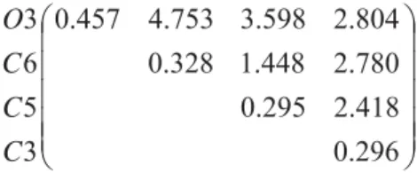
toxic compounds in the training set. Applying the procedure described earlier [8] assuming reasonable tolerances we got the following common 4x4 submatrix of activity:
(2) 296 . 0 418 . 2 295 . 0 780 . 2 448 . 1 328 . 0 804 . 2 598 . 3 753 . 4 457 . 0 3 5 6 3 C C C O
Analyzing the ECMC of the active compounds in the training set we found that the same ECSA obtained from
the four most active compounds satisfi es the next 17 compounds in some conformation assuming some reasonable
tolerances for the matrix elements (see below). These compounds and their corresponding ECSA are shown in Table 2 with the circled four toxicophore atoms corresponding to these 4x4 matrices.
Table 2
The electron-conformational submatrices of toxicity and molecular structures for 17 compounds containing the Tph.
The four toxicophore atoms are marked by circles.
21
342
.
0
663
.
2
296
.
0
053
.
3
098
.
1
340
.
0
060
.
3
956
.
3
304
.
5
347
.
0
2
5
3
1
Cl
C
Cl
Cl
22341
.
0
663
.
2
302
.
0
044
.
3
107
.
1
340
.
0
048
.
3
956
.
3
295
.
5
341
.
0
2
5
3
1
Cl
C
Cl
Cl
24355
.
0
059
.
3
351
.
0
522
.
2
962
.
0
349
.
0
050
.
3
407
.
4
726
.
4
365
.
0
6
9
8
3
C
C
C
C
25355
.
0
060
.
3
361
.
0
522
.
2
963
.
0
349
.
0
050
.
3
388
.
4
716
.
4
369
.
0
5
2
3
8
C
C
C
C
Averaging the corresponding matrix elements in these 17 sub-matrices we obtained the following averaged
matrix of toxicity
T
ˆ
av :318 . 0 481 . 2 302 . 0 743 . 2 207 . 1 350 . 0 053 . 3 053 . 4 803 . 4 395 . 0 4 3 2 1 ˆ T T T T
Tav (3)
with the matrix of tolerances (in relative values of the matrix elements):
116
.
0
233
.
0
160
.
0
121
.
0
202
.
0
133
.
0
131
.
0
144
.
0
105
.
0
162
.
0
ˆ
relT
(4)resulting in following ECSA, the digital toxicophore for the 17 compounds with the toxicity in interval -1.32 < -log LC50 < 0.30:
Moving to the compounds with lower activity one may fi nd out that this ECSA (revealed for the most active compounds) does not work for them, but they can be accommodated within this ECSA by allowing larger tolerances. In
this way one can get slightly different ECSA’s and hence pharmacophores/toxicophores for different levels of activity.
The activity is thus quantitatively a function of the tolerances. The dynamics of change of the tolerances of different
atoms when moving from more active to less active compounds reveals also the role of their fl exibility in the change
of activity.
Let us illustrate this important feature by an example. The compound benzaldehyde with logLC50= 1.57 has the
following ECSA:
31
.
0
41
.
2
31
.
0
79
.
2
45
.
1
35
.
0
89
.
2
06
.
5
79
.
4
51
.
0
6
4
3
1
C
C
C
O
(6)
We see that although this matrix does not fi t to the above ECSA for the most active compounds (5), it fails just
by some increase of the tolerances, mainly in the distance 5.06 Ả between the oxygen atom and the carbon C4, which
is out of the limits of the corresponding distance 4.05±0.58 in the most active compounds. This feature comprises the
next 7 less active compounds with toxicities 1.9 > logLC50>1.4. It demonstrates the important role of the most active
heteroatom (chlorine, oxygen, etc.) in the toxicity. The falling toxicity with the increase of the distance of this active
atom to the main skeleton of the molecule may be due to either its out of the limits of effi cient docking to the bioreceptor
or its poor bonding and hence decay through the metabolic processes. The dependence of the quantitative activity on the tolerances is of general importance in the EC method, and it is used to estimate the activity quantitatively (see below).
As mentioned above, this picture of activity presented by ECSA as the pharmacophore or toxicophore and the dependence of the quantitative activity on the tolerances may be complicated by the presence of out-of-pharmacophore
groups that infl uence the activity either by shielding of or competing with the pharmacophore in its interaction with
the bioreceptor [8]. Therefore the presence of the pharmacophore should be considered as just a necessary condition of
activity, but not a suffi cient one. Since it is based on fi rst principles (quantum-chemical calculations) and not involving statistics, the prediction of the pharmacophore in the EC method has a potential of the same reliability as that of the experimental data of the training set, meaning 100% reliability if based on experimental data of highest accuracy. The latter are thus most important in getting the absolutely reliable pharmacophore.
Another important feature that infl uences the pharmacophore in the EC method is the diversity of the training
set. The ECSA (the pharmacophore/ toxicophore) is valid for levels of activity and classes of molecules the ECMC of which were used in its calculation. For the same mechanism of substrate-receptor interaction different classes of compounds should have the same pharmacophore, but if the latter is obtained from one (or a limited number) of classes with active compounds, it may happen that it includes more active sites (atoms) than the minimal necessary, which thus may be absent in active molecules from other classes.
To screen new compounds for the activity under consideration, their ECMC should be calculated, and then the presence of the ECSA should be checked taking into account the above class and activity level limits. If only very active compounds are looked for, the ECSA (5) of the highly active ones should be tried, while for less active compounds
another ECSA (with enlarged tolerances) should be involved. We thus have a very fl exible system of searching for leads
of active compounds. Presently these procedures of matrix comparison are very fast.
The high reliability of pharmacophore identifi cation by the EC method may serve as a basis for revealing novel
knowledge [7]. Indeed, if the pharmacophore is a necessary condition of activity, it should be present in all the active compounds. What if there are active compounds that have no pharmacophore (as in some of the above compounds in Table 1)? If the structure of the molecules, as well as the experimental measurement of activity are beyond doubt and the compound under consideration is within the diversity of the training set, the situation calls for a special investigation.
The fi rst time we encountered such a case was in the search for the pharmacophore in musk odorant activity [24]. The
pharmacophore that was present in several hundred compounds with musk odor from a variety of rather different classes failed in the case of a patented musk tibeten (a benzene derivative). The solution of this controversy was found in the suggestion that this molecule exhibits its odor properties in the form of a dimer formed by two stacking substituted benzene rings; the dimer has the pharmacophore.
In fact bimolecular activity does not require a priori dimerization before the interaction with the bioreceptor. Indeed, it is widely accepted that the drug-receptor interaction, like any other substrate(S)-enzyme(E) interaction, follows the Michaelis-Menten mechanism [25] with pre-equilibrium in the formation of the complex SE before the transformation to the product P:
This mechanism can be extended to the simultaneous action of two molecules. Indeed, if the fi rst molecule S has no pharmacophore, there will not be transformation to the product. Then the equilibrium (Boltzman distribution) allows for the second molecule to enter the intermediate complex:
S + E
⇔
SESE + S
⇔
SSE⇒
P (8)If two molecules in a bimolecular docking to the receptor posses the pharmacophore, they may produce the necessary action that triggers the drug activity, albeit with lower probability than a single molecule with the pharmacophore. In the problem of aquatic toxicity to fi sh under consideration, for instance, the molecule allyl chloride (II) with logLC50 = 1.20
has no pharmacophore, while a 1,4-dichloro-2-butene (I) with logLC50 = -0.16 has the pharmacophore. The structure of
these two molecules in Fig. 2 shows how two molecules of II produce the structure III which is similar to I and has the
pharmacophore; hence the activity of II can be explained in this way.
I
Cl
Cl
II
Cl
III Cl
Cl
Fig. 2. Illustration of bimolecular activity: two active molecules of allyl chloride (II), which separately have no pharmacophore, by stacking along the double bond produce a bimolecular structure (III) which is similar to
1,4-dichloro-2-butene (I) and has the pharmacophore.
Step 6. In accordance with the EC method techniques [8], the presence of toxicophore is only a necessary condition of activity; the evaluation of activity quantitatively involves the regression analysis mentioned above. For the 17 most active molecules the following relations for biological activity was employed [7, 8]:
]
[
30259
.
2
)
log(
)
log(
50 50S
R
kT
E
E
LC
LC
i ref i ref i (9)where (LC50), and (LC50)refstand for numerical values of activity of the i-th compound and the reference compound,
respectively, Eiis the relative energy of the lowest energy conformer of the i-th compound that contains toxicophore,
and Si[R] is a function of the electronic and geometric parameters of the substrate molecule.
The parameters in the function Si[R] in Eq. (9) should be obtained from the condition of minimum difference
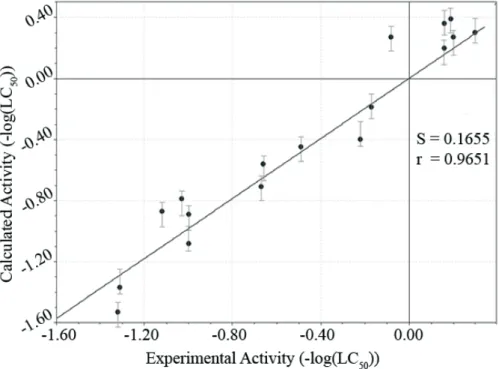
between the calculated toxicity LC50)i and the experimental values (LC50)expi . The regression analysis was performed
over the 17 toxicophore-containing molecules and the correlation coeffi cient as good as R2 = 0.94 and R2 = 0.92 in a
cross-validated procedure was obtained with just three parameters of their electronic structure:
S
i[
R
]
k
1(
II
maxiII
maxref)
k
2(
q
iq
ref)
k
3(
C
13iC
13ref),
(10)where IImaxi is the largest “interaction index” II in i-th molecule, qi is a Mulliken charge of the most electron-negative
atom near to the toxicophore atom T2i (within the radius of R < 2Å of the typical chemical bond length) and Ci
13 is
the matrix element of the ECSA corresponding to the distance (in Å) between the toxicophore atoms T1i and T3i; T1,
T2, T3, and T4 denote consecutively the four atoms in their ECSA (see eq. 3). The following values of the coeffi cients
minimize the expression (10):
k
17
.
58
;
k
214
.
74
;
k
31
.
36
.
(11)The theoretical values of toxicities calculated with this set of coeffi cients and those received in the leave-one-out
Fig. 3. Calculated vs. experimental -log(LC50) ratio for 17 most active compounds of the training set
Table 3
Calculated with and without cross-validation (C-V) and experimental values of -log(LC50)
of 17 most active compounds
-log(LC50)exp
-log(LC50)calc
Without C-V With C-V
1(REFER) 0.30 0.30
2 0,20 0.27 0.28
3 0.19 0.39 0.39
4 0.16 0.36 0.37
5 0.16 0.21 0.21
6 -0.08 0.28 0.29
7 -0.17 -0.20 -0.23
8 -0.22 -0.40 -0.43
9 -0.49 -0.45 -0.42
10 -0.66 -0.56 -0.53
11 -0.67 -0.71 -0.72
17 -1.00 -0.89 -0.85
18 -1.00 -1.08 -1.10
21 -1.03 -0.79 -0.75
22 -1.12 -0.87 -0.83
24 -1.31 -1.38 -1.41
25 -1.32 -1.53 -1.62
With the parameters of the weakly active compounds included a quantitative relationships between the toxicophore parameters and the toxicity can be obtained from a regression calculation that yields the following new correlation formula:
i i
i i
i
a
a
II
a
q
a
C
a
C
LC
50)
0 1 max 2 max 3 13 4 12log(
(12)where ai are the fi tting coeffi cients, IIimax and qimax are the largest values of the ECSA atomic interaction index and
atomic charge, respectively, and C1ij are the corresponding fi rst row matrix elements of the ECSA in Table 2 (the
The results are presented in Table 4: I and II are thefi ttings obtained over the 17 most active compounds with
three and fi ve regression parameters, respectively, and III is a fi tting received over 21 compounds that include the
weakly active ones. While correlation II is somewhat better than I due to the additional two parameters, correlation
III allows one to screen and predict also weak toxicities with logLC50 > 1.4. For strong toxicity with logLC50 < 1.4
correlation I is quite reasonable. This indicates the signifi cance of the parameters of the active atom 1 in reducing the
toxicity.
Table 4
Regression coeffi cients for three types of correlation (eqs. 10, 12)
Regression a0 a1 a2 a3 a4 R2
I - 7.58 14.74 1.36 - 0.94
II 5.18 2.83 5.82 0.39 0.31 0.97
III 1.6 3.29 6.49 0.46 0.54 0.86
Conclusions
Using the electron-conformational method we have shown that aquatic toxicity to fi sh is due to a special
functional group, the toxicophore, a combination of four atomic sites with special electronic and topology characteristics represented numerically as the sub-matrix of activity, ECSA. An important feature of the latter is the set of tolerances in the values of the matrix elements which control the quantitative value of activity. It was shown that by varying the tolerances one can obtain different toxicophores for different levels of activity. A distinguished feature of this approach
is that it does not employ arbitrary descriptors and statistics at the level of toxicophore identifi cation, thus making the
latter void of artifacts and chance correlations and fully reliable (at the level of reliability of the experimental data in the training set). This led us to suggest a bimolecular mechanism of activity for allyl chloride. The toxicophore for 17
most active compounds yields a good correlation (R2 = 0.94) with the experimental activities using only three electronic
structure parameters.
References
Guner O.F. Ed. Pharmacophore Perception, Development, and Use in Drug Design, International University [1]
Line: La Jolla, 2000.
Kubinyi, H.; Folkers, G.; Martin, Y. C. 3D QSAR in Drug Design. Volume 3, Recent Advances; Kluwer Academic [2]
Publishers: New York, 2002.
Marshall G.R. In: Wolff M.E., Ed. Burger’s medicinal chemistry and drug discovery: principles and practice. 5th [3]
ed. Vol. 1. New York: John Wiley, 1995, 573-659. O. Ivanciuc, Internet Electron. J. Mol. Design,
[4] 2, 195–208 (2003).
Zhong–Sheng Yi, Shu–Shen Liu, Internet Electron. J. Mol. Design,
[5] 4, 835–849 (2005).
Verhaar, H.J.M.. Leeuwen, C.J, Hemiens. J.L.M., Chemosphere
[6] , 25, 471 -491(1992).
I. B. Bersuker, J. Comp.-Aid. Mol. Des
[7] ., submitted for publication.
I. B. Bersuker. Current Pharmaceutical Design.,
[8] 9, 1575-1606 (2003).
Bersuker I. B., Dimoglo A. S., In Reviews in Computational Chemistry; Lipkowitz K.B., Boyd D.B. Eds.; VCH: [9]
New York, 1991; vol.2, pp.423-60.
Bersuker, I. B.; Bahceci, S.; Boggs, J. E.; Pearlman, R. S. , J. Computer-aided Mol. Design
[10] 13, 419–434
(1999).
Bersuker, I. B.; Bahceci, S.; Boggs, J. E., J. Chem. Inf. Comput. Sci
[11] ., 40 (2000)1363–1376.
Rosines, E.; Bersuker, I. B.; Boggs, J. E., Quan. Struct.-Act. Relat.
[12] 2001, 20, 327–334.
A. H. Makkouk, I. B. Bersuker and J. E. Boggs, Int. J. Pharm. Med
[13] ., 18, 81-89 (2004).
A.V. Marenich, Pei-Han Yong, I. B. Bersuker and J. E. Boggs, J. Chem. Inf. Mod
[14] ., submitted.
Katritzky, A.R., Tatham, D.B. and Maran U., J. Chem. Inf. Comput. Sci
[15] .,41, 1162-1176 (2001).
Dearden. J.C. and Raevsky, O.A., SAR and QSAR in Environm. Research,
[16] 15, 433-448 (2004).
Halgren T. A., J. Comp. Chem
[17] . 1996, 17, 490–519.
Chambers, C. C.; Hawkins, G. D.; Cramer, C. J.; Truhlar, D. G. J. Phys. Chem
[18] . 1996, 100, 16385–16398.
Spartan’02
[19] ; Wavefunction, Inc.: Irvin, CA. Except for molecular mechanics and semi-empirical models, the
M.; Hirata, S.; Hsu, C.-P.; Ishikawa, N.; Florian, J.; Warshel, A.; Johnson, B. G.; Gill, P. M. W.; Head-Gordon, M.; Pople, J. A. Q-Chem 2.0: A High-Performance Ab Initio Electronic Structure Program Package. J. Comput. Chem. 2000, 21, 1532–1548.
Stewart, J. J. P. , J. Comp. Chem
[20] . 1989, 10, 209–220.
Basch, H.; Viste, A.; Gray, H. B., Theoret. chim. Acta
[21] (Berl.) 1965, 3, 458–464.
Bondi, A., J. Phys. Chem
[22] . 1964, 68, 441–451).
Slater, J. C., J. Chem. Phys
[23] . 1964, 41, 3199–3204).
Bersuker I.B., Dimoglo A.S., Gorbachov M.Yu., Vlad P.F., Pesaro M.
[24] New J. Chem.,15, 307-20 (1991).