Short Report
S
J. Braz. Chem. Soc., Vol. 22, No. 8, 1610-1615, 2011. Printed in Brazil - ©2011 Sociedade Brasileira de Química 0103 - 5053 $6.00+0.00*e-mail: [email protected]
Three New Compounds from
Piper montealegreanum
Yuncker (Piperaceae)
Harley da S. Alves, Maria de F. V. de Souza and Maria C. de O. Chaves*
Laboratório de Tecnologia Farmacêutica, Universidade Federal da Paraíba, CP 5009, 58051-970 João Pessoa-PB, Brazil
Três novos compostos: dois lavonóides [(S)-8-formil-3’,5-diidroxi-7-metoxi-6-metillavanona (1) e 3’-formil-3,4’,6’-triidroxi-2’-metoxi-5’-metilchalcona (2)] e um fenilpropanóide [3,4-metilenodioxi-5-metoxi-7,8-diidrocinamato de etila (3)] foram isolados dos ramos secos de
Piper montealegreanum. As estruturas desses compostos foram estabelecidas através das técnicas espectroscópicas UV, IV, EM e RMN (1H e 13C, 1D e 2D), além da interpretação dos dados de
RMN de 1H e 13C dos derivados metilados dos compostos 1 e 2.
Three new compounds: two flavonoids [(S )-8-formyl-3’,5-dihydroxy-7-methoxy-6-methyllavanone (1) and 3’-formyl-3,4’,6’-trihydroxy-2’-methoxy-5’-methylchalcone (2)] and one phenylpropanoid [ethyl 3,4-methylenedioxy-5-methoxy-7,8-dihydrocinnamate (3)] were isolated from dried branches of Piper montealegreanum. Their structures were established by UV, IR, MS, 1D and 2D (1H and 13C) NMR spectroscopic techniques, besides interpretation of spectral data
(1H and 13C NMR)of methylated derivativesof 1 and 2 compounds.
Keywords:Piper montealegreanum, lavanone, chalcone, phenylalcanoid
Introduction
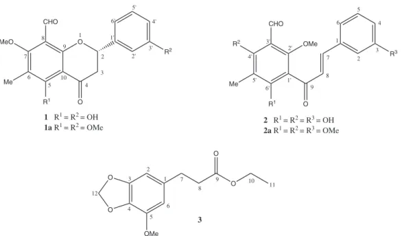
Piper montealegreanum Yuncker (Piperaceae) is a shrub, native to the north Brazil1 and has no previous chemical studies reported. A continuing search on the chemistry and bioactive agents from Brazilian north-northeast Piperaceae species have resulted in the isolation of amides,2-5 aristolactams6,7 and propenylphenols.8-12 In this paper, we report the isolation and structure elucidation of (S )-8-formyl-3’,5-dihydroxy-7-methoxy-6-methyllavanone (1), 3’-formyl-3,4’,6’-trihydroxy-2’-methoxy-5’-methylchalcone (2), and ethyl 3,4-methylenedioxy-5-methoxy-7,8-dihydrocinnamate (3) from the branches of P.montealegreanum (Figure1). The structures of the compounds were determined by interpretation of the spectral data analysis of UV, IR, MS, 1H and 13C NMR, including 2D NMR HMQC (heteronuclear multiple quantum coherence), HMBC (heteronuclear multiple bond correlation), and by comparison with those reported in the literature.13
Results and Discussion
Compound 1 was obtained as orange-yellow crystals. The MS spectrum presented a molecular ion peak at m/z 327.0887 (M-1)−, in LC-MS-IT-TOF apparatus (ion trap-time of light liquid chromatography mass spectrometry). The 1H NMR spectrum showed the presence of four singlets at dH 12.63 (1H), 10.15 (1H), 3.99 (3H) and 2.05 (3H) consistent with the presence of chelated hydroxyls, aldehyde, methoxyl and methyl groups, respectively. The presence of three signals at dH, 5.43 (dd,1H, J 10.8 and 4.6 Hz), 2.97 (dd, 1H, J 17.0 and 10.8 Hz), 2.85 (dd,1H, J 4.6 and 17.0 Hz), and also signals for four coupled aromatic protons at dH, 7.26 (t, 1H, J 8.0 Hz), 6.85 (brd, 1H, J 8.0 Hz) and 6.93 (m, 2H), suggested a lavanone nucleus14 with a 3’-monosubstituted B ring, deduced by analysis of the multiplicity and coupling constants of the aromatic protons.15 The above data and UV spectrum, with λ
max at 266 nm, reinforced a lavanone nature for compound 1.13 Since no further aromatic protons were evident, ring A should be fully substituted. The presence of the signal to methine carbon at dC 193.8 in the
Alves et al. 1611 Vol. 22, No. 8, 2011
12.63 (s, 1H)] evidenced the chelated hydroxyl at C-5 with the C=O of the α,β-unsaturated carbonyl group.13
In the HMBC spectrum, the presence of cross peaks at
dH 12.63 with dC 166.3 and 109.3, besides the cross peak at dH 2.05 with dC 166.3, 166.1 and109.3, evidenced the existence of a methyl group at C-6 and also suggested the attachment position of the methoxyl group at C-7. This was conirmed by correlation of the peak at dH 10.15 (aldehyde hydrogen) and dH 3.99 (methoxyl hydrogen) with dC 166.1, and consequently, the placement of the formyl group at C-8 (Figure 2).
The compound 1a (Figure 1), a methylated derivative of 1, showed correlations in HMQC spectrum between dH 3.95 / dC 64.3 and dH 3.82 / δC 55.3. The HMBC spectrum displayed correlations between dH 3.95 and 2.14 / dC 164.6 conirming the methoxyl group at C-5. The presence of cross peaks between signals in dH 3.82 / dC 159.9 supported the methoxyl group at the C-3’.
Compound 2 was obtained as yellow crystals. The MS spectrum gave a molecular ion peak at m/z 327.0870 (M-1)−, in LC-MS-IT-TOF apparatus. The 1H NMR spectrum indicated signals at dH 7.78 and 7.84 suggesting the presence of protons on a α,β-unsaturated ketone moiety.13,16 The above data and UV absorption bands (λmax) at 317 and 282 nm suggested a chalcone structure for compound 2.13 Signals for four coupled aromatic protons: a triplet at dH 7.33 (1H,J 7.8 Hz), a multiplet at dH 7.28-7.23 (m, 2H) and a double double doublet at dH 6.97 (1H, J 7.8, 2.0 and 1.6 Hz) suggested a 3’-substituted B ring.15 Additionally, the 1H NMR spectrum showed tree singlets at d
H, 2.01 (3H), 4.01 (3H) and 10.17 (1H), consistent with the presence of methyl, methoxyl and aldehyde groups, respectively, besides two hydroxyl group singlets at dH 12.83 and 14.21. The two low-ield hydroxyl protons evidenced a formyl group at C-3’. This conclusion is achieved since the down-ield shift of OH-4’ (dH 12.83) can be explained by hydrogen-bonding with the oxygen atom of the formyl substituent at aneighboring carbon atom and at the same time that the down-field shift of OH-6’ (dH 14.21) is caused by chelation between the 6’-hydroxyl proton and the carbonyl oxygen of the α,β-unsaturated carbonyl group function.13 This intramolecular hydrogen bonding corroborated the assignments of the signals at d 7.78 (d, 1H) and d 7.84 (d, 1H) to α and β positions, respectively.16 Spectral analysis of 2a showed Hα and Hβ signals with a marked difference in chemical shifts [d 7.29 (d, 1H, J 16.0 Hz, H-7) and d 6.98 (d, 1H, J 16.0 Hz, H-8)] related with the absence of the intramolecular hydrogen bonding with the C=O of the α,β-unsaturated carbonyl group. The
O O CHO MeO Me R1 R2 1 2 3 4 5 6 7 8 9 10 1' 2' 3' 4' 5' 6'
1 R1 = R2 = OH 1a R1 = R2 = OMe
O CHO R2 Me R1 R3 1 2 3 4 5 6 8 7 9 1' 2' 3' 4' 5' 6'
2 R1 = R2 = R3 = OH
2a R1 = R2 = R3 = OMe
OMe O O O OMe O 1 2 3 4 5 6 7 8 9 10 11 12 3
Figure 1. Structures of the isolated compounds 1-3 from Piper montealegreanum.
O O CHO MeO Me OH OH 1 2 3 4 5 6 7 8 9 10 1' 2' 3' 4' 5' 6'
Three New Compounds from Piper montealegreanum Yuncker (Piperaceae) J. Braz. Chem. Soc.
1612
coupling constant (16.0 Hz) observed in 2a indicated the E-isomer for the double bond.16 The placement of the other groups in the A-ring was made on the basis of the HMBC correlations (Figure 3): dH 14.21 (s, 1H, OH-6’) / dC 169.8 and 109.0; dH 12.83 (s, 1H, OH-4’) / dC 166.6, 109.2 and 109.5; dH 2.01 (s, 3H) / dC 109.2; dH 10.17 (s, 1H) / dC 166.6 and dC 109.5; dH 4.01 / dC 168.5 that evidenced the hydroxyl, formyl, methyl and methoxyl groups at C-6’-4’, C-3’, C-5’ and C-2’, respectively.
The hydroxyl group in the C-3 was conirmed for the methylated derivative of 2 (Figure 1) which showed correlation between dH 3.80 / dC 55.3 in the HMQC spectrum and dH 3.80 / dC 159.9 (C-3) in the HMBC spectrum. Other correlations observed in the HMQC spectrum of 2a were dH 3.86 / dC 62.7 and dH 3.75 / dC 61.9. In the HMBC spectrum, the signals at dH 3.86 and 3.75 showed correlations with dC 163.0 and 161.8, respectively, providing support for the presence of the methoxyl groups at the C-6’ and C-4’ position.
Formyl flavonoids have been reported from a few species in the plant kingdom. Early reports included a description of 2’,4-dihydroxy-4’-methoxy-5’-formylchalcone from Psoralea corylifolia (Fabaceae).17 Its isomeric compound, with methoxy group at the 2’-position, has been reported from the same species.18 2’,4’,6’- trihydroxy-3’-formylchalcone has been reported from Psidium acutangulum (Myrtaceae)19 and its retrochalcone derivative was obtained from Anredera scandens (Basellaceae).20 3’-formyl-4’,6’-dihydroxy-2’-methoxy-5’-methylchalcone and (2S )-8-formyl-5-hydroxy-7-methoxy-6-methyllavanone were isolated from Cleistocalyx operculatus (Myrtaceae).13
The compound 3 was obtained as yellow powder. The 1H spectrum showed two doublets in d
H 6.37 (J 1.4 Hz, 1H) and dH 6.34 (J 1.4 Hz, 1H) typical of meta aromatics hydrogens in addition to two singlets in dH 5.91 (2H) and 3.86 (3H) typical of methylenedioxy and methoxyl groups, respectively. These data suggested the presence of tetrasubstituted aromatic ring. The signals in dH 4.11
(q, 2H, J 7.0 Hz) and 1.23 (t, 3H, J 7.0 Hz) supported the presence of ethyl group attached to heteroatom and also two signals at dH 2.84 (t, 2H, J 7.5 Hz) and 2.55 (t, 2H,
J 7.5 Hz) typical of methylene hydrogens.
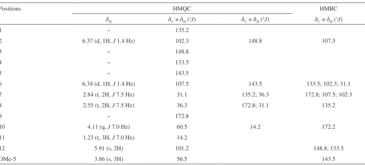
The 13C NMR spectrum of 3 showed 13 signals. The signals at dH 2.84, 2.55 and 4.11 are, in the HMBC spectrum, correlated with a signal characteristic of carbonyl ester at dC 172.8, conirming the assignment of the chemical shifts for H-7, H-8 and H-10, respectively, and dC 172.8 for C-9 of a dihydrocinnamoyl group. The HMBC spectrum also showed correlations between dH 3.86 / dC 148.8, dH 5.91 / dC 143.5 and 133.5, conirming the methoxyl and methylenedioxy groups at C-5 and C-3-4, respectively.
Experimental
General procedures
Melting points (mp) were determined on a MQAPF-302 melting point digital apparatus. UV spectra were recorded on a Vankel-50 UV-Vis spectrophotometer. IR spectra were obtained on an FT-IR-1750 spectrophotometer, Perkin-Elmer apparatus. The spectra mass were obtained on a SHIMADZU LCMS-IT-TOF (225-07100-34) equipped with a Z-spray ESI (electrospray) source and operated in negative mode.
1H and 13C NMR (1D and 2D) spectra were recorded on a Varian Mercury 200 spectrometer in CDCl3 and (CD3)2CO, with TMS as internal standard. Sephadex LH-20 and silica gel 60 (PF254 art. 7749 and art. 7731) were purchased from Merck.
The methylated lavonoids were obtained by treatment of the sample, dissolved in dry propanone, with 1.1 equiv. of dimethyl sulphate (Me2SO4) and 1.1 equiv. of potassium carbonate (K2CO3) to each free hydroxyl. The reactions were carried at room temperature during 12 h. After removal of the solvent in vacuum, the residue was suspended in H2O (50 mL), treated with 5 mL of ammonia and extracted with CHCl3 (3 × 15 mL). The CHCl3 solution was dried with Na2SO4,iltrated and concentrated to dryness.13
Plant material
Branches of Piper montealegreanum Yuncker was collected in Belém (Pará State, Brazil), in December 2002 and identiied in the Botanical Garden, Rio de Janeiro (Rio de Janeiro State, Brazil). A voucher specimen (MSP-010) was deposited at Emilio Goeldi Museum, Belém.
O CHO HO Me OH OH 1 2 3 4 5 6 8 7 9 1' 2' 3' 4' 5' 6' OMe
Alves et al. 1613 Vol. 22, No. 8, 2011
Extraction and isolation
The powered material of P. montealegreanum (1.3 kg) was exhaustively extracted with EtOH (4 × 2.0 L), the solvent removed under reducted pressure furnished a green residue (115.0 g). The crude extract amount of 13.5 g was chromatographed over Sephadex gel LH-20 and eluted with methanol (column 1) yielding 43 fractions. Fraction 17 was further fractionated over Sephadex gel LH-20 column providing 5 fractions. Fraction 3 after submitted to recristalization with a chloroform and methanol mixture yielding (S )-8-formyl-3’,5-dihydroxy-7-methoxy-6-methyl-lavanone (1) (30 mg).
Fraction 18 (column 1) was chromatographed over Sephadex LH-20 and yielded five fractions. Fraction 4 gave 3’-formyl-3,4’,6’-trihydroxy-2’-methoxy-5’-methylchalcone (2) (40 mg), after submitted to recristalization with a chloroform and methanol mixture.
Fractions 8-11 (column 1) were also fractionated over Sephadex gel yielding 29 fractions. Fraction 8-14, after recrystallization with chloroform, gave ethyl
3,4-methylenedioxy-5-methoxy-7,8-dihydrocinnamate (3) (12 mg).
Compound 1.Orange yellow crystals (CDCl3:MeOH); mp 162 °C; [α]D25 −20 (MeOH, 0.025); UV λmax/nm (MeOH): 266; UV λmax/nm (AlCl3): 286, 323 (sh); IR
νmax/cm-1 (KBr): 3425, 1688, 1620, 1570, 1464; MS [M-1]− 327.0887; 1H and 13C NMR spectral data, see Table 1.
Compound 2. Yellow crystals (CHCl3:MeOH); mp 164 °C; UV λmax/nm (MeOH): 317, 282; UV λmax/nm (AlCl3): 349, 306; IR (KBr) νmax/cm
-1: 3445, 1616, 1581, 1422; MS [M-1]− 327.0870; 1H and 13C NMR spectral data, see Table 1.
Compound 1a and 2a.1H and 13C NMR spectral data, see Table 2.
Compound 3.Amorphous green powder; IR (KBr)
νmax/cm
-1: 2924, 2853, 1734; MS [M+] 252; 1H and 13C NMR spectral data, see Table 3.
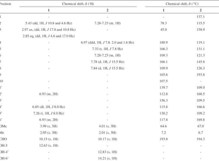
Table 1.1H and 13C NMR data for compounds 1 and 2
Position Chemical shift, d (1H) Chemical shift, d (13C)
1 2 1 2
1 - - - 137.1
2 5.43 (dd, 1H, J 10.8 and 4.6 Hz) 7.28-7.23 (m, 1H) 78.3 115.5
3 2.97 ax, (dd, 1H, J 17.0 and 10.8 Hz) - 45.0 158.9
2.85 eq, (dd, 1H, J 4.6 and 17.0 Hz)
4 - 6.97 (ddd, 1H, J 7.8. 2.0 and 1.6 Hz) 188.9 119.1
5 - 7.33 (t, 1H, J 7.8 Hz) 166.3 131.1
6 - 7.28-7.23 (m, 1H) 109.3 121.3
7 - 7.78 (d, 1H, J 15.5 Hz) 166.1 145.8
8 - 7.84 (d, 1H, J 15.5 Hz) 109.9 126.3
9 - 165.6 193.8
10 - - 107.5
-1’ - - 139.7 109.0
2’ 6.93 (m, 2H) - 112.8 168.5
3’ - - 156.3 109.5
4’ 6.85 (dl, 1H, J 8.0 Hz) - 115.8 166.6
5’ 7.26 (t, 1H, J 8.0 Hz) - 130.2 109.2
6’ 6.93 (m, 2H) - 117.8 169.8
OMe 3.99 (s, 3H) 4.01 (s, 3H) 64.6 67.0
Me 2.05 (s, 3H) 2.01 (s, 3H) 7.2 6.7
CHO 10.15 (s, 1H) 10.17 (s, 1H) 193.8 194.3
OH-5 12.63 (s, 1H) - -
-OH-4’ - 12.83 (s, 1H) -
-Three New Compounds from Piper montealegreanum Yuncker (Piperaceae) J. Braz. Chem. Soc.
1614
Table 2.1H and 13C NMR data for methylated compounds 1a and 2a
Position Chemical shift, d (1H) Chemical shift, d (13C)
1a 2a 1a 2a
1 - - - 135.5
2 5.48 (dd, 1H, J 12.0 and 4.0 Hz) 7.02 (m, 1H) 78.9 113.2
3 3.02 ax (dd, 1H, J 16.7 and 12.0 Hz); 2.87 eq (dd,1H, J 16.7 and 4.0 Hz)
- 45.3 159.9
4 - 6.91 (m, 1H) 188.8 116.9
5 - 7.27 (t, 1H, J 7.8 Hz) 164.6 129.9
6 - 7.12 (m, 1H) 117.3 121.4
7 - 7.29 (d, 1H, J 16.0 Hz) 164.8 146.4
8 - 6.98 (d, 1H, J 16.0 Hz) 117.9 128.3
9 - - 165.6 193.7
10 - - 115.6
-1’ - - 139.6 122.1
2’ 7.03-6.99 (m, 2H) - 111.8 158.9
3’ - - 159.9 119.23
4’ 6.94-6.88 (m, 1H) - 113.9 161.8
5’ 7.35 (t, 1H, J 8.2 Hz) - 130.0 125.1
6’ 7.03-6.99 (m, 2H) - 118.0 163.0
OMe-3’ 3.82 (s, 3H) - 55.3
-OMe-5 3.95 (s, 3H) - 64.3
-OMe-7 3.83 (s, 3H) - 62.3
-Me 2.14 (s, 3H) 2.19 (s, 3H) 8.4 8.9
CHO 10.33 (s, 1H) 10.30 (s, 1H) 188.2 188.2
OMe-2’ - 3.78 (s, 3H) - 64.5
OMe-4’ - 3.75 (s, 3H) - 61.9
OMe-6’ - 3.86 (s, 3H) - 62.7
OMe-3 - 3.80 (s, 3H) - 55.3
Table 3.1H (200 MHz) and 13C (50 MHz) NMR data for compound 3 and correlations obtained in HMQC and HMBC experiments, J (Hz) in parenthesis
Positions HMQC HMBC
dH dC × dH (1J) dC × dH (2J) dC × dH (3J)
1 - 135.2
2 6.37 (d, 1H, J 1.4 Hz) 102.3 148.8 107.5
3 - 148.8
4 - 133.5
5 - 143.5
6 6.34 (d, 1H, J 1.4 Hz) 107.5 143.5 133.5; 102.3; 31.1
7 2.84 (t, 2H, J 7.5 Hz) 31.1 135.2; 36.3 172.8; 107.5; 102.3
8 2.55 (t, 2H, J 7.5 Hz) 36.3 172.8; 31.1 135.2
9 - 172.8
10 4.11 (q, J 7.0 Hz) 60.5 14.2 172.2
11 1.23 (t, 3H, J 7.0 Hz) 14.2
12 5.91 (s, 2H) 101.2 148.8; 133.5
Alves et al. 1615 Vol. 22, No. 8, 2011
Acknowledgements
The authors thank CNPq (Conselho Nacional de Desenvolvimento Cientíico e Tecnológico) for inancial support, Laboratório de Espectrometria de Massas do Nordeste (LEMANOR) for MS spectra and Maria das Graças Bichara Zoghbi for plant material.
Supplementary Information
Supplementary information (1H and 13C NMR data for compounds 1, 2, 3, 1a and 2a, Figures S1-S15) is available free of charge at http://jbcs.org.br as a PDF ile.
References
1. Yuncker, T. G.; Hoehnea1972, 2, 36.
2. Araújo-Jr, J. X.; Chaves, M. C. O.; Da Cunha, E. V. L.; Gray, A. I.; Phytochemistry1997, 44, 559.
3. Chaves, M. C. O.; Santos, B. V. O.; Biochem. Syst. Ecol. 1998,
27, 113.
4. Chaves, M. C. O.; Da Cunha, E. V. L.; Fitoterapia2001, 72, 197.
5. Chaves, M. C. O.; Figueiredo-Jr, A. G.; Santos, B. V. O.;
Fitoterapia2003, 74, 181.
6. Araújo-Jr, J. X.; Chaves, M. C. O.; Da Cunha, E. V. L.; Gray, A. I.; Biochem. Syst. Ecol. 1999, 27, 325.
7. Cardozo-Júnior, E. L.; Chaves, M. C. O.; Pharm. Biol. 2003,
41, 216.
8. Oliveira, L. C. P.; Mause, R.; Nunomura, S. M.; J. Braz. Chem. Soc.2005, 16, 1439.
9. Chaves, M. C. O.; Santos, B. V. O.; Biochem. Syst. Ecol. 1999,
27, 539.
10. Chaves, M. C. O.; Santos, B. V. O.; Fitoterapia2002, 73, 547. 11. Santos, B. V. O.; Chaves, M. C. O.; da Cunha, E. V. L.;
Gray, A.I.; Biochem. Syst. Ecol. 1997, 25, 471.
12. Santos, B. V. O.; Chaves, M. C. O.; da Cunha, E. V. L.; Gray, A. I.; Phytochemistry 1998, 49, 1381.
13. Ye, Chun L.; Yan-Hua, L.; Dong-Zhi, W.; Phytochemistry2004,
65, 445.
14. Asai, T.; Hara, N.; Kobayashi, S.; Fujimoto, Y.; Phytochemistry
2008, 69, 1234.
15. Alavez-Solano, D.; Reyes-Chilpa, R.; Jiménez-Estrada, M.; Gómez-Garibay, F.; Chavez-Uribe, I.; Sousa-Sánchez, M.;
Phytochemistry2000, 55, 953.
16. Quintin, J.; Desrivot, J.; Thoret, S.; Le Menez, P.; Cresteil, T.; Lewin, G.; Bioorg. Med. Chem. Lett. 2009, 19, 167.
17. Gupta, S. R.; Seshadri, T. R.; Good, G. R.; Indian J. Chem.
1975, 13B, 632.
18. Gupta, B. K.; Gupta, G. K.; Dhar, K. L.; Atal, C. K.;
Phytochemistry1980, 19, 2034.
19. Miles, D. H.; de Medeiros, J. M. R.; Chittawong, V.; Hedin, P. A.; Swithenbank, C.; Lidert, Z.; Phytochemistry1991,
30, 1131.
20. Calzada, F.; Mata, R.; Bye, R.; Linares, E.; Phytochemistry
1990, 29, 2737.
21. Silva, T. M. S.; Carvalho, M. G.; Braz-Filho, R.; Quim. Nova
2009,32, 1119.
Submitted: December 8, 2009
Supplementary Information
S
I
J. Braz. Chem. Soc., Vol. 22, No. 8, S1-S8, 2011.Printed in Brazil - ©2011 Sociedade Brasileira de Química 0103 - 5053 $6.00+0.00
*e-mail: [email protected]
Three New Compounds from
Piper montealegreanum
Yuncker (Piperaceae)
Harley da S. Alves, Maria de F. V. de Souza and Maria C. de O. Chaves*
Laboratório de Tecnologia Farmacêutica, Universidade Federal da Paraíba, CP 5009, 58051-970 João Pessoa-PB, Brazil
Figure S1.1H NMR spectrum (d, CDCl
Three New Compounds from Piper montealegreanum Yuncker (Piperaceae) J. Braz. Chem. Soc.
S2
Figure S2.1H NMR spectrum (d 8.0-2.8 ppm, CDCl
3, 200 MHz) of 1.
Figure S3. 13C NMR (APT) spectrum (d, CDCl
Alves et al. S3 Vol. 22, No. 8, 2011
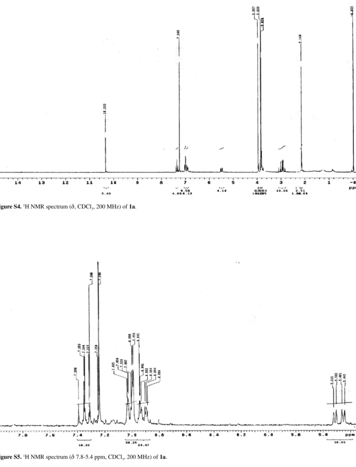
Figure S4.1H NMR spectrum (d, CDCl
3, 200 MHz) of 1a.
Figure S5.1H NMR spectrum (d 7.8-5.4 ppm, CDCl
Three New Compounds from Piper montealegreanum Yuncker (Piperaceae) J. Braz. Chem. Soc.
S4
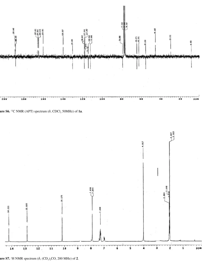
Figure S6. 13C NMR (APT) spectrum (d, CDCl
3 50MHz) of 1a.
Figure S7.1H NMR spectrum (d, (CD
Alves et al. S5 Vol. 22, No. 8, 2011
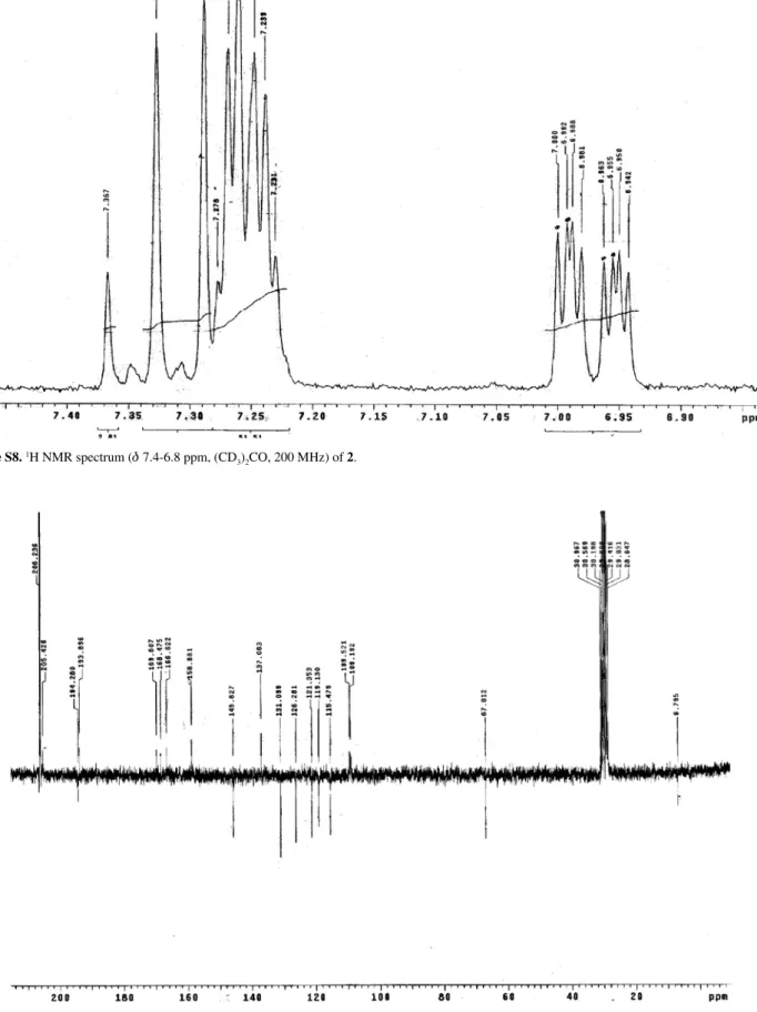
Figure S8.1H NMR spectrum (d 7.4-6.8 ppm, (CD
3)2CO, 200 MHz) of 2.
Figure S9.13C NMR (APT) spectrum (d, (CD
Three New Compounds from Piper montealegreanum Yuncker (Piperaceae) J. Braz. Chem. Soc.
S6
Figure S10. 1H NMR spectrum (d, CDCl
3, 200 MHz) of 2a.
Figure S11.1H NMR spectrum (d 7.4-6.8 ppm, CDCl
Alves et al. S7 Vol. 22, No. 8, 2011
Figure S12.13C NMR (APT) spectrum (d, CDCl
3 50MHz) of 2a.
Figure S13.1H NMR spectrum (d, CDCl
Three New Compounds from Piper montealegreanum Yuncker (Piperaceae) J. Braz. Chem. Soc.
S8
Figure S14.1H NMR spectrum (d 6.5-1.0 ppm, CDCl
3, 200 MHz) of 3.
Figure S15.13C NMR (APT) spectrum (d, CDCl