Short Report
Printed in Brazil - ©2013 Sociedade Brasileira de Química0103 - 5053 $6.00+0.00S
*e-mail: [email protected]
N
-(4-((
E
)-3-Arylacryloyl)phenyl)acetamide Derivatives and
their Antileishmanial Activity
Dency J. Pacheco,a Jorge Trilleras,*,a Jairo Quiroga,b Jennifer Gutiérrez,c Luis Prent,a
Tobinson Coavas,a Juan C. Marínd and Gabriela Delgadoc
aGrupo de Investigación en Compuestos Heterocíclicos, Programa de Química,
Facultad de Ciencias Básicas, Universidad del Atlántico, km 7 Antigua vía Puerto Colombia, Barranquilla-Atlántico, Colombia
bGrupo de Investigación de Compuestos Heterocíclicos, Departamento de Química,
Universidad del Valle, A. A. 25360, Cali, Colombia
cGrupo de Investigación en Inmunotoxicología and dGrupo de Investigación Farmacognosia y
Fitoquímica Universidad Nacional, Departamento de Farmacia, Facultad de Ciencias, Universidad Nacional de Colombia, A. A. 14490, Bogotá, D. C., Colombia
A atividade leishmanicida de uma série de derivados enônicos (chalconas) sintetizados via reações de condensação de Claisen-Schmidt assistidas por radiação ultrassom foi caracterizada através da análise de suas citotoxicidades contra formas promastigotas de Leishmania (Viannia) panamensis, espécie responsável por mais de 90% dos casos de Leishmania na Colômbia. Dois compostos foram ativos contra Leishmania com índices de seletividade de LC50 EC50-1 (concentração letalmédia e
concentração efetiva média) maiores que 27 e 3, respectivamente. Estes resultados sugerem que a substituição de um dos dois anéis de chalcona (anel A aromático) com oxigênio é apropriada. Um composto deve ser pesquisado quanto à sua atividade leishmanicida, especialmente por ser fácil de obter em elevados rendimentos, tornando possível a produção de medicamentos para o tratamento de leishmaniose cutânea.
The antileishmanial activity of a series of enonic derivatives (chalcones) synthesized via Claisen-Schmidt condensation reactions assisted by ultrasonic radiation was characterized by analyzing their cytotoxicity against Leishmania (Viannia) panamensis promastigotes, a species responsible for over 90% of Leishmania cases in Colombia. Two compounds were active against Leishmania with selectivity indexes of LC50 EC50
-1 (lethal concentration 50 and effective
concentration 50) higher than 27 and 3, respectively. These results suggest that a substitution on one of the two chalcone rings (aromatic ring A) with oxygen is convenient. Compound 3g should be further investigated for its antileishmanial activity, especially for being easy to obtain in high yields, making it possible to produce drugs for the treatment of cutaneous leishmaniasis.
Keywords: chalcone, sonochemistry, antileishmanial activity, Leishmania (Viannia) panamensis
Introduction
Leishmania spp is a protozoan parasite of the
Trypanosomatidae family that infects approximately 12 million people worldwide. This parasite causes a wide variety of diseases, ranging from cutaneous manifestations to clinical pictures with visceral leishmaniasis.1 This
parasitic infection has become endemic in tropical and
subtropical areas around the world due to the incidence of new cases in which Leishmania spp is common among
patients with human immunodeficiency virus (HIV).2
new alternatives to control this parasitosis. A large number of natural and synthetic compounds with diverse chemical structures have been analyzed in vitro. However, synthetic difficulties and low yields represent the limitations for the development of new antileishmanials. Within the group of compounds with promising antileishmanial activity are nitrogenated heterocycles, such as quinozolines,9-12
benzodiazepines,13 indolylglyoxylamides derivatives,14
pyrimidines and triazines.15-16 These α,β-unsaturated
systems are convenient and easily available as materials or intermediates for the synthesis of a wide variety of heterocyclic compounds.17 Compounds with α,β
-unsaturated-based structures have been reported to exhibit a range of pharmacological activities, including cytotoxicity toward neoplastic,18,19 antimitotic,20 antiviral,21 antibacterial22,23 and
antileishmanial cells.24 With regards to leishmanial activity,
many compounds have been studied, but only a few prototypes have made it to preclinical stages, due to, among other reasons, the difficulty of scaling up their production to industrial levels. Strategies that utilize simple syntheses and guarantee the production of sufficient quantities of the compound with the required quality for a pharmochemical activity are needed to provide new therapeutic alternatives to control infectious diseases, such as leishmaniasis. The synthesis of heterocyclic compounds with enonic fragments can be performed via the non-conventional procedures of green chemistry, in which large amounts of compound can be obtained using methodologies compatible with the environment. In this study, the antileishmanial activity of a series of synthetic chalcones was characterized by screening for the viability of macrophages and promastigotes of Leishmania (Viannia) panamensis, the specie responsible for over 90% of cutaneous leishmaniasis cases in Colombia.25
Results and Discussion
Chemistry
An ultrasonic-enhanced synthesis of N-(4-((E )-3-arylacryloyl)phenyl)acetamides (3a-k) was performed under Claisen-Schmidt conditions using the various substituted aromatic aldehydes shown in Figure 1.
The synthesis of N-(4-((E)-3-arylacryloyl)phenyl) acetamide derivatives was performed by dissolving
N-(4-acetylphenyl)acetamide 1 (1.0 mmol) and aromatic aldehydes (1.0 mmol) in ethanol (5.0 mL) with a KOH catalytic amount. The reaction mixture was placed in an ultrasonic bath at room temperature for 5-20 min, cooled to room temperature and then poured onto crushed ice to obtain the products. The precipitate obtained was filtered, washed with distilled water and recrystallized from ethanol. Although the yields are similar to those reported in the literature for conventional method (stirred at room temperature), the reaction times were significantly shorter (Table 1).26 Thus, it was clear from the data that, ultrasound
could accelerate the reaction times to obtain chalcones affording more yield than conventional conditions as can be appreciated from Table 1.
After purification, all synthesized chalcones were characterized by spectroscopic methods, such as infrared and nuclear magnetic resonance (NMR) spectroscopies, mass spectrometry and elemental analysis. In the infrared spectra of compounds 3(d, g, h, i), absorptions were observed
between 1662-1676, 1667-1681 and 1532-1598 cm-1,
which were related to HN–C=O, carbonyl and −C=C
stretching, respectively. The 1H NMR spectra for all
chalcones displayed signals between 7.45 and 7.52 ppm, corresponding to hydrogens attached to nitrogen. The signals corresponding to aromatic hydrogens were
displayed between 6.84 and 8.15 ppm. The 1H NMR
spectra of compounds (3d, 3g, 3h and 3i) showed a doublet between 7.35 and 7.69 ppm, as well as 7.39 and 7.98 ppm, corresponding to Hα and Hβ, respectively. The 1H NMR
spectra indicated that the chalcones were geometrically pure and of the trans configuration (J 15.6 Hz), which is in agreement with data previously described.26
Biological activity assays
Recent studies have focused on searching whether modifications as substitutions and additions, located specifically on the ring A of the molecule, differentially modulate chalcones biological activity. In this study, we examined the leishmanicidal activity of various chalcones with structural modifications and identified two compounds with antileishmanial activity against promastigotes. Results of the bioassays with the synthesized compounds are summarized in Table 2. Compounds 3g and 3h (selectivity
index of ca. 27 and ca. 3, respectively) were the most active against Leishmania parasites with respect to other derivatives. Notably, compound 3g exhibited superior parasiticidal activity compared to pentamidine isethionate (positive control). These compounds are substituted with oxygen on the ring A, enhancing their leishmanicidal capacity over flagellate forms. Similar findings have been reported for other synthetic chalcones with antileishmanial activity, demonstrating that chalcone molecules acting as growth inhibitors of promastigotes and amastigotes are preferable if they contain oxygenated substitution patterns and are hydrophilic in character.27
The pharmacophore for antileishmanial chalcones has been proposed to be composed of two aromatic rings and a spacing chain (propanone). In addition, the importance
of the substituents on the ring A has been demonstrated, as well as the low impact of the ring B substitution pattern on the antileishmanial activity.28 It has been reported the
notable influence of the hydrophilic/lipophilic radius of substituents on both aromatic rings.29 In the present
study, all compounds have the p-acetamide substituent on the ring B. Thus, the differences in cytotoxic activity are strictly a result of the influence of the substituent on the ring A. Due to the presence of the α,β-unsaturated carbonyl system, molecules could possibly react with nucleophilic species through a Michael addition, which has already been reported for chalcones with antimalarial activity.30 However, a study has demonstrated the low
influence of this system on antiparasitic activity, indicating that propanone only functions as a spacer between the aromatic rings.29 None of the synthesized analogs, such
as the 4-halogenated and heteroaromatic rings, exhibited significant cytotoxic activity against promastigotes. Notably, the 3f analog (4-H3CO), although oxygenated,
does not show significant inhibitory activity, which may be due to the lack of a more voluminous substituent which is present in compounds 3g and 3h. Finally, with our data and the number of compounds synthesized, it is premature to conclude appropriately about the influence of electron-donating or -withdrawing substituents in the activity.
Our results also show that the methylenedioxy derivative exhibits strong inhibitory activity on promastigote growth with an appreciable selectivity index. The fact that compound 3g does not show citotoxicity is probably consequence that the methylenedioxy bridge prevents its oxidation to a reactive quinone molecule, being in that way less citotoxic.31 Thus, this molecule acts on some
molecular target of the parasite. However, it is necessary to complement this data with biological activity studies on amastigotes, as well as in vivo studies, to determine
Table 1. Reaction conditions of synthesis of N-(4-((E)-3-arylacryloyl)phenyl)acetamide derivatives
entry Ar Reaction condition
time / min Yield / % mp / °C
3a C6H5 20/overnighta 71/80a 161-163/161.7-162.2a
3b 4-H3CC6H4 8/overnighta 84/79a 198-201/197.5-199a
3c 4-ClC6H4 17/overnighta 75/83a 215-217/ 215-215.7a
3d 4-FC6H4 10 80 119-121
3e 4-O2NC6H4 7/overnighta 87/89a 228-230/239.7-241.0a
3f 4-H3COC6H4 5/overnighta 85/84a 198-200/206.5-207.0a
3g 3,4-OCH2OC6H2 6 92 175 (dec)
3h 3,4,5-tri-H3COC6H2 5 83 150 (dec)
3i 2-pirryl 17 90 216-218
3j 2-furyl 20/overnighta 87/95a 99-103/118.1-119.0a
3k 2-thienyl 20/overnighta 90/90a 143-147/147.8-149.3a
aData to N-(4-((E)-3-arylacryloyl)phenyl)acetamide derivatives reported in the literature.26
Table 2.In vitro cytotoxic activity against Phagocyte J774 cell lines and leishmanicidal activity against Leishmania panamensis promastigotes
Compound
In vitro activity against
Selectivity index Phagocyte J774 cell line Leishmania panamensis Promastigote
LC50 / µmol EC50 / µmol LC50 EC50-1
3a 23.8 ± 1.8 55.7 ± 3.5 ca. 0
3b 58.1 ± 1.0 > 715.99 ± 0 ca. 0
3c 8.0 ± 0.9 49.7 ± 0.77 ca. 0
3d > 705.97 ± 0 > 706 ± 0 ca. 1
3e > 644.5 ± 0 > 644.5 ± 0 ca. 1 3f > 677.2 ± 0 > 677.2 ± 0 ca. 1 3g > 646.5 ± 0 < 23.9 ± 0 ca. 27
3h 72.82 < 20.9 ± 0 ca. 3
3i > 786.52 ± 0 > 592.3 ± 0 ca. 1 3j > 783.48 ± 0 > 783.5 ± 0 ca. 1 3k > 737.09 ± 0 > 737.1 ± 0 ca. 1
Pentamidine ca. 12 < 1.18 ca. 10.25
if this activity is conserved. The mechanism of action of this chemical compound needs to be elucidated for the development of novel therapeutic agents to treat cutaneous leishmaniasis or parasiticidal compounds against promastigote forms.
Conclusions
In the present work, we report the preparation in two steps of N-(4-((E)-3-arylacryloyl)phenyl)acetamide derivatives under ultrasonic irradiation. This mild, convenient and improved protocol for the ultrasound-promoted preparation of chalcones indicates that, unlike conventional conditions, ultrasonic irradiation results in higher yields, shorter reaction times and milder conditions. Due to the broad spectrum of biological activities of chalcones, specifically N-(4-((E)-3-arylacryloyl)phenyl)acetamide derivatives were selected to biological evaluation against Leishmania
panamensis promastigotes (in an initial screening to
analyze the antileishmanial potential).
Higher cytotoxic activity against promastigotes was observed with compounds with oxygenated functions on the ring A. Due to its significant antileishmanial activity and high selectivity index, compound 3g (methylenedioxy derivative) appears to be a potential candidate for more detailed future studies for the development of new drugs against leishmaniasis, specifically, cutaneous forms associated with the infection caused by Leishmania (Viannia) panamensis.
Experimental
General
All the chemicals and solvents were purchased from Merck or Sigma Aldrich and used without purification. Melting points were determined using a Thermo Scientific Fluke 51 II, model IA 9100 melting point apparatus and are reported uncorrected. 1H and 13C NMR (400 and
100 MHz, respectively) spectra were recorded at room temperature on a Bruker Ultra Shield 400 using TMS (tetramethylsilane) as internal standard and deuterated chloroform (CDCl3) as solvent. High-resolution mass
spectrometry ESI-MS analyses were conducted in a high-resolution hybrid quadrupole (Q) and orthogonal time-of-flight (TOF) mass spectrometer (Waters/ Micromass Q-TOF micro, Manchester, UK) with a constant nebulizer temperature of 100 °C. The experiments were carried out in positive ion mode, and the cone and extractor potentials were set at 10 and 3.0 V, respectively, with a scan range of m/z 100-600. MS/MS experiments were carried out by mass selection of a specific ion
in Q1, which was then submitted to collision-induced dissociation (CID) with helium in the collision chamber. The product ion MS analysis was accomplished with the high-resolution orthogonal TOF analyzer. The samples were directly infused into the ESI source, via a syringe pump, at flow rates of 5 µL min-1, via the instrument
injection valve. EIMS spectra were run on a Shimadzu GC-MS 2010 spectrometer, which was operating at 70 eV. IR spectra were recorded as KBr pellets on a Shimadzu FTIR-8400 instrument. The ultrasonic irradiation was performed by using a Branson ultrasonic cleaner bath, model 1510, 115 V, 1.9 L with mechanical timer (60 min with continuous hold) and heater switch, 47 KHz.
Typical procedure for the synthesis of 4-acetylamino-chalcones (3)
A mixture of 4-acetamidoacetophenone (5 mmol), appropriate aromatic aldehyde (5 mmol), KOH (1 mmol) and ethanol (2 mL) was sonicated for 5-20 min in the water bath of an ultrasonic cleaner bath. The progress of the reaction was monitored by thin layer chromatography (TLC) using dichlorometane:ethyl acetate (9:1 v/v) as eluent. The reaction mixture was cooled in ice-water bath. The formed precipitate was filtered, washed with cool water and purified by recrystallization from ethanol to give the target compounds in high yields of 71-92%. The spectral data and melting point of compounds 3a-c, 3e-f and 3j-k
were consistent with literature values.26 The authenticity of
the products (3d, 3g, 3h and 3i) was established by their
1H NMR, IR and MS data.
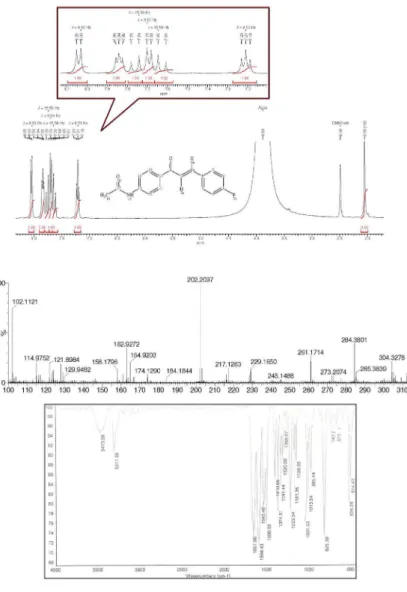
(2E )-1-(4-Acetylaminophenyl)-3-(4-fluorphenyl)prop-2-enone (3d): 80% yield; mp 119-121 ºC; FTIR ν/cm-1
3473 (H−NCO, st), 1657 (NC=O, st), 1596 (−C=O, st), 1508 (−C=C, st); 1H NMR d8.04 (d, 2H, J 8.5 Hz, 1-Aryl),
7.84 (t, 2H, 3-Aryl), 7.76 (d, 1H, J 15.6 Hz, Hβ), 7.69 (d, 2H, J 8.5 Hz, 1-Aryl), 7.63 (d, 1H, J 15.6 Hz, Hα), 7.21 (t, 2H, J 8.5 Hz, 3-Aryl), 2.06 (s, 3H, CH3); MS (ESI, positive
scan) m/z 306.1270 [M + Na]+.
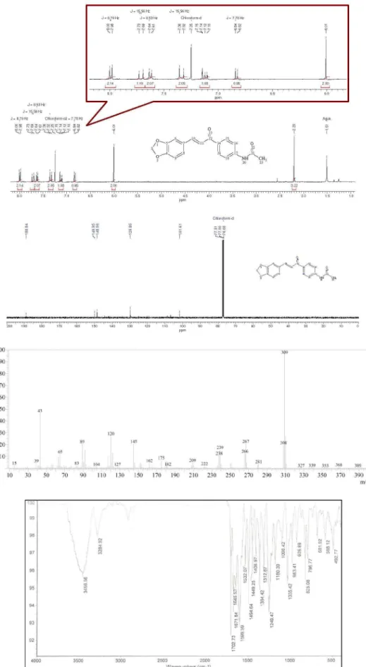
(2E )-1-(4-Acetylaminophenyl)-3-(3,4-metilendioxy-phenyl)prop-2-enone (3g): 92% yield; mp 175 ºC (dec); FTIR ν/cm-1 3455 (H−NCO, st), 1657 (NC=O, st), 1599
(−C=O, st), 1532 (−C=C, st); 1H NMR d 8.00 (d, 2H,
J 8.7 Hz, Hm, 1-Aryl), 7.72 (d, 1H, J 15.6 Hz, Hβ), 7.63 (d, 2H, J 8.6 Hz, 1-Aryl), 7.35 (d, 1H, J 15.5 Hz, Hα), 7.15 (d, 1H, J 1.5 Hz, Ho, 3-Aryl), 7.12 (dd, 1H, J 8.03, 1.5 Hz, Ho, 3-Aryl), 6.84 (d, 1H, J 8.03 Hz, Hm, 3-Aryl), 6.01 (d, 1H, J 8.0 Hz, CH2), 2.21 (s, 3H, CH3);
13C NMR d 188.8,
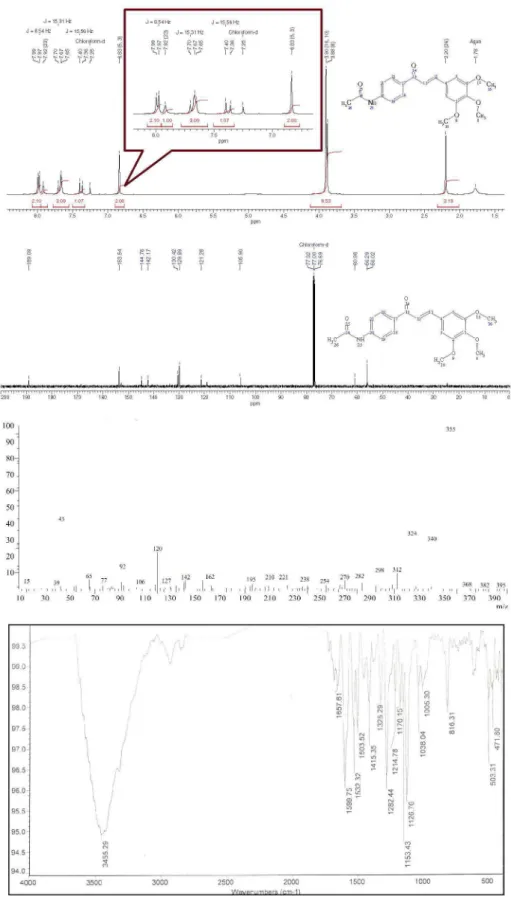
(2E)-1-(4-Acetylaminophenyl)-3-(3,4,5-trimetoxyphenyl) prop-2-enone (3h): 83% yield; mp 150 ºC (dec); FTIR ν/cm-1 3456 (H-NCO, st), 1671 (NC=O, st), 1599 (−C=O,
st), 1532 (−C=C, st); 1H NMR d 7.99 (d, 2H, J 8.5 Hz, Hm,
1-Aryl), 7.93 (s, 1H, N−H), 7.68 (m, 3H, Ho, 1-Aryl y Hα),
7.39 (d, 1H, J15.6 Hz, Hβ), 6.83 (s, 2H, Ho, 3-Aryl), 3.90 (s, 6H, m (OCH3)2), 3.88 (s, 3H, p-OCH3), 2.20 (s, 3H, CH3); 13C NMR d 189.0, 153.5, 144.7, 142.1, 130.4, 129.8, 121.2,
105.9, 60.9, 56.2, 56.0; MS m/z 355 (100, M+).
(2E)-1-(4-Acetylaminophenyl)-3-(2-pirryl)prop-2-enone (3i): 90% yield; mp 216-218 ºC; FTIR ν/cm-1 3301 (H−NCO,
st), 1669 (NC=O, st), 1647 (−C=O, st), 1527(−C=C, st);
1H NMR d 11.65 (s, 1H, NH), 9.91 (s, 1H, NH), 7.98 (d,
2H, J 8.3 Hz, 1-Aryl), 7.73 (d, 2H, J 8.3 Hz, 1-Aryl), 7.54 (broad signal, 2H, Hα and Hβ), 7.10 (broad signal, 1H, 2-pirryl), 6.69 (broad signal,1H, 2-pirryl), 6.20 (broad signal, 1H, 2-pirryl), 2.08 (s, 3H, CH3); MS (ESI, positive
scan) m/z 277.1696 [M + Na]+.
Cell cultures
Murine macrophages of the J774 cell line
Macrophages of the J774 cell line were stored in 25 cm2 polystyrene boxes (Techno Plastic Products AG,
Switzerland) in RPMI 1640 medium (Gibco BRL-Life Technologies Inc, Grand Island, NY) supplemented with 5% of fetal bovine serum and incubated at 37 ºC with 5% CO2. The culture medium was replaced every other day.32
Parasites
P r o m a s t i g o t e s o f L e i s h m a n i a p a n a m e n s i s (MHom/CO/87/UA140) were generously donated by Dr. Sara María Robledo from the Universidad de Antioquia (Colombia) and cultivated in 25 cm2 polystyrene boxes
(Techno Plastic Products AG, Switzerland) in RPMI 1640 medium (Gibco BRL-life Technologies Inc, Grand Island, NY) supplemented (cRPMI) with 5% fetal bovine serum (FBS, Microgen, Bogota, Colombia) and 1% L-glutamine (Gibco BRL-Life Technologies, Inc, Grand Island, NY) at 26 ºC. The parasites were incubated in 5 to 6 mL of medium for 6 days. To change the media, the parasite culture was placed in 15 mL polystyrene tubes (BD Biosciences, Falcon) and centrifuged at 1500 rpm for 5 min. Fresh medium was subsequently added.32
Evaluation of the cytotoxicity of chalcones against the J774 cell line
An aliquot of cell suspension (100 µL, 1 × 10⁴ cells well-1)
was used to seed 96 well flat bottom boxes (Techno Plastic
Products AG, Switzerland) for a 24 h incubation period to allow adhesion. Cells were later exposed to 100 µL of 4 different compounds prepared by a series of 1:4 dilutions in curved bottom plates of 96 wells (Techno Plastic Products AG, Switzerland) at the following concentrations: 200, 50, 12.5 and 3.25 µg mL-1. Next, the cells were incubated at
37 ºC with 5% CO2 for 72 h. Cells that were not exposed
to these compounds and wells containing only the culture medium served as controls. For positive controls for cytotoxicity, cells were exposed to the following concentrations of pentamidine isethionate (Pentacarinat®,
Sanofi-aventiz, UK): 10, 2.5, 0.625 and 0.156 µg mL-1. The
cells were later exposed to resazurin at a final concentration of 44 µmol L-1 well-1 and incubated again for 4 h under the
same conditions. Subsequently, the reduction of resazurin to resorufin was evaluated using a plate reader, Tecan GENios Microplate Reader (Tecan Trading AG, Switzerland).32,33
Evaluation of the efficiency of chalcones on Leishmania
(Viannia) panamensis promastigotes
Leishmania (Viannia) panamensis promastigotes
(2 × 10⁵ parasite well-1) were placed in a 96 well plate
containing different concentrations of compounds (200, 66.6, 22.2 and 7.4 µg mL-1) and incubated at 26 ºC for
72 h. Subsequently, 44 µmol L-1 well-1 of resazurin were
added to parasites and incubated for 48 h. Resorufin derived from viable cells was quantified using the Tecan GENios Microplate Reader (Tecan Trading AG, Switzerland). Wells with either only promastigotes or culture medium served as controls. As antileishmanial positive control, the promastigotes were exposed to pentamidine isethionate (leishmanicidal drug) in a series of four 1:3 dilutions to an initial concentration of 10 µg mL-1.32,33
Variable incubation time in the exposition to resazurin for murine and parasite cells is based in previous assays in which we evidenced a differential metabolism that depends of the kind of cellular population. Specifically J774 macrophages are faster in metabolizing (activity of mitochondria) the resazurin than the promastigotes and a period of 4 and 48 h, respectively, is necessary to obtain the maximum production of resorufin (derived from wells with viable cells without treatment).
Analysis of results
The results were analyzed using Graph Pad Prism version 5.00 statistical software (Graph Pad Software, USA) to calculate the lethal concentration 50 (LC50)
population is observed after being exposed to compounds
3a-k in cytotoxicity assays against J774. The effective concentration 50 (EC50) corresponds to the concentration
at which there is a 50% inhibition of promastigote viability with respect to that of the control. These data were obtained in efficiency assays in which compounds (3a-k) were incubated with promastigotes.
Supplementary Information
Supplementary information (Figures S1-S12) is available free of charge at http://jbcs.sbq.org.br as PDF file.
Acknowledgments
The authors thank the members of their respective research groups from the Universidad Nacional de Colombia and Universidad del Atlántico. Part of this project was financed by Bogotá Research Division (DIB) at the Universidad Nacional de Colombia (project No. 16015).
References
1. Sizova, O. V.; Ross, A. J.; Ivanova, I. A.; Borodkin, V. S.; Ferguson, M. A.; Nikolaev, A. V.; ACS Chem. Biol.2011, 6, 648.
2. Desjeux, P.; Comp. Immunol. Microbiol. Infect. Dis.2004, 27, 305.
3. Seifert, K.; Open Med. Chem. J.2011, 5, 31.
4. Barat, C.; Zhao, C.; Ouellette, M.; Tremblay, M. J.; J. Infect. Dis.2007, 195, 236.
5. De Souza, R. O.; Pereira, V. L.; Muzitano, M. F.; Falcão, C. A.; Rossi-Bergmann, B.; Filho, E. B.; Vasconcellos, M. L.; Eur. J. Med. Chem.2007, 42, 99.
6. Alvar, J.; Croft, S.; Olliaro, P.; Adv. Parasitol.2006, 61, 223. 7. Valderrama, J. A.; Zamorano, C.; González, M. F.; Prina, E.;
Fournet, A.; Bioorg. Med. Chem.2005, 13, 4153.
8. Olliaro, P. L.; Guerin, P. J.; Gerstl, S.; Haaskjold, A. A.; Rottingen, J. A.; Sundar, S.; Lancet Infect Dis.2005, 5, 763. 9. Kumar, R.; Khan, S.; Verma, A.; Srivastava, S.; Viswakarma, P.;
Gupta, S.; Meena, S.; Singh, N.; Sarkar, J.; Chauhan, P. M.; Eur. J. Med. Chem.2010, 45, 3274.
10. Kumar, S.; Shakya, N.; Gupta, S.; Sarkar, J.; Sahu, D. P.; Bioorg. Med. Chem. Lett.2009, 19, 2542.
11. Agarwal, K. C.; Sharma, V.; Shakya, N.; Gupta, S.; Bioorg. Med. Chem. Lett.2009, 19, 5474.
12. Kabri, Y.; Azas, N.; Dumètre, A.; Hutter, S.; Laget, M.; Verhaeghe, P.; Gellis, A.; Vanelle, P.; Eur. J. Med. Chem.2010, 45, 616.
13. Clark, R. L.; Carter, K. C.; Mullen, A. B.; Coxon, G. D.; Owusu-Dapaah, G.; McFarlane, E.; Duong Thi, M. D.; Grant, M. H.; Tettey, J. N.; Mackay, S. P.; Bioorg. Med. Chem. Lett. 2007, 17, 624.
14. Chauhan, S. S.; Gupta, L.; Mittal, M.; Vishwakarma, P.; Gupta, S.; Chauhan, P. M.; Bioorg. Med. Chem. Lett. 2010, 20, 6191. 15. Gupta, L.; Sunduru, N.; Verma, A.; Srivastava, S.; Gupta, S.;
Goyal, N.; Chauhan, P. M.; Eur. J. Med. Chem.2010, 45, 2359. 16. Sunduru, N.; Nishi, P. S.; Chauhan, P. M.; Gupta, S.; Eur. J.
Med. Chem.2009, 44, 2473.
17. Rane, R. A.; Telvekar, V. N.; Bioorg. Med. Chem. Lett.2010, 20, 5681.
18. Konieczny, M. T.; Konieczny, W.; Sabisz, M.; Skladanowski, A.; Wakieć, R.; Augustynowicz-Kopeć, E.; Zwolska, Z.; Eur. J. Med. Chem.2007, 42, 729.
19. Kumar, D.; Kumar, N. M.; Akamatsu, K.; Kusaka, E.; Harada, H.; Ito, T.; Bioorg. Med. Chem. Lett.2010, 20, 3916. 20. Ducki, S.; Forrest, R.; Hadfield, J. A.; Kendall, A.; Lawrence,
N. J.; McGown, A. T.; Rennison, D.; Bioorg. Med. Chem. Lett. 1998, 8, 1051.
21. Deng, J.; Sánchez, T.; Al-Mawsawi, L. Q.; Dayam, R.; Yunes, R. A.; Garofalo, A.; Bolger, M. B.; Neamati, N.; Bioorg. Med. Chem.2007, 15, 4985.
22. Kraus, G. A.; Kumar, G.; Phillips, G.; Michalson, K.; Mangano, M.; Bioorg. Med. Chem. Lett.2008, 18, 2329. 23. Sivakumar, P. M.; Seenivasan, S. P.; Kumar, V.; Doble, M.;
Bioorg. Med. Chem. Lett. 2007, 17, 1695.
24. Nielsen, S. F.; Kharazmi, A.; Christensen, S. B.; Bioorg. Med. Chem.1998, 6, 937.
25. Saravia, N. G.; Weigle, K.; Navas, C.; Segura, I.; Valderrama, L.; Valencia, A. Z.; Escorcia, B.; McMahon-Pratt, D.; Am. J. Trop. Med. Hyg.2002, 66, 738.
26. De Campos-Buzzi, F.; Padaratz, P.; Meira, A. V.; Corrêa, R.; Nunes, R. J.; Cechinel-Filho, V.; Molecules2007, 12, 896. 27. Liu, M.; Wilairat, P.; Croft, S. L.; Tan, A. L.; Go, M. L.; Bioorg.
Med. Chem. 2003, 11, 2729.
28. Nowakowska, Z.; Eur. J. Med. Chem. 2007, 42, 125. 29. Kayser, O.; Kiderlen, A. F.; Phytother. Res. 2001, 15, 148. 30. Nielsen, S. F.; Christensen, S. B.; Cruciani, G.; Kharazmi, A.;
Liljefors, T.; J. Med. Chem. 1998, 41, 4819.
31. Stokes, A.; Hastings, T.; Vrana, K.; J. Neurosci. Res. 1999, 55, 659.
32. Sánchez-Suárez, J.; Coy-Barrera, E.; Cuca, L. E.; Delgado, G.; Nat. Prod. Commun. 2011, 6, 231.
33. Escobar, L.; Rivera, A.; Aristizabal, F.; Vitae2010, 17, 67.
Submitted: April 19, 2013
Supplementary Information
Printed in Brazil - ©2013 Sociedade Brasileira de Química0103 - 5053 $6.00+0.00S
I
*e-mail: [email protected]
N
-(4-((
E
)-3-Arylacryloyl)phenyl)acetamide Derivatives and
their Antileishmanial Activity
Dency J. Pacheco,a Jorge Trilleras,*,a Jairo Quiroga,b Jennifer Gutiérrez,c Luis Prent,a
Tobinson Coavas,a Juan C. Marínd and Gabriela Delgadoc
aGrupo de Investigación en Compuestos Heterocíclicos, Programa de Química,
Facultad de Ciencias Básicas, Universidad del Atlántico, km 7 Antigua vía Puerto Colombia, Barranquilla-Atlántico, Colombia
bGrupo de Investigación de Compuestos Heterocíclicos, Departamento de Química,
Universidad del Valle, A. A. 25360, Cali, Colombia
cGrupo de Investigación en Inmunotoxicología and dGIFFUN, Departamento de Farmacia,
Facultad de Ciencias, Universidad Nacional de Colombia, A. A. 14490, Bogotá, D. C., Colombia