Printed in Brazil - ©2005 Sociedade Brasileira de Química 0103 - 5053 $6.00+0.00
A
r
ti
c
le
* e-mail: wgarcez@nin.ufms.br
Indole Alkaloid and other Constituents from
Ocotea minarum
Walmir S. Garcez*, Fernanda R. Garcez, Lillian May G. E. da Silva and Angela A. Shimabukuro
Departamento de Química, Universidade Federal de Mato Grosso do Sul, 79070-900 Campo Grande-MS, Brazil
Dos frutos de Ocotea minarum foram isolados um novo alcalóide indólico, triptofol-5-O-β -D-glicopiranosídeo, a cumarina escopoletina e os flavonóides taxifolina, quercetina-7-O-β -D-glicopiranosídeo, eriodictiol-3’-O-β-D-glicopiranosídeo e naringenina-7-O-β-D-glicopiranosídeo. Do cerne foi isolado um novo alquil fenol, 3-(1,4-diidroxipentil)-metoxifenol, além de 5-propilresorcinol, trans-asarona, lioniresinol, 3-O-β-D-glicopiranosil estigmasterol e estigmasta-4,22-dien-3-ona. Da casca do caule foram obtidos o sesquiterpeno ácido lanceólico (como seu derivado éster metílico após metilação com diazometano) e β-sitosterol. O ácido lanceólico e 5-propilresorcinol estão sendo relatados pela primeira vez como produtos naturais.
From the fruits of Ocotea minarum a new indole alkaloid, tryptophol-5-O-β-D-glucopyranoside, was isolated in addition to the coumarin scopoletin and the flavonoids taxifolin, quercetin-7-O-β -D-glucopyranoside, eriodictyol-3’-O-β-D-glucopyranoside and naringenin-7-O-β-D-glucopyranoside. A new alkyl phenol, 3-(1,4-dihydroxypentyl)-5-methoxyphenol, was obtained from the heartwood in addition to 5-propylresorcinol, trans-asarone, lyonyresinol, 3-O-β-D-glucopyranosyl stigmasterol and stigmasta-4,22-dien-3-one, whereas from the trunk bark the sesquiterpene lanceolic acid (as its methyl ester derivative after methylation procedures) and β-sitosterol were isolated. 5-propylresorcinol and lanceolic acid are reported for the first time as natural products.
Keywords: Ocotea minarum, Lauraceae, indole alkaloid, lanceolic acid, alkyl-phenols
Introduction
In continuation of our program on chemical investi-gation of Lauraceous plants occurring in the “Pantanal” and “Cerrado” of Mato Grosso do Sul, Brazil, the fruits,
heartwood and trunk bark components of Ocotea minarum
(Meissn.) Mez., an endemic plant which has been found up to now only in the “Cerrado”, have been examined.
A previous work on the leaves of a specimen identified as O. minarum which was collected in Minas Gerais, Brazil,
resulted in the isolation of fourteen aporphinic alkaloids.1
However, no alkaloids have been detected in the leaves of the specimen investigated in the present work. The great morphological resemblance among the species belonging to the Lauraceae is well known and sometimes this may lead to misidentification of the specimen under investigation.
We describe herein the isolation of an indole alkaloid
(1), a coumarin (2) and four flavonoids (3 – 6) from
the fruits, a lignan (7), two alkyl phenols (8, 9), a aryl propene derivative (10) and two steroids (11, 12) from the
heartwood and a sesquiterpene (13) and a steroid (14) from
the trunk bark of a specimen of Ocotea minarum collected
in Campo Grande, MS State.
Results and Discussion
After a series of partition procedures and a combination of column chromatography on silica gel, gel filtration and reversed phase HPLC separations of the ethanol extract
from the fruits, the indole alkaloid tryptophol-5-O-β
-D-glucopyranoside (1) was isolated, together with the
coumarin scopoletin (2) and the flavonoids taxifolin (3),
quercetin-7-O-β-D-glucopyranoside (4),
eriodictyol-3’-O-β-D-glucopyranoside (5) and prunin (naringenin-7-O-β
-D-glucopyranoside, 6).
The positive ESI-mass spectrum of 1 showed a quasi
-molecular ion at m/z 362 (M + Na)+ accordingly to a
molecular formula C16H21NO7. Its 1H NMR spectrum
displayed signals attributable to a 1,2,4-trisubstituted aromatic ring (δ 7.21, d, J 2.0 Hz; δ 6.85, dd, J 8.0 and 2.0 Hz; δ 7.12, d, J 8.0 Hz) in addition to a broad singlet at δ
Hz) and signals ascribed to a sugar moiety (δ 4.85 – 3.20)
were observed. The sugar moiety was assigned as β
-D-glucopyranose on the basis of 1H and 13C NMR data and
the J value for the anomeric hydrogen at δ 4.85 (d, J 7.5
Hz). The presence of a disubstituted indole moiety was
suggested by the chemical shifts of the eight sp2 carbons
in the 13C NMR spectrum (Table 1).2 A downfield signal at
δ 152.7 indicated that the O-glucosyl residue was located
para to the nitrogen atom of the indole nucleus, at the
C-5 position. This assumption was also supported by the upfield signals in this spectrum attributed to C-4 and C-6
(δ 106.8 and 114.2, respectively) and the long-range HMBC
correlations of H-4, H-6, H-7 and H-1’ of the glucosyl
moiety with C-5 (Table 1). The presence of a β
-hydroxy-ethyl substituent at C-3 could be inferred by the two
methylene carbon signals at δ 29.7 and 62.6 which in turn
showed cross-peak correlations with the two
afore-mentioned triplets at δ 2.82 and 3.80, respectively, in the
HMQC spectrum. This information was corroborated by the HMBC correlations between the signals of H-10 and that of C-2 and between the signals of H-4 and H-10 and
that of C-3. Therefore, compound 1 is thus unambiguously
shown to be the 5-O-glucosyl derivative of tryptophol (indole-3-ethanol). Tryptophol has been isolated as a constituent of higher plants, fungi and marine organisms
and is considered to be a plant auxin,3 however its
5-O-glucosyl derivative 1 was hitherto unreported in the
literature.
Column chromatography on silica gel of a crude
ethanol extract obtained from the heartwood of Ocotea
minarum followed by Sephadex LH-20 separations
afforded the lignan lyonyresinol (7), the alkyl phenols
5-propylresorcinol (8) and
3-(1,4-dihydroxypentyl)-5-methoxyphenol (9), the aryl propene derivative trans
-asarone (10) and the steroids 3-O-β-D-glucopyranosyl
stigmasterol (11) and stigmasta-4,22-dien-3-one (12).
The 1H NMR spectrum of 8 showed characteristic
signals of a resorcinol derivative (two broad singlets at δ
6.28 and 6.16, the latter integrating for two hydrogens) containing an n-propyl group at C-5 (two triplets at δ 0.83,
J 6.6 Hz and 2.39, J 7.5 Hz and one multiplet at δ 1.50).
The signal of the phenol hydrogens, which disappeared
after addition of D2O, was observed at δ 5.86. Accordingly,
in the 13C NMR spectrum of 8, four signals at δ 146.1,
107.9, 156.7 and 100.2 accounted for a symmetrically substituted resorcinol ring, while resonances of two
methylene and one methyl carbons at δ 32.0, 36.0 and
14.1, respectively, were indicative of an n-propyl
substituent at C-5. Compound 8 was thus identified as
5-propylresorcinol, which has been previously detected as one of the constituents of the secretion of the ant
Crematogaster deformis 4 and not reported in higher plants
before now.
The molecular formula of 9 was deduced as C12H18O4
on the basis of the quasi-molecular ion peak at m/z 249
[M+Na]+ observed in its positive ESI-mass spectrum and
information provided by its 1H and 13C (including DEPT)
NMR spectra. In the 1H NMR spectrum of 9, the presence
of an 1,3,5-trisubstituted aromatic ring was inferred by the
three broad singlets at δ 6.41, 6.38 and 6.24 (Table 2). This
spectrum also showed signals attributable to one methoxyl
(δ 3.73) and an aliphatic chain bearing two carbinolic
hydrogens, as revealed by a triplet at δ 4.48 (1H, J 7.1 Hz)
and a multiplet partly obscured by the OMe singlet (δ
3.73). In the HMQC spectrum, cross-peak correlations between the signals of these carbinolic hydrogens and
carbon resonances at δ 75.2 and 68.5, respectively,
Table 2.1H (300 MHz) and 13C (75 MHz) NMR spectral data for 9
(δ, CDCl3)
C/H δ Ha δ C
1 - 159.5
2 6.41 br s 101.0
3 - 148.9
4 6.38 br s 104.0
5 - 162.3
6 6.24 br s 106.6
1 ’ 4.48 t (7.1) 75.2
2 ’ 1.72 m 36.30
3 ’ 1.31 m 36.33
4 ’ 3.73 m 68.5
5 ’ 1.22 d (6.2) 23.5
6 ’ 3.73 s 55.6
a Coupling constants (J in Hz) are given in parentheses.
Table 1. 1H (300 MHz) and 13C (75 MHz) NMR spectral data for 1
(δ, CD3OD)
C/H δ Ha δ C
6.96 s 124.7
3 _ 112.8
4 7.21 d (2.0) 106.8
5 _ 152.7
6 6.85 dd (8.0, 2.0) 114.2
7 7.12 d (8.0) 112.5
8 _ 134.5
9 _ 124.2
1 0 2.82 t (7.2) 29.7
1 1 3.80 t (7.2) 62.6
1 ’ 4.85 d (7.5) 104.2
2 ’ 3.47b 75.1
3 ’ 3.45b 78.1
4 ’ 3.41b 71.6
5 ’ 3.28b 78.1
6 ’ 3.49b3.64b 63.7
a Coupling constants (J in Hz) are given in parentheses; b
confirmed these assignments. The 13C NMR spectrum of 9
displayed then signals for twelve carbons, six of which could be attributed to the aromatic carbons, one to the OMe group and the five remaining to the aliphatic carbons of the aromatic ring side-chain (Table 2). The chemical
shift values of δ 162.3 and 159.5 were consistent with the
presence of a methoxyl and a hydroxyl respectively, in a
meta orientation with respect to each other in the
1,3,5-trisubstituted aromatic ring, while the signal at δ 148.9
was ascribed to the aromatic carbon bearing the alkyl chain. One of the methine carbons of this side-chain was shown to be directly attached to the aromatic ring on the basis of the correlations observed in the HMBC spectrum
between its corresponding carbinol hydrogen (δ 4.48) and
the aromatic carbons C-2, C-3 and C-4. Likewise, the location of the second hydroxyl group at C-4’ was inferred
by the appearance of the C-5’ methyl as a doublet at δ 1.22
(J = 6.2 Hz), which in turn showed one-bond 1H-13C
connectivity with the carbon signal at δ 23.5 (C-5’) and
by the long-range correlations of the carbinolic hydrogen
H-4’ with C-2’, C-3’ and C-5’. The structure of 9, which is
being described for the first time in the literature, was thus established as 3-(1,4-dihydroxypentyl)-5-methoxyphenol.
A crude ethanol extract of the trunk bark of O. minarum
was partitioned between methanol-H2O (9:1) and hexane.
Sesquiterpene 13 (Lanceolic acid) and β-sitosterol (14)
were obtained from the hexane soluble fraction after
column chromatography on silica gel. Compound 13 was
isolated and characterized after methylation with
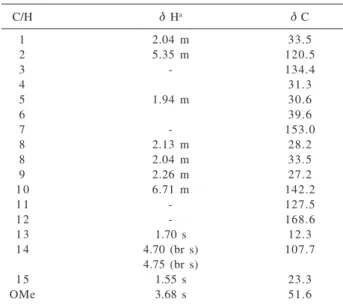
diazomethane as its methyl ester derivative 13a. The 1H
NMR spectrum of 13a showed, in addition to a singlet at δ
3.68 assignable to a carbomethoxy group, signals for two
vinylic hydrogens (two multiplets at δ 5.35 and 6.71), an
exocyclic methylene group (two broad singlets at δ 4.70
and 4.75) and two olefinic methyl groups at δ 1.55 and
1.70 (Table 3). The 13C NMR data (including DEPT) of
13a resembled closely those of the sesquiterpene β
-bisabolene,5 except for the deshielding and shielding shifts
of the C-10 and C-11 resonances, respectively, caused by
the presence of a carbomethoxy function in 13a in place
of the C-12 methyl group (Table 3). The stereochemical assignments for the 10,11-trisubstituted double bond in
13a were confirmed as (E) on the basis of the proton and
carbon resonances of methyl-13 (δ 1.70 and 12.3,
respectively), which were indicative of its cis relationship
with C-9.6 Further evidence for the structure 13a was
provided by two- and three-bond correlations discernible in the HMBC spectrum (Table 3). Since the optical rotation
of 13a showed a negative value, the (S) stereochemistry at
C-6 is proposed, similarly to other analogous
(S)-(E)-bisabolene derivatives.7,8 Therefore, compound 13a was
characterized as the methyl ester of lanceolic acid 13.
These two compounds were previously obtained by
synthesis and only incomplete 1H NMR assignments have
been published for 13a.7, 9 Therefore, 13 is reported for the
first time as a genuine natural product.
The structures of the known compounds 2 - 7, 10 - 12
and 14 were identified by comparison of their 1H and 13C
NMR data with those found in the literature and/or with
authentic samples.10
The isolation of 1 from Ocotea minarum is noteworthy
for its chemosystematic relevance, since members of the Lauraceae have been reported to contain aporphine and
benzyltetrahydroisoquinoline but not indole alkaloids.11
It is also worth of mention in this work the compart-mentalized accumulation of the secondary metabolites
present in O. minarum: the indole alkaloid and the
flavonoid derivatives are accumulated in the fruits, whereas the alkyl phenols, the aryl propene derivative and the lignan were isolated from the heartwood and the sesquiterpene from the trunk bark.
Experimental
General experimental procedures
IR spectra were recorded as KBr pellets on a Bomem-Hartmann & Braun FT IR spectrometer. The
uni-dimensional 1H and 13C and the two dimensional 1H-1H
COSY, HMQC and HMBC NMR spectra were recorded at
300 MHz (1H) and 75 MHz (13C) on a Bruker DPX-300
spectrometer. Standard pulse sequences were used for
Table 3.1H (300 MHz) and 13C (75 MHz) NMR spectral data for 13a
(δ, CDCl3)
C/H δ Ha δ C
1 2.04 m 33.5
2 5.35 m 120.5
3 - 134.4
4 31.3
5 1.94 m 30.6
6 39.6
7 - 153.0
8 2.13 m 28.2
8 2.04 m 33.5
9 2.26 m 27.2
1 0 6.71 m 142.2
1 1 - 127.5
1 2 - 168.6
1 3 1.70 s 12.3
1 4 4.70 (br s) 107.7
4.75 (br s)
1 5 1.55 s 23.3
homo- and heteronuclear correlation experiments. ESIMS spectra were obtained using a Micromass Platform II single quadrupole mass spectrometer (Faculdade de Ciências Farmacêuticas, USP, Ribeirão Preto, SP, Brazil). Optical rotations were determined on a Perkin-Elmer 341 polarimeter. Silica gel 60 (70-230 and 230-400 mesh) and Sephadex LH-20 were used for column chromatography. Reversed phase semi-preparative HPLC separations were performed with a Shimadzu LC-6AD pump, using a
RP-18, 25x250 mm, 5μm particle size, Shim-Pack
PREP-ODS(H) column, with a flow rate of 10 mL min-1 and
monitoring at 254 nm.
Plant material
Fruits, heartwood and trunk bark of Ocotea minarum
(Meissn.) Mez. were collected in Campo Grande, Mato Grosso do Sul, Brazil, in December 2001. The plant material was identified by Dr. João Batista Baitello (Horto Florestal – São Paulo, SP, Brazil) and a voucher specimen (No. 11467) was deposited at the CGMS Herbarium, Universidade Federal de Mato Grosso do Sul, MS, Brazil.
Extraction and isolation of chemical constituents
Ground fruits (2.5 kg) were extracted at room temperature with EtOH. The residue obtained from the
EtOH extract was partitioned between hexane-CH3
CN-CHCl3-H2O (20:34:10:10) originating two phases: the
organic upper layer and the hydro-organic lower layer. The first contained only fatty material which was not further investigated. The hydro-organic phase was
concentrated in vacuo and the residue extracted with
EtOAc and then with n-BuOH. The EtOAc extract (6.5 g)
was subjected to CC on silica gel (230-400 mesh, 50 g,
CHCl3-MeOH gradient) to afford 2 (3.8 mg) and 3 (16.9
mg). The n-BuOH extract (6.4 g) was chromatographed on
a Sephadex LH-20 column (30 g, four portions of 1.6 g each, MeOH) to provide fifty fractions of 5 mL each. The fractions showing similar spots by TLC were combined to
give eight fractions (A→H). Fraction E was further
subjected to CC on Sephadex LH-20 (30 g, MeOH) to yield fifty fractions of 5 mL each. From these, fractions
8-14 consisted of 1 (25.0 mg), fractions 19-27 yielded 6 (4.5
mg) after semi-preparative HPLC (MeOH-H2O 1:1),
fractions 38-42 afforded 4 (9.8 mg) and fractions 47-49
gave 5 (5.3 mg).
Air dried and powdered heartwood (900 g) was extracted at room temperature with EtOH. After
concentration in vacuo, the residue (29.0 g) was applied
to a silica gel CC (70-230 mesh, 100 g, hexane-EtOAc and
EtOAc-MeOH gradients) to give nine fractions (A→I) of
100 mL each. Fraction D (683 mg) afforded 10 (6.4 mg)
and 11 (8.3 mg) after CC on silica gel (230-400 mesh, 50
g, hexane-EtOAc 7:3), followed by a second CC (silica gel
230-400 mesh, 30 g, CHCl3-CH2Cl2 99.5:0.5). Fractions
E-G (1.3 g) were subjected to CC on Sephadex LH 20 (50
g, CHCl3-MeOH 4:1) to provide forty-two fractions of 10
mL each. Fractions 4-7 were rechromatographed on a silica
gel column (230-400 mesh, 50 g, CHCl3-MeOH 1:1) to
yield 8 (7.3 mg) and 9 (5.3 mg). Fraction H (900 mg) was
again separated by CC on Sephadex LH-20 (30 g,
EtOAc-MeOH 1:1) to give 7 (11.5 mg) and 12 (10.6 mg).
Air-dried and powdered trunk bark (2.7 kg) were extracted at room temperature with EtOH. After
concentration in vacuo, the residue was partitioned
between MeOH-H2O 9:1 and hexane. The hexane phase
(5.0 g) was separated on a silica gel column (230-400 mesh, 45 g) eluted with a gradient of hexane-EtOAc 8:2 and hexane-EtOAc-MeOH 9:1:0.2, 8:2:1 and 7:3:1 yielding one hundred fractions of 10 mL each. Fraction
20 consisted of 14 (465.0 mg). Fraction 8 (600 mg)
consisted of a complex mixture from which 13 was
isolated as the corresponding methyl ester derivative 13a
(10 mg) after treatment of part of this fraction (300 mg) with an ethereal solution of diazomethane followed by successive CC separations on silica gel (230-400 mesh, 50 g, hexane-EtOAc gradient).
Tryptophol-5-O-β-D-glucopyranoside (1). Colorless amorphous solid. [α]: - 29.3o (MeOH; c 0.3). IR (KBr) ν
max/
cm-1: 3400, 2927, 1628, 1482, 1202, 1074, 1041, 803. UV
λmax/nm (log ε) (MeOH): 225 (2.7), 280 (sh). ESIMS m/z
362 [M+Na]+. 1H and 13C NMR: see Table 1.
5-propylresorcinol (8). Brownish oil. 1H NMR (CDCl 3):
δ 0.83 (3H, t, J 6.6 Hz, H-3’), 1.50 ( 2H,m, H-2’), 2.39 ( 2H, t, J 7.5 Hz, 1’), 5.86 (2H, br s, 2 x OH), 6.16 (2H, br s,
H-2, H-4), 6.28 (1H, br s, H-2).13C NMR (CDCl
3): δ 14.1
(C-3’), 32.0 (C-2’), 36.0 (C-1’), 100.2 (C-4), 107.9 (C-2, 6), 146.1 (C-1), 156.7 (C-3, 5).
3-(1,4-dihydroxypentyl)-5-methoxyphenol (9). Colorless amorphous solid. [α]: - 10.0o (MeOH; c 0.17). IR
(KBr) νmax/ cm
-1: 3401, 2926, 2854, 1598, 1456, 1437,
1337, 1301, 1154, 1054, 840, 699. ESIMS m/z 249
[M+Na]+. 1H and 13C NMR: see Table 2.
Methyl lanceolate (13a). Colorless oil. [a]: - 58.0o
(MeOH; c 0.25). IR (KBr) νmax/ cm
-1: 1720, 1652, 1135,
Acknowledgments
The authors are grateful to Fundect-MS, CPq-PROPP-UFMS and PROAP-CAPES for their financial support and to CAPES and CNPq for the scholarships awards. Thanks are also due to Dr. Norberto P. Lopes (Faculdade de Ciências Farmacêuticas, USP, Ribeirão Preto, Brazil) for the ESIMS spectra.
References
1. Vecchietti, V.; Casagrande, C.; Ferrari, G.; Severini Ricca, G.;
Farmaco-Ed. Sci.1979, 34, 829.
2. Pouchert, C. J.; Behnke, J.; The Aldrich Library of 13C and 1H
FT NMR Spectra, Aldrich Chemical Co.:Milwaukee, 1993, vol. 3.
3. Rayle, D. R.; Purves, W. K.; Plant Physiol.1967, 42, 520; Erdogan, I.; Sener, B., Higa T.; Biochem. Syst. Ecol.2000, 28, 793; Sugawara, F.; Strobel, G. A.; Phytochemistry1987, 26, 1349.
4. Attygalle, A. B.; Siegel, B.; Vostrowsky, O.; Bestmann, H. J.; Maschwitz, U.; J. Chem. Ecol.1989, 15, 317.
5. Rahman, A.; Ahmad, V. U.; 13C NMR of Natural Products Vol. 1, Monoterpenes and Sesquiterpenes, Plenum Press: New York, 1992.
6. Breitmaier, E.; Voelter, W.; 13C NMR Spectroscopy, 2nd ed.,
Verlag Chemie: New York, 1978.
7. Manjarrez, A.; Ríos, T.; Guzmán, A.; Tetrahedron1954, 20, 333.
8. Crawford, R. J.; Erman, W. F.; Broaddus, C. D.; J. Am. Chem. Soc.1972, 94, 4298.
9. Katzenellenbogen, J. A.; Crumrine, A. L.; J. Am. Chem. Soc
1976, 98, 4925.
10. Agrawal, P. K.; Thakur, R. S.; Bansal, M. C. In Carbon-13 NMR of Flavonoids; Agrawal, P. K., ed.; Elsevier: Amsterdam, 1989, ch. 3; Agrawal, P. K.; Bansal, M. C. In Carbon-13 NMR of Flavonoids; Agrawal, P. K., ed.; Elsevier: Amsterdam, 1989, ch. 6; Agrawal, P. K.; Thakur, R. S.; Magn. Reson. Chem.
1985, 23, 389; González, M. C.; Sentandreu, M. A.; Rao, K. S.; Zafra-Polo, M. C.; Cortes, D.; Phytochemistry 1996, 43, 1361; Kojima, H.; Sato, N.; Hatano, A.; Ogura, H.;
Phytochemistry 1990, 29, 235; Almeida, M. L. S.; Kocovský, P.; Bäckvall, J-E.; J. Org. Chem. 1996, 61, 6587.
11. Garcez, W. S.; Yoshida, M.; Gottlieb, O. R.; Phytochemistry
1995, 39, 815.
Received: June 8, 2005