Printed in Brazil - ©2005 Sociedade Brasileira de Química 0103 - 5053 $6.00+0.00
Article
* e-mail: [email protected]
New Biflavonoid and Other Flavonoids from the Leaves of
Chimarrhis turbinata
and their Antioxidant Activities
Carmem L. Cardoso, Dulce H. S. Silva, Ian Castro-Gamboa and Vanderlan da S. Bolzani*
Instituto de Química, Universidade Estadual Paulista CP 355, 14800-900 Araraquara-SP, Brazil
Um novo biflavonol denominado chimarrosídeo (1)e oito flavonóis glicosilados adicionais (2-9) foram isolados das folhas de Chimarrhis turbinata. Suas estruturas foram elucidadas com base nos dados dos experimentos de RMN 1D e 2D, como: 3-O-rutinosilquercetina (2), 3-O -rutinosil-kaempferol (3), 3-O-galactopiranosil-(6→1)-ramnopiranosil kaempferol (4), 3-O-β
-galactopiranosil-(6→1)-α-ramnopiranosil-quercetina (5), 6-hidroxirutina (6), 3-
O-galactopiranosil-kaempferol (7), 3-O-glucopiranosil-kaempferol (8) e 3-O- ramnopiranosil-(6→1)-glucopiranosil-(4→1)-ramnopiranosil-kaempferol (9). Adicionalmente, a catequina (10)
e a procianidina B-3 (catequina-(4α→8)-catequina) (11) também foram isoladas. O extrato
bruto, frações e compostos isolados foram avaliados quanto às suas propriedades antioxidantes no teste em CCD aspergida com solução de β-caroteno, e teste espectrofotométrico utilizando o radical livre 1,1-difenil-2-picrilidrazila (DPPH). Os flavonóides 2, 5, 6, 10 e 11 apresentaram forte atividade antioxidante quando comparados com os padrões BHT e rutina.
A new biflavonol, named chimarrhoside (1), and eight known flavonol glycosides (2-9), were isolated from the leaves of Chimarrhis turbinata. Their structures were established on the basis of 1D and 2D NMR experiments as quercetin-3-O-rutinoside (2), kaempferol-3-O-rutinoside (3), kaempferol-3-O-α-L-rhamnopyranosyl-(1→6)-β-D-galactopyranoside (4), quercetin-3-O
-α-L-rhamnopyranosyl-(1→6)-β-D-galactopyranoside (5), 6-hydroxy-rutin (6), kaempferol-3-O-D-galactopyranoside (7), kaempferol-3-O-D-glucopyranoside (8) and kaempferol-3-O-α -L-rhamnopyranosyl-(1→6)-α-L-rhamnopyranosyl-(1→4)-β-D-glucopyranoside (9). In addition,
catechin (10)and catechin-(4α→8)-catechin-procyanidin B-3) (11) were isolated. The crude
extract, fractions and isolated compounds were evaluated for their antioxidative properties using an autographic assay based on β-carotene bleaching on TLC plates, and spectrophotometric detection by reduction of the stable 1,1-diphenyl-2-picrylhydrazyl (DPPH) free radical. Flavonoids 2, 5, 6, 10 and 11 displayed strong free radical scavenging activity, when compared with the standards BHT and rutin.
Keywords: Rubiaceae, chimarrhoside, DPPH, β-carotene
Introduction
In recent years, flavonoids have been widely recognized as a major class of secondary metabolites with antioxidant properties due to their ability to scavenge free radicals.1-3 Antioxidant is a broad classification for
molecules that may act prior to, or during, a free radical chain reaction at initiation, propagation, termination, decomposition, or subsequent reaction of oxidation products as sensitive targets.4 There are several diseases,
whose causes and severity are linked to oxidation, mainly those associated with oxygen free radicals, which have
been implicated as mediators of degenerative and chronic deteriorative, inflammatory, and autoimmune diseases,5
such as rheumatoid arthritis,6 diabetes, vascular disease
and hypertension, cancer and hyperplasic diseases,7
cataract formation and aging processes.8 Radical-mediated
pathologies such as ischemia reperfusion, asthma and many others involving an imbalance in pro-oxidant-antioxidant processes.9 In our search for antioxidant
compounds from Amazonian plant species, we examined constituents of Chimarrhis turbinata DC Rodr.. (Rubiaceae) leaves, collected in the city of Belém, Pará State, Brazil. The chemical composition of C. turbinata
of a new biflavonoid, named chimarrhoside (1), and eight known flavonoids, as well as the antioxidant effects, which were deduced by electrochemical detection coupled to an HPLC system, bleaching of β-carotene on TLC plates, and parameters related to the evaluation of antioxidant activity measured by free radical scavenging ability towards DPPH (1,1-diphenyl-2- picrylhydrazyl).
Results and Discussion
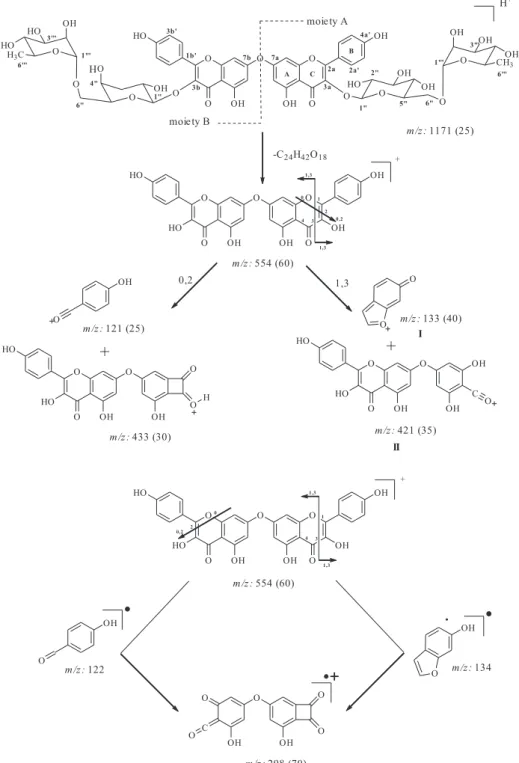
Chimarrhoside (1) was obtained as an amorphous pale yellow powder from the EtOH extract and exhibited in its mass spectrum a molecular ion peak at m/z 1171 [M+H]+ obtained from ESIMS. The IR and UV spectra of 1
revealed the presence of hydroxyl (3410 cm-1) and phenolic
groups (270, 295, 344 nm, 1690 cm-1). Part of the 1H and 13C NMR signals of compound 1 were observed in
duplicate, suggesting a dimer (Table 1). Comparison of the 13C NMR values of 1 with literature data13 evidenced
the kaempferol aglycone and additional similar spectral features to those of 3, also isolated in this work, except for the duplication of some signals for carbons arising from a dimer structure. In the 1H NMR spectrum of 1,
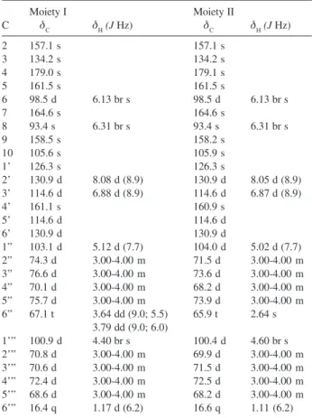
signals for two kaempferol units as two meta-coupled A-ring hydrogens (H-6/H-8) and two pairs of coupled B-ring hydrogens (H-3'/H-5' and H-2'/H-6') for each aglycone were observed, in addition to four anomeric hydrogens at δ 5.12 (1H, d, J 7.7 Hz), 5.02 (1H, d, J 7.7 Hz), 4.40 (1H, br s), 4.60 (1H, br s), 20 hydroxymethine and 2 hydroxymethylene hydrogens. The 13C NMR
spectrum of 1 revealed the presence of two kaempferol moieties and 24 hydroxymethine and hydroxymethylene signals which, analyzed together with 1H NMR data (Table
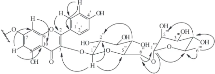
1) were consistent with the presence of two diglycoside moieties identified as rutinosyl and α -L-rhamnopyranosyl-(1→6)-β-D-galactopyranoside. This assumption was supported by signals at δ 103.1 / 67.1 and δ 104.0 / 65.9, assigned to C-1'’ C-6'’ of the glucopyranosyl and of the galactopyranosyl moiety respectively. Those signals also showed HMQC correlations with H-1'’ (anomeric) and H-6'’ (Table 1); as well as signals at δ 100.9/16.4 and δ 100.4/16.6, assigned to C-1’’’/C-6’’’ of the two rhamnopyranosil moieties, respectively, which also showed HMQC correlations with H-1'’’ (δ 4.4 0 and 4.60) and H-6’’’ (δ 1.17 and 1.11). On the basis of the above data, the chemical shifts assigned for each sugar: the glucosyl and galactosyl moieties were confirmed taking into account the values at δ 103.1, 76.6, 75.7, 74.3, 70.1 and 67.1 as well as δ 104.0, 73.9, 73.6, 71.5, 68.2 and 65.9 attributed to glucose and galactose, respectively.15
According to literature data, using the same solvent, the
chemical shifts of hydroxymethines carbons in glucose are quite deshielded when compared to those of galactose. The glucosyl/galactosyl groups are linked to the rhamnosyl residue at C-6”, because the signal assigned to this carbon, in both sugar moieties, was markedly displaced downfield at δ 67.1/65.9 (∆δ +6), respectively when compared with the 13C NMR spectrum of free hydroxymethylenes in
glucose/galactose. This also was confirmed by the observation of a HMBC long-range correlation of the anomeric hydrogen signal of the rhamnosyl group at δ 4.40 / 4.60 with C-6” of the glucosyl moieties at 67.1 and 65.9, respectively.
Additionally, the site of glycosylation at C-3 of each kaempferol moiety was evidenced from downfield shifts of C-2 (∆δ +9) and C-4 (∆δ +2) and upfield shift of C-3 (∆δ -3) when compared to the aglycone,15 as well as from
HMBC correlations of H-1'’ to C-3a/3b within each kaempferol diglycoside moiety (Figure 1). Further HMBC correlations of H-6 and H-8 with C-7 (δ 164.6) confirmed the assignments for ring A and suggested the interflavonoidic linkage at C-7 due to the observed downfield shift of its signal when compared to the monomer.15 Moreover, ESIMS analysis showed peaks of
m/z 298 (70) and 133 (40) resulting from Retro Diels-Alder (RDA) fragmentations. From moiety A wherein bonds 1 and 3 undergo scission, lead to formation of ions I and II as well as scission of bonds 0 and 2 led to the formation of fragment m/z 433 (30). The fragment m/z
298 (70), was generated by both RDA suffered by moiety A and moiety B (Figure 2). Additional fragments observed in the mass spectra are in accordance with those reported for kaempferol.16 These data, combined with HPLC
analysis, which showed a lower retention time for compound 1 (5.7 min) when compared to monomeric flavonol diglycosides 2 (9.8 min) and 3 (13.8 min.) (Figure 3), confirmed the proposed structure for compound 1 as kaempferol 3-O-β-rutinosyl-(7α→O→7β
)-kaempferol-3-O-β-L-rhamnopyranosyl-(1→6)-β-D-galactopyranoside. Further studies of the EtOAc extract from leaves of C. turbinata resulted in 10 known flavonoids already reported in the literature: Quercetin-3-O-rutinoside (2), kaempferol-3-O-rutinoside (3), kaempferol-3-O
Chimarrhis turbinata
Figure 2. Selected fragment ions from compound 1 by ESIMS.
pyranosyl-(1→6)-β-D-galactopyranoside (4), quercetin-3-O-α-L-rhamnopyranosyl-(1→6)-β-D-galactopyranoside (5), 6-hydroxy-rutin (6), kaempferol-3-O-D- galacto-pyranoside (7), kaempferol-3-O-D-glucopyranoside (8) and kaempferol-3-O-α-L-rhamnopyranosyl-(1→6)-α -L-rhamnopyranosyl-(1→4)-β-D glucopyranoside (9), catechin (10) and catechin-(4α→8)-catechin (procyanidin B-3) (11). These compounds were identified and their
structures were established on the basis of 1D and 2D NMR experiments and compared with literature data (Figure 4).13-15, 18, 19-22, 26-28
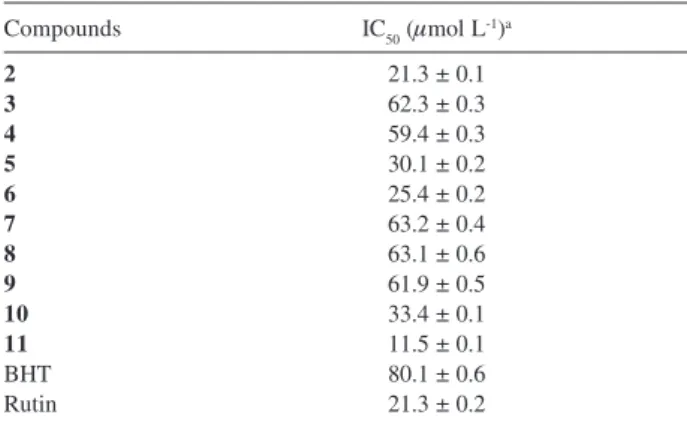
The radical scavenging effects obtained for compounds
2-11 assayed with DPPH are shown in (Table 2) using as reference the antioxidant standard rutin (IC50 21.34 μg mL-1) and BHT (IC
50 62.50 μg mL
-1). These results indicate
micromolecules is due to its hydrogen-donating ability, provided by the ease of stabilization of the phenoxyl radical after reduction of DPPH and is enhanced by the presence of catechol groups and the α,β-unsaturated carbonyl moiety, as evidenced by the IC50 values for compounds 2, 5, 6, 10, and 11.24
It is also evident from Table 2, that ortho-dihydroxy moiety plays more important role for this type of activity than the α,β-unsaturated carbonyl, as the lack of the latter, e.g. in compound 10, contributed in a lesser extent to the loss of activity, as observed for compounds 3, 4 and 7-9, due to the missing catechol group. On the other side, the
Table 1. 1H and 13C NMR data for Dimmer 1 in methanol-d 4
a
Moiety I Moiety II
C δC δH(J Hz) δC δH(J Hz)
2 157.1 s 157.1 s
3 134.2 s 134.2 s
4 179.0 s 179.1 s
5 161.5 s 161.5 s
6 98.5 d 6.13 br s 98.5 d 6.13 br s
7 164.6 s 164.6 s
8 93.4 s 6.31 br s 93.4 s 6.31 br s
9 158.5 s 158.2 s
10 105.6 s 105.9 s
1’ 126.3 s 126.3 s
2’ 130.9 d 8.08 d (8.9) 130.9 d 8.05 d (8.9) 3’ 114.6 d 6.88 d (8.9) 114.6 d 6.87 d (8.9)
4’ 161.1 s 160.9 s
5’ 114.6 d 114.6 d
6’ 130.9 d 130.9 d
1” 103.1 d 5.12 d (7.7) 104.0 d 5.02 d (7.7) 2” 74.3 d 3.00-4.00 m 71.5 d 3.00-4.00 m 3” 76.6 d 3.00-4.00 m 73.6 d 3.00-4.00 m 4” 70.1 d 3.00-4.00 m 68.2 d 3.00-4.00 m 5” 75.7 d 3.00-4.00 m 73.9 d 3.00-4.00 m 6” 67.1 t 3.64 dd (9.0; 5.5) 65.9 t 2.64 s
3.79 dd (9.0; 6.0)
1’” 100.9 d 4.40 br s 100.4 d 4.60 br s 2’” 70.8 d 3.00-4.00 m 69.9 d 3.00-4.00 m 3’” 70.6 d 3.00-4.00 m 71.5 d 3.00-4.00 m 4’” 72.4 d 3.00-4.00 m 72.5 d 3.00-4.00 m 5’” 68.6 d 3.00-4.00 m 68.2 d 3.00-4.00 m 6’” 16.4 q 1.17 d (6.2) 16.6 q 1.11 (6.2) a 500 MHz for 1H NMR and 125 MHz for 13C NMR Assignments con-firmed by 1D-TOCSY, DQ-COSY, HMQC, and HMBC experiments. Figure 3. Chromatograms of compounds 1-3. (ODS, Phenomenex-Luna
C-18; 250 x 4.6 mm x 5 mm MeOH:H2O 60:40; l= 280 nm; 1.0 mL min-1).
Chimarrhis turbinata
presence of an additional catechol group as in compound
11, rendered as the most effective in scavenging free radicals, evidenced by its IC50 value, lower that that for rutin used as standard compound.
Experimental
General experimental procedures
Commercial β-carotene (Aldrich) and DPPH (Aldrich) were used in the antioxidant assays. NMR spectra were recorded on a Varian Unity 500 MHz spectrometer at 25 °C and referenced to the residual proton solvent resonance (CD3OD at δ 3.33 and 49.0 for 1H and 13C NMR,
respectively). IR spectra were recorded on an FT-IR-Nicolet, model EMACT-40 Perkin Elmer 1600 FT-IR spectrophotometer, in the range 500-4000 cm-1. ESIMS
spectra were acquired using an ESI capillary voltage of 3 kV and a cone voltage of 10-20 eV with argon. Silica gel 60H (230-400 μ), (60-230 μ) (Merck), Sephadex LH-20 (Pharmacia Biotech) and XAD-16 (Sigma) were used in column chromatography. TLC plates were illuminated under UV light at 254 and 366 nm. For preparative HPLC, Varian Star Dynamax model SD-1 pump, ODS Phenomenex,, Luna column (250 x 21.20 mm, 5 μm), and pre-Column (50 x 10.00 mm) were used in this study. For semi-preparative HPLC, a Supelco ODS column 8 (250 mm x 10 mm x 5 μm) was used. Peaks were detected using Varian model 320-chromatointegrator connected to a UV detector. All EIMS spectra were obtained by direct insertion of the samples, using an electric cone potential of +70 eV.
Plant material
Leaves from C. turbinata DC Rodr. were collected at
the Viro Reserve, in the State of Pará, in October 1996 and February 2000. Dr. Inês Cordeiro identified the specimen, and a voucher No. Cord-2367 was deposited in the Herbarium of the São Paulo Botanic Garden, São Paulo, Brazil.
Extraction and isolation
C. turbinata were collected twice, and the plant material obtained (1.0 kg), were dried, powdered and extracted with CHCl3:MeOH (2:1, v/v) and EtOH, successively, affording extracts A and B, respectively, after solvent evaporation under reduced pressure. Extract B was dissolved in MeOH:H2O (80:20) and partitioned with hexane. The hydroalcoholic fraction was partially evaporated to MeOH:H2O (60:40), and then extracted successively with CH2Cl2, EtOAc, and n-BuOH. All fractions were preliminarily screened with β-carotene solution on TLC for antioxidant compounds. The EtOAc fraction (1.0 g after solvent evaporation) was dissolved in MeOH (5.0 mL) and submitted to gel filtration over Sephadex LH-20 eluted with MeOH. The subfractions obtained were compared by TLC analysis and pooled into fractions A-1 to A-12. Fraction A-5 (160.0 mg) was submitted to gel filtration over Sephadex LH-20, eluted with MeOH and the obtained subfractions were compared by TLC analysis and pooled into fractions A-5-I, A-5-II and A-5-III.
Fraction A-5-II was purified by HPLC [Phenomenex ODS, 250 x 21.2 mm, 10 μm; eluent: MeOH-H2O (2:3,
v/v); 12.0 mL min-1; UV detection at 280 nm] affording
compounds 2 (7.0 mg), 3 (16.0 mg) and 4 (12.0 mg). Fraction A-6 (63.1 mg) was submitted to gel filtration over9 Sephadex LH-20 eluted with MeOH. The
subfractions obtained were compared by TLC analysis and pooled into fractions A-6-I, A-6-II and A-6-III. Fraction A-6-I (25.0 mg) was purified by LC over polivinylpirrolidone (PVPP)25 eluted with MeOH affording
the new compound 1 (2.6 mg).
Leaves of C. turbinata (powdered and air-dried material 1.2 kg) obtained from the second collection were extracted exhaustively with EtOH at room temperature. The EtOH solutions were evaporated under vacuum to give a residue (57.1 g). This residue was dissolved in EtOH:H2O (90:10), and then extracted with n-hexane to give a n-hexane fraction (28.9 g). The EtOH:H2O fraction was extracted with CH2Cl2, EtOAc and n-BuOH successively, to give a CH2Cl2 fraction (4.0 g), EtOAc fraction (4.1 g), n-BuOH fraction (12.4 g) and H2O-soluble residues (7.6 g), respectively, which were screened by β-carotene test on TLC.
Table 2. Radical scavenging activity for DPPH radical for electrochemially active compounds 2 - 11
Compounds IC50 (μmol L-1)a
2 21.3 ± 0.1
3 62.3 ± 0.3
4 59.4 ± 0.3
5 30.1 ± 0.2
6 25.4 ± 0.2
7 63.2 ± 0.4
8 63.1 ± 0.6
9 61.9 ± 0.5
10 33.4 ± 0.1
11 11.5 ± 0.1
BHT 80.1 ± 0.6
Rutin 21.3 ± 0.2
The EtOAc (4.1 g) underwent to gel filtration on Sephadex LH-20 eluted with MeOH, affording 25 fractions (A-1 to A-25). Fraction A-8 (157.0 mg) was further separated HPLC (ODS, MeOH: H2O (45: 55), 10.0 mL min-1, UV detection at 280 nm) affording 11 sub-fractions.
Sub-fraction A-8-8 (10.4 mg) corresponded to compound
2. Sub-fraction A-8-(10-11) (17.5 mg), was purified by semi-preparative HPLC (MeOH:H2O) (40:60), 2.0 mL min-1, UV detection at 280 nm), giving compound 3 (4.5
mg). Fraction A-9 (77.6 mg), after purification by preparative HPLC (ODS, MeOH: H2O (47:53), 10.0 mL min-1, UV detection at 280 nm) afforded compounds 2
(7.4 mg), 4 (6.1 mg) and 5 (1.9 mg). Fractions A-10 and A-11 (319.6 mg) were combined and purified by preparative HPLC (ODS, MeOH: H2O (20:80), 10.0 mL min-1, UV detection at 280 nm) affording compound 10
(39.0 mg). Fraction A-12 (144.1 mg) was purified by preparative HPLC (ODS, MeOH:H2O (15:85), 10.0 mL min-1, UV detection at 280 nm), affording compound 11
(10.7 mg). Fractions A-6 and A-7 were pooled resulting in fraction A-6-7 (599.0 mg), which was submitted to gel filtration over Sephadex LH-20 eluted with MeOH affording 36 subfractions.Subfraction A-6-7-II (369.5 mg) was purified by preparative HPLC [ODS MeOH:H2O (18:82), 10.0 mL min-1, UV detection at 280 nm], affording
10 subfractions. Subfraction 8 was identified as compound
4 (80.0 mg). Subfraction 9 (10.0 mg) was purified by semipreparative HPLC [MeCN:H2O 20:80), 1.0 mL min-1,
UV detection at 280 nm] to afford compound 7 (6.1 mg). Subfraction A-6-7-II-5 (12.7 mg) was purified by semipre-parative HPLC [MeOH:H2O (35:65), 2.0 mL min-1, UV
detection 280 nm] and afforded compound 8 (4.4 mg). The n-BuOH fraction (12.3 g) was fractionated by LC over XAD-4, using H2O, MeOH:H2O gradient 20-100%, acetone, MeOH:CH2Cl2 (1:1, v/v), CH2Cl2 and Et2O (750.0 mL each) as eluents and afforded fractions XAD-1 to XAD-11. Fraction XAD-2 (700.0 mg) was submitted to gel filtration over Sephadex LH-20 using MeOH as eluent and afforded 23 fractions. Fraction XAD-2-II (90.0 mg), was purified by preparative HPLC [ODS, MeCN:H2O:AcOH (15:84.95:0.05), 12.0 mL min-1, UV
detection at 280 nm], and resulted in the isolation of flavonoid 9 (1.8 mg).
β-Carotene bleaching experiments
TLC plates, after elution and drying, were sprayed with a solution of β-carotene (Aldrich) (0.02%) in CH2Cl2.
Plates were placed under natural light until discoloration of background was observed. The persisting yellow spots indicated the presence of antioxidant substances.17
Determination of the radical scavenging activity
1,1-Diphenyl-2-picrylhydrazyl (DPPH) radical was used in methanol (100 μmol L-1). 2. 0 mL of the reagent
was added to a 1.0 mL aliquot of the compounds, previously dissolved in methanol, with yield final concentrations of 100, 80, 40, 20, 10 and 5 μmol L-1. Each
mixture was shaken and maintained for 30 min at room temperature, in the dark. Rutin and BHT were used as standard compounds. DPPH solution (2.0 mL) in methanol (1.0 mL) served as control. Absorbances of the resulting solutions were measured using a Milton Roy 20 D spectrophotometer at 517 nm and the percent inhibition was determined by comparison with a MeOH treated control group.
Chimarrhoside (1). Amorphous yellow powder; UV (MeOH) λmax/nm:270 sh, 295 sh and 344; IR νmax/cm
-1
(KBr): 20 (OH), 1650 (C=O), 1H e 13C NMR (see Table
1); ESIMS [M+ H]+ m/z 1171, [M-C24H42O18]+ m/z 554,
which were compatible with the molecular formula C54H58O29.
Quercetin-3-O-rutinoside (rutin), (2). Yellow crystals; UV (MeOH) λmax/nm:280 (ε 6025); 340 (ε 10500). IR
νmax/cm
-1 (KBr): 3406 (OH), 1650 (C=O). The 1H and 13C
NMR and ESIMS were comparable with literature values.15-28
Kaempferol-3-O-rutinoside (3). Yellow rhombic crystals, UV (EtOH) ) λmax/nm: (log µ) 239 (4.58), 336
(3.70), 349 (3.71). IR νmax/cm
-1 (KBr): 3424 (OH), 1655
(C=O). The 1H and 13C NMR and ESIMS were comparable
with literature values.13-15, 28
Kaempferol-3-O- α-L-rhamnopyranosyl-(1→6)- β
-D-galactopyranoside (4). Yellow sharp crystals. UV (EtOH) λmax/nm: (log ε) 239 (4.58), 336 (3.70), 349 (3.71). IR
νmax/cm
-1 (KBr): 3395 (OH), 1656 (C=O). 1H and 13C NMR
and EIMS were comparable with literature values.15, 19-22, 28
Quercetin-3-O-α-L-rhamnopyranosyl(1→6)-β
-D-galactopyranoside (5). Yellow crystals. IR νmax/cm -1 (KBr):
3406 (OH), 1650 (C=O). 1H and 13C NMR and ESIMS
are in agreement with reported literature values.15, 22, 28
6-Hydroxy-rutin (6). Yellow needles, UV λmax/nm: (log
µ) 350 (4.14); IR νmax/cm
-1 (KBr): 3435 (OH), 1657 (C=O).
The 1H and 13C NMR and ESIMS data are in accordance
Chimarrhis turbinata
Kaempferol-3-O-galactopyranoside (7). Amorphous yellow powder. IR νmax/cm
-1 (KBr): 3438 (OH), 1655
(C=O). 1H and 13C NMR and ESIMS data are in accordance
with literature values.15, 18, 28
Kaempferol-3-O-glucopyranoside (8). Amorphous yellow powder. IR νmax/cm
-1: (KBr) 3438 (OH), 1655
(C=O). 1H and 13C NMR and ESIMS data are in accordance
with literature values.15, 18, 28
K a e m p f e ro l - 3 - O - g l u c o p y r a n o s y l - ( 4→1 ) -α
-rhamnopyranosyl-(6→1)-rhamnopyranoside (9). Amorphous yellow powder, IR νmax/cm
-1 (KBr): 3438 (OH),
1655 (C=O). 1H and 13C NMR and ESIMS data are in
accordance with literature values.15, 26, 28
Catechin (10). Amorphous yellow powder, IR νmax/cm -1
(KBr): 3396 (OH), 1617, 1519, 1457, 1373 (C=C) from aromatic ring. 1H and 13C NMR and ESIMS data are in
accordance with literature values.15, 27-28
Procyanidin B-3 (11). Amorphous yellow powder, UV (EtOH) λmax/nm (log ε) 239 (4.58), 336 (3.70), 349 (3.71),
IR νmax/cm
-1 (KBr): 3396 (OH), 1618, 1518, 1450, 1382
(C=C) from aromatic ring. 1H and 13C NMR and ESIMS
data are in accordance with literature values.15, 28
Acknowledgments
This work was funded by grants from the Fundação de Amparo à Pesquisa do Estado de São (FAPESP) as part of the Biota-FAPESP – The Biodiversity Virtual Institute Program (www.biotasp.org.br); Grant No. 03/02176-7 awarded to Dr. Bolzani, principal investigator. V. da S. B., D.H.S.S., I. C-G and C.L.C also acknowledge CNPq and FAPESP for researcher and student fellowships.
References
1. Havsteen, B. H.; Pharmacol. Therap. 2002, 96, 67. 2. Middleton E. Jr.; Pharmacol. Rev. 2000, 52, 674.
3. Cuzzocrea, S.; Riley, D.P.; Caputi, A. P.; Salvemini, D.;
Pharmacol. Rev. 2001, 53, 135.
4. Buettner, G.R.; Arch. Biochem. Biophys. 1993, 300, 535. 5. Miesel, R.; Zuber, M.; Inflammation 1993, 17, 18.
6. Heliovaara, M.; Knekt, P.; Aho, K.; Aaran, R. K.; Ann. Rheum. Dis. 1994, 5, 51.
7. Ferguson, L. R.; Mutat. Res. 1994, 307, 395.
8. Ames, B.N.; Shigenaga, M. K.; Hagen, T. M.; Proc. Natl. Acad. Sci. USA 1993, 90, 7915.
9. Bast, A.; Haenen, G. R.; Doelman, C. J.; Am. J. Med. 1991, 91,
2S.
10. Cardoso, C.L.; Silva, D.H.S.; Tomazela, D.M.; Young, M.C.M.; Eberlin M.N.; Verli, H.; Bolzani, V. da S.; J. Nat. Prod. 2003,
66, 1017.
11. Cardoso, C.L.; Castro-Gamboa, I.; Silva, D.H.S.; Furlan, M., Epifânio, R.A., Pinto, A.C.; Rezende, C.M.; Lima, J.A.; Bolzani, V. S.; J. Nat. Prod. 2004, 67, 1882.
12. Castro-Gamboa, I.; Cardoso C. L.; da Silva, D. H. S.; Cavalheiro J. A.; Furlan, M.; Bolzani, V. S.; J. Braz. Chem. Soc. 2003, 14, 771.
13. Gamez, E. J.; C.; Luyengi, L.; Lee, S. K.; Zhu L. F.; Zhou, B. N.; Fong, H. H. S.; Pezzuto, J. M.; Kinghorn, A. D.; J. Nat. Prod. 1998, 61, 706.
14. Harbone, J. B. In The Flavonoids: Advances in Research since 1996; Harborne, J. B., ed., Chapman & Hall: London, 1996, ch. 9, p. 464.
15. Agrawal, P. K.; Thakur, R. S.; Bansal, M. C. In Carbon –13 NMR of Flavonoids; Agrawal, P. K., ed., Elsevier: New York, 1989, ch. 6, v. 39, p. 287-293.
16. March, R. E.; Miao, X-S.; Internat. J. Mass Spectrom. 2004,
231, 157.
17. Pratt, D. E.; Miller, E. E.; J. Am. Oil Chem. Soc. 1984, 61, 1064.
18. Wenkert, E.; Gottlieb, H. E.; Phytochemistry 1977, 16, 1811. 19. Bashir, A.; Hamburger, M.; Gupta M. P.; Solis P. N.;
Hostettmann K.; Phytochemistry 1991, 30, 3781. 20. Brasseur, T.; Angenot, L; Phytochemistry 1986, 25, 563. 21. Yasukawa, K.; Takido, M.; Phytochemistry 1987, 26, 1224 22. Agrawal, P. K.; Phytochemistry 1992, 31, 3307.
23. Ribeiro, A. B.; Bolzani, V. da S.; Yoshida, M.; Santos, L. S.; Eberlin, M. N.; Silva, D. H. S.; J. Braz. Chem. Soc. 2005, 16, 526.
24. Potterat, O.; Curr. Org. Chem. 1997, 1, 415. 25. Clifford, M. N.; J. Chromatogr. A 1974, 94, 261.
26. Webby R. F.; Markham K. R.; Phytochemistry 1990, 29, 289. 27. Thompson, R. S.; Jacques, D.; Haslam, E.; J. Chem. Soc., Perkin
Trans.1 1972, 1387.
28. Agrawal, P. K.; Bansal, M. C. In Carbon –13 NMR of Flavonoids; Agrawal, P. K., ed., Elsevier: New York, 1989, ch. 8, p. 450-453.
Received: October 26, 2005 Published on the web: December 5, 2005