0103 - 5053 $6.00+0.00
Article
* e-mail: [email protected], [email protected]
Quantitative HPLC Analysis of Sesquiterpene Lactones and Determination of Chemotypes
in Eremanthus seidelii MacLeish & Schumacher (Asteraceae)
Humberto T. Sakamotoa, Leonardo Gobbo-Netob, Alberto J. Cavalheiroc, Norberto P. Lopes*,b and João L. C. Lopes*,b
a
Departamento de Química, Faculdade de Filosofia, Ciências e Letras de Ribeirão Preto, Universidade de São Paulo, 14040-901 Ribeirão Preto – SP, Brazil
b
Departamento de Física e Química, Faculdade de Ciências Farmacêuticas de Ribeirão Preto, Universidade de São Paulo, 14040-903 Ribeirão Preto – SP, Brazil
c
Instituto de Química, Universidade Estadual Paulista, 14800-900 Araraquara – SP, Brazil
Eremanthus seidelii MacLeish & Schumacher tem ocorrência restrita ao cerrado, em torno do reservatório de Furnas (MG), em habitats que vêm sendo seriamente deteriorados pela ação humana. A presente investigação fitoquímica mostrou, como metabólitos secundários majoritários das folhas de E. seidelii, as lactonas sesquiterpênicas (LS) 4β ,5-diidro-2’,3’-diidroxi-15-desoxi-goiazensolídeo (1) e 4β,5-diidro-1’,2’-epoxi-eremantolídeo-C (2); uma metodologia foi desenvolvida em CLAE para sua análise quantitativa. A análise por CLAE mostrou que não há variações sazonais significativas nas concentrações de ambas LS. Não foram encontradas diferenças qualitativas nos perfis de LS nos indivíduos amostrados. Entretanto, há diferenças no perfil quantitativo entre os indivíduos analisados, apontando para a existência de três quimiotipos quantitativos desta espécie, com diferenças possivelmente originadas na atividade enzimática das enzimas que ciclizam uma LS tipo goiazensolídeo (1) em uma LS tipo eremantolídeo (2).
Eremanthus seidelii MacLeish & Schumacher has a restricted occurrence to the Brazilian “cerrado” surrounding the Furnas (MG) reservoir, in environments that have been seriously damaged by human activity. The present phytochemical investigation reveals that the sesquiterpene lactones (SL) 4β,5-dihydro-2’,3’-dihydroxy-15-desoxy-goyazensolide (1) and 4β ,5-dihydro-1’,2’-epoxy-eremantholide-C (2) are the major secondary metabolites in E. seidelii leaves, and an HPLC method was developed for their quantitative analysis. HPLC analysis showed no significant seasonal variation in the concentrations of both SL. No qualitative differences were found in the SL patterns of all individuals sampled.However, there is a different SL quantitative pattern among the plants analyzed, pointing to the existence of three quantitative chemotypes of this species, with differences possibly originating from the activity of the enzymes that cyclize the goyazensolide type SL (1) to a eremantholide type SL (2).
Keywords: HPLC, sesquiterpene lactones, chemotypes, populational variation, Eremanthus seidelii
Introduction
The genus Eremanthus Less (Asteraceae: Vernonieae)
comprises 22 species that are restricted to the Brazilian “cerrado”.1,2 Previous phytochemical investigations with
this genus led to the isolation of flavonoids, triterpenes, poliacetylenes and revealed sesquiterpene lactones (SL) as the main secondary metabolites.3-9 Some of the SL
isolated from the Eremanthus genus posses important
biological activities,3,6-8,10 like the antiproliferative effects
on some cancer cell lines11 and the modulation of the
inflammatory process in vitro, by inhibiting the NF-κβ
transcriptor factor.12
Seasonal,13-16 circadian17-19 and populational20-24
variations in the content of almost all classes of secondary metabolites, including SL,25-29 were reported. The
specially for the determination of qualitative and quantitative chemical markers that could be applied to establish chemotypes and as additional taxonomic characters for the definition of complex taxa.22,30,31In this
way, SL occurrence has been used as taxonomic support to determine the limits of the tribes and sub-tribes in Asteraceae, including the sub-tribe Lychnophorinae, in which Eremanthus belongs.1,32,33
Eremanthus seidelii MacLeish & Schumacher has a
restricted ocurrence to the “cerrado” surrounding the Furnas reservoir in southwestern Minas Gerais state, in microenvironments that have been seriously damaged by human activity.1,2 Previous phytochemical investigation
of this species afforded SL of goyazensolide and eremantholide types.9 The lack of studies regarding
possible temporal or intra-specific variations in E. seidelii
secondary metabolism, as well as the lack of studies in this way with plants from the Brazilian “cerrado”, led us to develop a method for the quantitative analysis of the major secondary metabolites of this plant.
Experimental
Chemicals
The solvents employed for isolation of standards were AR grade, and those used in HPLC and sample preparation were HPLC grade (Mallinckrodt). HPLC grade water (18 mΩ) was prepared using a Milli-Q system (Millipore). Coumarin used as the HPLC internal standard was from Merck.
Equipment
HPLC analysis was performed on a Shimadzu LC-6A apparatus with a UV detector SPD-6AV (set at 280 nm) coupled with an auto injector (SIL-10ADvp, Shimadzu) or a Shimadzu LC-6AD apparatus with a Diode Array Detector (SPD-M10Avp, Shimadzu), coupled with an auto injector (SIL-10AF, Shimadzu), both using the software CLASS-VP 6.14. For quantitative analysis, a Spherisorb ODS-2 column (5 μm, 4.6/250 mm; Sigma-Aldrich) coupled with a guard-column (Supelguard 2 cm – Supelco) were used. For preparative HPLC a Shimpack ODS column (5 μm, 20/250 mm; Shimadzu) was used.
Plant material
Plants from three populations of E. seidelii were
identified by Prof. Dr. João Semir, UNICAMP, where voucher material were deposited (NPL 223, NPL 226 and
NPL 228; herbarium UEC). Population A: São João Batista do Glória - MG (NPL 223; S 20° 37.540’, W 046° 19.391’; altitude 900 m); population B: Furnas - MG (NPL 228; S 20° 42.107’, W 046° 17.336’; altitude 1090 m) and population C: Furnas - MG (NPL 226; S 20° 38.316’, W 046° 15.318’; altitude 1010 m). Ten plants in each population were randomly marked for collection.
For 25 months (April/2000 – April/2002), leaves of each marked plant were collected at 12:00 pm (±30 min) in intervals of one month for seasonal studies. Every three months interval, starting on June/2000, plants from population A were also sampled at 12:00 pm, 4:00 pm, 9:00 pm, 2:00 am and 7:00 am (±15 min) for circadian analysis. All plant material was dried, as soon as possible, at 40 °C under forced ventilation for 48 h, and then stored in a freezer.
Isolation and identification of standards
Milled leaves of E. seidelii (1080 g) were exhaustively
extracted with ethanol by maceration. 50.0 g of the crude extract was suspended in 800 mL of hexane and filtered, affording 40.0 g of precipitate. 21.6 g of the precipitate were suspended in 500 mL of H2O and extracted with
n-BuOH (150 mL x 3), yielding 13.6 g of n-BuOH fraction.
A 4.0 g aliquot of the n-BuOH fraction was applied to a
Sephadex LH-20 (Sigma, 400 g) column using MeOH as eluent to afford 210 fractions (13 mL each). These fractions were pooled by similarity in thin layer chromatography (TLC) on silica gel plates using n-BuOH/
acetic acid/H2O (60:15:25) as developing solvent and UV light and cerium sulfate in H2SO4 as detection system, yielding 14 sub-fractions.
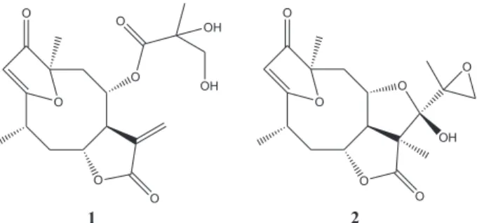
Preparative HPLC analysis (isocratic elution with MeOH/H2O 9:1; λ = 265 nm; flow 9.5 mL/min) of the
n-BuOH sub-fraction 45-48 (400 mg) afforded the 4β ,5-dihydro-1’,2’-epoxy-eremantholide-C (2, Figure 1). The
n-BuOH sub-fraction 37-40 (756 mg) was submitted to CC
in 40.0 g of polyamide (polyamide CC-6; Macherey-Nagel) eluted with mixtures of increasing polarity of hexane, AcOEt and MeOH. The new fractions were pooled as described above and sub-fraction 13-14 was identified as 4β,5-dihydro-2’,3’-dihydroxy-15-desoxy-goyazensolide (1, Figure 1). These two SL were identified by comparison of their 1H and 13C NMR and ESI-MS data with those
previously published.9,34
Sample preparation for chromatographic analysis
Powdered leaves of E. seidelii were weighed (10 mg)
containing the internal standard coumarin (50 μg mL-1).
400 μL of the extract was transferred to a centrifuge tube (1.5 mL), followed by the addition of 600 μL of hexane. This mixture was stirred in a vortex and then centrifuged at 1200 g for 10 min. An aliquot of 200 μL was taken from hydro-alcoholic phase, filtered on a 0.45 μm cellulose acetate membrane and submitted to HPLC analysis, by injection of 20 μL.
Analytical HPLC method
Solvents: A = aqueous acetic acid 2% (v/v); B = MeCN with 2% acetic acid (v/v). Elution profile, 0 to 10 min: 10 to 25% B (linear gradient); 10 to 15 min: 25% B (isocratic); 15 to 20 min: 25 to 45% B (linear gradient); 20 to 25 min: 45% B (isocratic); 25 to 30 min: 45 to 80% B (linear gradient); 30 to 35 min: 80% B (isocratic); 35 to 40 min: 80 to 10% B (linear gradient); 40 to 50 min: 10% B (isocratic). Flow rate: 1 mL min-1. UV detection set at 280 nm.
Qualitative and quantitative determination
The identity of the peaks relative to 1, 2 or coumarin was established by comparison of retention time, UV spectra and co-injection of reference standards purified as described above. Furthermore, a relative retention time considering coumarin as internal reference was determined for each peak. A quantitative analysis was done by the internal standard method, plotting calibration curves for 1 and 2 at concentrations of 1, 2, 5, 10, 20, 40 and 80μg mL-1 in MeOH/
H2O 9:1. Each determination was carried out in triplicate. The purity of the standards was confirmed by HPLC analysis performed under the described chromatographic conditions and, for purposes of calculation, it was assumed that each standard was 100% pure.
For quantification of SL in E. seidelii leaves,
chromatographic analyses were performed in duplicate and the SL content was estimated by a ratio of the two analysed peak areas averaged to the area of internal standard. Concentration is shown as percentage of SL in dry weight of leaves of E. seidelii.
Results and Discussion
This phytochemical investigation of E. seidelii leaves
revealed the SL 4β ,5-dihydro-2’,3’-dihydroxy-15-desoxy-goyazensolide (1) and 4β ,5-dihydro-1’,2’-epoxy-eremantholide-C (2) (Figure 1), both already isolated from this species and E. goyazensis,9 as its major
secondary metabolites. Together, these SL can be responsible for up to 7.5% of the weight of the dry leaves
(data from plant 6, population A). This remarkable predominance, taken together with the SL utility as chemotaxonomic markers24,26,29,32,33 and biological
activities3,6-8,10-12 led us to develop the present HPLC
method for their quantification.
HPLC method
Analysis carried out in triplicate with three consecutive extractions of the same material (powdered leaves of E. seidelii) revealed that 95.2% of 1 and 97.5% of 2 were extracted in the first one, which led us to use the one step extraction procedure described. The overall recovery of the compounds using the method of extraction and determination described was 97% for 1 and 98% for 2, and were based on 5 extractions (addition of 4 different concentrations of 1 and 2 and a control without addition of SL) done using powdered leaves of E. glomerulatus, a
similar species lacking these two compounds, as matrix. Calibration curves were made by plotting the ratio of SL peak areas to the area of coumarin as internal standard versus
concentrations of each SL, and good linearity was obtained with the standard solutions of 1 and 2 in concentrations between 1 - 80 μg mL-1. The regression equations for 1 and
2 were, respectively, y=0.6144x + 0.0258 (r=0.9991) and y=0.8630x + 0.0327 (r=0.9992). The retention times in the system developed were 26.28 min for 1, 24.87 min for 2 and 23.25 min for the coumarin internal standard (Figure 2).
The HPLC method developed was carefully validated. For the highest concentration the accuracy for both SL was 97.7%. The relative standard deviation obtained from nine analyses performed on different days was 2.05% for 1 and 2.31% for 2, which gives a very high level of reproducibility. Extracts were also analyzed immediately and 24h after their acquisition, and no significant differences were detected in peak areas. The limit of detection (signal-to-noise ratio of 3:1) was 0.30 and 0.22 μg mL-1 for 1 and 2 respectively. The maximum limit of
quantification for the detector and UV wavelength
Figure 1. Chemical structures of the sesquiterpene lactones 4β
employed was 422.0 μg mL-1 for 1 and 449.3 μg mL-1 for
2. Extracts were also done with samples stored for up to one year to evaluate their stability. No significant alteration in concentrations were found in samples stored for up to 11 months. The largest difference detected was 1.9% for leaves stored for one year, which shows good stability during storage.
Analysis of E. seidelii leaves
No qualitative differences were found in the SL patterns of all plants sampled. However, significant differences were found in the quantitative proportions of the main compounds (SL 1 and 2).
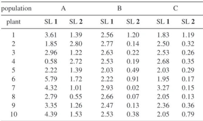
The concentrations of 1 and 2 in leaves of E. seidelii
obtained for each individual in samples collected at the same time and day (19/March/2001, 12:00 pm ± 30min, Table 1) led us to group the studied individuals in three clusters with different ratios of compounds 1 and 2.
HPLC analysis carried out with plants selected to represent each of these three clusters, showed no significant seasonal variation in the concentration of 1 and 2 in none of them over the two years sampled (Figure 3). Individuals analyzed for seasonal variation were: 1, 2, 4, 5 and 8 from population A; 1, 3 and 7 from population B; and 1, 3 and 7 from population C. Furthermore, SL concentration in leaves of E. seidelii showed no significant correlation with the
plant stem diameter or height, which were proposed as variables indicatives of the age of the plants. It is well known that in some tribes of Asteraceae, including Vernonieae and Heliantheae amongst others, SL are predominantly (if not
Figure 2. Chromatogram obtained for individual 5 from population A with the described method. Retention times are 26.28 min for 4β
,5-dihydro-2’,3’-dihydroxy-15-desoxy-goyazensolide(1), 24.87 min for 4β,5-dihydro-1’,2-epoxy-eremantholide-C (2) and 23.25 min for the internal standard coumarin (IS).
Figure 3. Examples of seasonal quantitative analyses of individuals with
predominance of the goyazensolide type sesquiterpene lactone (4β ,5-dihydro-2’,3’-dihydroxy-15-desoxy-goyazensolide,SL1, graph in top)
and eremantholide type sesquiterpene lactone (4β ,5-dihydro-1’,2-epoxy-eremantholide-C, SL2, bottom).
Table 1. Sesquiterpene lactone concentrations in leaves of the 30
Eremanthus seidelii individuals sampled in 19/March/2001 at 12:00pm ± 30min
Sesquiterpene lactones concentration in Eremanthus seidelii (% per leaf dry weight)
SL 1 = 4β,5-dihydro-2’,3’-dihydroxy-15-desoxy-goyazensolide SL 2 = 4β,5-dihydro-1’,2-epoxy-eremantholide-C
population A B C
plant SL 1 SL 2 SL 1 SL 2 SL 1 SL 2
1 3.61 1.39 2.56 1.20 1.83 1.19
2 1.85 2.80 2.77 0.14 2.50 0.32
3 2.96 1.22 2.63 0.22 2.53 0.26
4 0.58 2.72 2.53 0.19 2.68 0.35
5 2.22 1.39 2.03 0.49 2.03 0.29
6 5.79 1.72 2.22 0.91 1.95 0.17
7 4.32 1.01 2.93 0.02 3.27 0.15
8 2.79 0.55 2.66 0.07 2.05 0.13
9 3.35 1.26 2.47 0.13 2.36 0.36
exclusively) produced in glandular trichomes, which biosynthetic capacity is finished before leaves are expanded.35 Thus, no circadian variation could be expected
for these metabolites. Since the major secondary metabolites found in E. seidelii are SL, no analyses were carried out
with samples collected previously for circadian studies. Thus, any influence of environmental or developmental factors on the contents of 1 and 2 were excluded by the facts that neighboring trees can belong to different clusters, and that there are no seasonal variations in SL concentrations. Additionally, 1 (a goyazensolide type SL) is a biosynthetic precursor for 2 (a eremantholide type SL),3 which reveals genetic conditions to produce both
SL in the plants studied. Therefore, the difference possibly remains in a differential activity of the enzymes that cyclisation 1 to 2. These significant differences in the levels of biosynthetically related SL, taken together with the valuable property of SL as chemotaxonomic markers24,26,29,32,33 and the fact that 1 and 2 are the main
constituents from the secondary chemistry in leaves of E. seidelii, lead us to propose the following quantitative
chemotypes: (i) plants in this cluster are the only ones
that present 2 in higher concentrations than 1. In fact, individuals in this cluster present the highest concen-trations of 2 (more than 2.7% of the dried leaf weight in the plants studied) and the lowest concentrations of 1 (below 1.7% in the plants studied). Individuals 2 and 4 from population A, which corresponds to 6.7% of the plants studied, represent this chemotype, denominated
high E for its high relative contents of the Eremantholide
type SL 2; (ii) in the other extreme are grouped individuals
with very low relative contents of 2, showing a ratio between 1 and 2 above 10. Individuals in this cluster show intermediate concentrations of 1 (between 1.9 and 3.3% in the plants studied) and the lowest concentrations of 2 (below 0.2% in the plants studied). Plants 2, 3, 4, 7, 8 and 9 from population B and plants 6, 7 and 8 from population C (in total 30.0% of the plants analyzed) are representative of this chemotype denominated low E, for its low relative
contents of the Eremantholide type SL 2; (iii) this last
cluster is the largest one comprising plants that present an intermediate ratio of compounds 1 and 2 (between 1.5 and 10). The plants not included in the clusters above (which represents 63.3% of the individuals analyzed) form this chemotype denominated medium GE, due to the
intermediate contents of both SL.
Similarly, quantitative chemotypes based on relative concentrations of biosynthetically related SL were, for example, established for Artemisia annua29 and Artemisia herba-alba.22
Acknowledgments
This work was supported by FAPESP, CAPES and CNPq. The authors acknowledge Prof. Dr. João Semir (Departamento de Morfologia e Sistemática Vegetal, Instituto de Biologia da Universidade Estadual de Campinas) for the plant identification and Dr Paul Gates (School of Chemistry, University of Bristol, UK) for the English language revision.
References
1. Robinson, H.; Smithsonian Contr. Bot.1999, 89. 2. MacLeish, N. F. F.; Ann. Mo. Bot. Gard.1987, 74, 265. 3. Le Quesne, P. W.; Levery, T. B.; Menachery, M. D.; Brennan,
T. F.; Raffauf, R. F.; J. Chem. Soc., Perkin Trans. 11978, 1572.
4. Lunardello, M. A.; Tomaz, J. C.; Vichnewski, W.; Lopes, J. L. C.; Gutierrez, A. B.; Diaz, J. G.; Herz, W.; J. Braz. Chem. Soc.
1995,6, 307.
5. Mauro, M. R. V.; Tucci, A. M.; Nasi, T.; Vichnewski, W.; Lopes, J. L.; Diaz, J. G.; Herz, W.; J. Braz. Chem. Soc.1993, 4, 30. 6. Raffauf, R. F.; Huang, P. C.; Le-Quesne, P. W.; Levery, S. B.;
Brennan, T. F.; J. Am. Chem. Soc.1975, 97, 6884.
7. Vichnewski, W.; Gilbert, B.; Phytochemistry1972, 11, 2563. 8. Vichnewski, W.; Sarti, S. J.; Gilbert, B.; Herz, W.;
Phytochemistry1976,15, 191.
9. Vichnewski, W.; Takahashi, A. M.; Nasi, A. M. T. T.; Gonçalves, D. C. R.; Dias, D. A.; Lopes, J. N. C.; Goedken, V. L.; Gutiérrez, A. B.; Herz, W.; Phytochemistry 1989, 28, 1441.
10. Picman, A. K.; Biochem. Syst. Ecol. 1986, 14, 255.
11. Santos, P. A.; Amarante, M. F. C.; Pereira, A. M. S.; Bertoni, B.; França, S. C.; Pessoa, C.; Moraes, M. O.; Costa-Lotufo, L. V.; Pereira, M. R. P.; Lopes, N. P.; Chem. Pharm. Bull.2004, 52, 1433.
12. Rüngeler, P.; Castro, V.; Mora, G.; Gören, N.; Vichnewski, W.; Pahl, H. L.; Merfort, I.; Schmidt, T. J.; Bioorg. Med. Chem.
1999, 7, 2343.
13. Agerbirk, N.; Olsen, C. E.; Nielsen, J. K.; Phytochemistry2001,
58, 91.
14. Angelopoulou, D.; Demetzos, C.; Perdetzoglou, D.; Biochem. Syst. Ecol.2002, 30, 189.
15. Menkovik, N.; Šavikin-Fodulovi, K.; Savin, K.; Planta Med.
2000, 66, 178.
16. Wilt, F. M.; Miller, G. C.; Biochem. Syst. Ecol.1992, 20, 53. 17. Høgedal, B. D.; Mølgaard, P.; Biochem. Syst. Ecol.2000, 28, 949. 18. Itenov, K.; Mølgaard, P.; Nyman, U.; Phytochemistry 1999,
52, 1229.
19. Lopes, N. P.; Kato, M. J.; Andrade, E. H. A.; Maia, J. G. S.; Yoshida, M.; Phytochemistry 1997, 46, 689.
21. Feuerstein, I.; Müller, D.; Hobert, K.; Danin, A.; Segal, R.; Phytochemistry 1986, 25, 2343.
22. Mabry, T. J.; Pure Appl. Chem. 1973, 34, 377.
23. Nehme, C. J.; Moraes, P. L. R.; Cavalheiro, A. J.; Biochem. Syst. Ecol. 2002, 30, 613.
24. Segal, R.; Feuerstein, I.; Danin, A.; Biochem. Syst. Ecol. 1987,
15, 411.
25. Passreiter, C. M.; Aldana, B. E. M.; Planta Med.1998, 64, 427.
26. Picman, A. K.; Towers, G. H. N.; Biochem. Syst. Ecol.1982,
10, 145.
27. Schmidt, T. J.; Bomme, U.; Alfermann, A. W.; Planta Med.
1998, 64, 268.
28. Zidorn, C.; Stuppner, H.; Taxon2001, 50, 115.
29. Wallaart, T. E.; Pras, N.; Beekman, A. C.; Quax, W. J.; Planta Med. 2000, 66, 57.
30. Williams, C. A.; Demissie, A.; Harborne, J. B.; Biochem. Syst. Ecol. 1983, 19, 81.
31. Wilt, F. M.; Geddes, J. D.; Tamma, R. V.; Miller, G. C.; Everett, R. L.; Biochem. Syst. Ecol.1992, 20, 41.
32. Seaman, F. C.; Bot. Rev.1982, 48, 121.
33. Zdero, C.; Bohlmann, F.; Plant Syst. Evol.1990, 171, 1. 34. Crotti, A. E. M.; Lopes, J. L. C.; Lopes, N. P.; J. Mass Spectrom.
2005, 40, 1030.
35. Spring, O.; Bienert, U.; J. Plant Physiol. 1987, 130, 441.
Received: May 20, 2005
Published on the web: November 9, 2005