Printed in Brazil - ©2005 Sociedade Brasileira de Química 0103 - 5053 $6.00+0.00
A
r
ti
c
le
* e-mail: [email protected]
Limonoids from
Spiranthera odoratissima
St. Hil
Tereza A. N. Ribeiroa, Eliane A. da Silva Ndiayea, Eudes da S. Velozob, Paulo C. Vieira*,c,
Javier Ellenad and Paulo T. de Sousa Júniora
a
Departamento de Química, Universidade Federal de Mato Grosso, Av. Fernando Correa s/n, 78060-900 Cuiabá - MT, Brazil
b
Faculdade de Farmácia, Universidade Federal da Bahia, Rua Barão de Geremoaba s/n, 40170-290 Salvador - BA, Brazil
c
Departamento de Química, Universidade Federal de São Carlos, CP 676, 13565-905 São Carlos - SP, Brazil
d
Instituto de Física, Universidade de São Paulo, CP 369, 13560-970 São Carlos - SP, Brazil
Onze substâncias foram isoladas das raízes de Spiranthera odoratissima: dois novos limonóides,
o limonóide já conhecido limonina, três alcalóides furoquinolínicos (dictamina, γ-fagarina e esquimianina), três alcalóides β-indoloquinazolínicos (rutaecarpina, evodiamina e 1-hidroxirutaecarpina), a cumarina aurapteno e β-sitosterol. A elucidação estrutural dessas substâncias foi realizada através de técnicas espectrais como IV e RMN em uma e duas dimensões; as estruturas novas foram confirmadas por difração de raios-X.
Eleven substances have been isolated from the roots of Spiranthera odoratissima, two new
limonoids, the known limonoid limonin, three furoquinoline alkaloids, dictamnine, γ-fagarine and skimmianine, three β-indoloquinazoline alkaloids, rutaecarpine, evodiamine and 1-hydroxyrutaecarpine, the coumarin aurapten and β-sitosterol. Structure elucidation has been carried out by IR as well as 1D and 2D NMR; the new structures were also confirmed by X-ray crystallographic analyses.
Keywords:Spiranthera odoratissima, Rutaceae, limonoids, alkaloids, X-ray diffraction
Introduction
Spiranthera odoratissma St. Hil., is a shrub found in the central Brazilian savannah, as well as in Bolivia.1 In Mato
Grosso state it is known by the vernacular name of “manacá”, being used in folk medicine to treat syphilis, rheumatism, kidney infections, urinary retention, abdominal pains, gout, acne and boil.2 This plant has already been investigated
from chemistry viewpoint. From a specimen collected in Bahia were isolated furoquinoline alkaloids; coumarins and terpenes.3 The Rutaceae family is characterized by the
abundance of anthranilic acid derived alkaloids coumarins, limonoids and flavonoids mainly.4
We have been interested in the chemistry of Rutaceae,5-8
and as part of ongoing work on this family, in this study we describe the isolation and the identification of two new limonoids, as well as the known limonin. Also described are
the isolation and identification of the furoquinoline alkaloids
γ-fagarine dictamnine, skimmianine, the β-indoloquinazoline alkaloids rutaecarpine, evodiamine and 1-hydroxy-rutaecarpine, the coumarin aurapten and β-sitosterol.
Experimental
General experimental procedures
Melting points were measured in a Mettler FP-80 apparatus and are uncorrected. Specific rotations were determined in a Perkin-Elmer 341 polarimeter. The IR spectra were obtained in a Bomem FT-IR MB100 equipment with the samples in KBr pellets. 1H and 13C
internal standard, as well as the residual hydrogen from the solvents (CDCl3 and DMSO-d6). Radial preparative chromatography (RPC) was carried out with a Chromatotron apparatus and were performed with Merck Kiesegel 60 PF254. Column chromatography (CC) was performed with silica gel 60 (Merck, 63-230 μm), and flash column chromatography with silica (Merck, 43-63 μm). Analytical thin layer chromatography (TLC) was carried out with Merck Kiesegel 60 F254 (0.25 mm) plates.
Plant material
The roots from S. odoratissima were collected at Cuiabá-Barão de Melgaço road (Km 1) in December 1999. A voucher specimen was deposited at Central Herbarium of Universidade Federal de Mato Grosso (registration # 24246).
Extraction and isolation of compounds
The roots of S. odoratissima (3.3 kg) were macerated at room temperature with dichloromethane (3 x 8L), with occasional stirring, during 7 days. The dichloromethane extract-DCE (126 g; 3.82%) was obtained after filtration and solvent removal in vacuo. Subsequent extraction was performed with methanol (3 x 8L) using the same procedure above, affording the methanol extract-ME (120 g; 3.64%). An aliquot of DCE (20.0 g) was submitted to conventional acid-base extraction (HCl 1%; 4 x 100 mL) and the alkaloid fraction (190 mg) was chromatographed by RPC, in a 4 mm radial plate, with hexane-CHCl3 (8:2; 100 mL); CHCl3 (250 mL) and CHCl3-MeOH (95:5; 100 mL), affording 6 fractions. The furoquinoline alkaloids γ-fagarine (15 mg), dictamnine (8 mg) and skimmianine (30 mg) were isolated after washing fractions 2, 3 and 4 with Et2O.
Another DCE fraction (42.5 g) was filtered in a silica column (400 g) with petrol ether (0.5 L), CH2Cl2 (2 L), CHCl3 (1 L), CHCl3-MeOH (99:1; 2 L), CHCl3-MeOH (9:1; 1 L), MeOH (1 L) and MeOH-0.1%HOAc (0.5 L), affording fractions A to G, respectively.
Column chromatography (CC) was carried out on fraction B (23.0 g) employing the gradient solvent system: hexane-CH2Cl2 (1:1; 450 mL), CH2Cl2 (200 mL), CH2Cl2 -MeOH (99:1; 200 mL), (95:5; 200 mL), (9:1; 600 mL) and MeOH (200 mL), affording 8 fractions after TLC analysis. Fraction 2 [from hexane-CH2Cl2 (1:1)] was chromatographed by RPC (hexane- CH2Cl2; 9:1; 7:3; 1:1), CH2Cl2 and CHCl3-MeOH (99:1), affording the coumarin aurapten (50 mg), after washing the crude hexane-CH2Cl2 (7:3) fraction with EtOH. Fraction 4 [from hexane-CH2Cl2 (99:1)] was submitted to medium pressure chromatography using the gradient solvent system: hexane-CH2Cl2 (2:8;
300 mL), (1:9; 70 mL), CH2Cl2 (150 mL), CH2Cl2-MeOH (9:1; 250 mL). Aurapten (109 mg) was obtained after washing with Et2O the combined hexane-CH2Cl2 (2:8) fractions; β-sitosterol was obtained after washing the combined hexane-CH2Cl2 (1:9) fractions with EtOH; 1 (30
mg) was obtained after washing the combined CH2Cl2 -MeOH (95:5 and 9:1) fractions with EtOH.
Fraction D (10 g) was submitted to medium pressure chromatography using the solvent system: CH2Cl2 (300 mL), CH2Cl2-CH3CN (98:2; 200 mL), (95:5; 300 mL), (7:3; 200 mL); (1:1; 300 mL) and MeOH (200 mL). The combined CH2Cl2-CH3CN (98:2 and 95:5) fractions (2.5 g) were re-submitted to medium pressure chromatography employing the gradient hexane-CHCl3 (1:9; 400 mL), CHCl3 (200 mL), CHCl3-MeOH (95:5; 300 mL), (9:1; 300 mL), (1:1; 200 mL) and MeOH (200 mL). Limonin (3)
(130 mg) precipitated after EtOH addition to the combined hexane-CHCl3 (1:1) and CHCl3 fractions. The supernatant liquid was chromatographed by CC in hexane-CHCl3 (2:8; 270 mL), CHCl3 (50 mL), CHCl3-CH3CN (8:2; 200 mL), (6:4; 150 mL), (3:7; 100 mL), CH3CN (50 mL), CH3 CN-EtOH (7:3; 100 mL) and (1:1; 150 mL). Skimmianine (30 mg) was obtained from the combined hexane-CHCl3 (2:8) and CHCl3 fractions after solvent removal and washing the solid with EtOH. The combined CHCl3-MeOH (95:5) fractions were chromatographed by CC using CH2Cl2 (50 mL), CH2Cl2-EtOAc (8:2; 300 mL), (7:3; 300 mL), (3:7; 200 mL), EtOAc (200 mL), EtOAc-MeOH (8:2; 200 mL) and MeOH (100 mL). The combined CH2Cl2-EtOAc (8:2) and (7:3) fractions were submitted to RPC with CHCl3, CHCl3-MeOH (95:5), (9:1) and EtOAc-MeOH (9:1). Limonin (3) (60 mg) was obtained after solvent removal
from fraction CHCl3-MeOH (9:1).
ME (30 g) was suspended in MeOH-H2O (7:3; 100 mL) and extracted with CH2Cl2 (3 x 100 mL), EtOAc (3 x 100 mL) and n-BuOH (3 x 100 mL). The CH2Cl2 fraction (2.7 g) was submitted to CC in CH2Cl2 (1.7 L), CH2Cl2-MeOH (99:1; 2.2 L), (98:2; 0.8 L), (95:5; 1.5 L); (9:1; 0.7 L), (7:3; 0.6 L), (1:1; 0.8 L) and MeOH (0.9 L). The combined CH2Cl2-MeOH (99:1) fractions (550 mg) were submitted to CC in CH2Cl2 (300 mL), CH2Cl2-MeOH (99:1; 150 mL), (97:3; 50 mL), (95:5; 350 mL), (9:1; 150 mL), (1:1; 50 mL) and MeOH (50 mL). A precipitate (80 mg) was obtained by adding EtOH to the combined CH2Cl2 fractions. Flash CC was performed with this precipitate, employing CH2Cl2 (150 mL), CH2Cl2-MeOH (99:1; 100 mL), (95:5; 100 mL), (1:1; 50 mL) and MeOH (50 mL). Rutaecarpine (4) (7 mg) was isolated after solvent removal
from the combined CH2Cl2 fractions; evodiamine (5) (5
-Spiranthera odoratissima
MeOH (95:5 and 1:1) fractions were submitted to flash CC in CH2Cl2 (40 mL) and CH2Cl2-MeOH (99:1; 30 mL). 1-hydroxyrutaecarpine (6) (4 mg) and 2 (7 mg) were
isolated from the second CH2Cl2 fraction and the third CH2Cl2-MeOH (99:1), respectively.
Compound (1) [α]25 (CHCl
3, c. 0.1): -26 oC. IR (KBr) νmax/ cm-1: 3405,
1767, 1741, 1714 . 1H and 13C NMR (CDCl
3): Tables 1 and 2.
Compound (2) [α]25 (CHCl
3, c. 0.05): -20 oC. IR (KBr) νmax/ cm-1: 1766,
1742, 1707. 1H and 13C NMR (CDCl
3): Tables 1 and 2.
Single crystal X-ray analysis
Low temperature X-ray diffraction data collections were performed at 120(2) K, on an Enraf-Nonius Kappa-CCD diffractometer equipped with an Oxford Cryosystem liquid N2 device, using graphite-monochromated MoK α radiation (0.71073 Å). Data were collected up to 50° in 2θ, with a redundancy of 4 in the phi scans and omega scans with kappa offsets modes. The final unit cell parameters were based on all reflections. Data collections were made using the COLLECT program;9 integration and scaling of the reflections were performed with the HKL Denzo-Scalepack system of programs.10 No absorption corrections were applied.
The structures were solved by direct methods with SHELXS 8611 and SHELXS-97.12 The models were refined by full-matrix least squares on F2 with SHELXL-97.13 All the
hydrogen atoms were stereochemically positioned and refined with the riding model.12 Hydrogen atoms of the CH and CH
2
groups were set isotropic with a thermal parameter 20% greater than the equivalent isotropic displacement parameter of the atom to which each one was bonded. This percentage was set to 50% for the hydrogen atoms of the CH3 groups. Data collections and experimental details for the complexes are summarized in Table 3. The programs SHELXL-97,13 and ORTEP-314 were used within WinGX15 to prepare materials for publication. Atomic coordinates, bond lengths and angles, and thermal parameters have been deposited at the Cambridge Crystallographic Data Centre (see below).
Results and Discussion
Limonoid (1) has been isolated from the
dichloromethane extract from the roots of S. odoratissima as a white amorphous solid (mp 174-177 oC), presenting
[α]D25 (CHCl
3): – 26º. The IR spectrum has shown
absorptions at 3405 (OH), 1767 (lactone), 1741 (α,β -unsaturated ester) and 1714 (ketone).
1H NMR spectrum presented signals related to the
β-substituted furan ring, where H-21 and H-23 appeared as multiplets at δ 7.41 and at δ 7.36, respectively. The
β-furan hydrogen H-22 appeared as a double doublet at δ
6.37 (J= 1.7, 0.8 Hz). The hydrogen H-17, from the δ-epoxylactone ring, was observed at δ 5.54 (s, 1H).
The 1H and 13C NMR spectra from (1) has shown
similarities with the correlated spectra of (3), also isolated
in this study, as shown in Tables 1 and 2. The absence of two doublets, associated to the geminal hydrogens at C-19, very common in structures related to (3), displaying
a A,D-seco ring,16 when linked with the low intensity,
quaternary carbon signal observed at δ 182.5, from the DEPT experiment, was a good indication for the presence of a carbonyl group at C-19 in 1. HMBC spectrum has
shown long distance coupling (J3) between H-1, H-9 and C-19, confirming that C-19 in 1 is oxidized.
The α,β-unsaturated ester moiety, detected by the IR spectrum, was further confirmed by 1H NMR (δ 3.71, s, 3H;
-CO2CH3), as well as by the signals at δ 165.5 (-CO2CH3) and δ 52.3 (-CO2CH3) in 13C NMR. The olefinic hydrogens
H-1 and H-2 were detected as two doublets at δ 6.65 and δ
6.12 respectively; the cis configuration of the double bond could be determined by the coupling constant value (J1,2 = 12.4 Hz). HSQC spectrum showed correlation between these two protons and the carbons at δ 151.6 (C-1) and δ
123.2 (C-2), respectively.
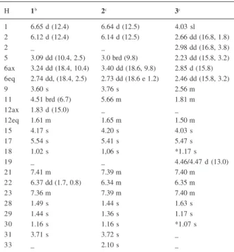
Table 1. 1H NMR spectral data for compounds 1-3
H 1b 2c 3c
1 6.65 d (12.4) 6.64 d (12.5) 4.03 sl 2 6.12 d (12.4) 6.14 d (12.5) 2.66 dd (16.8, 1.8)
2 _ _ 2.98 dd (16.8, 3.8)
5 3.09 dd (10.4, 2.5) 3.0 brd (9.8) 2.23 dd (15.8, 3.2) 6ax 3.24 dd (18.4, 10.4) 3.40 dd (18.6, 9.8) 2.85 d (15.8) 6eq 2.74 dd, (18.4, 2.5) 2.73 dd (18.6 e 1.2) 2.46 dd (15.8, 3.2)
9 3.60 s 3.76 s 2.56 m
11 4.51 brd (6.7) 5.66 m 1.81 m
12ax 1.83 d (15.0) _ _
12eq 1.61 m 1.65 m 1.50 m
15 4.17 s 4.20 s 4.03 s
17 5.54 s 5.41 s 5.47 s
18 1.02 s 1,06 s *1.17 s
19 _ _ 4.46/4.47 d (13.0)
21 7.41 m 7.39 m 7.40 m
22 6.37 dd (1.7, 0.8) 6.34 m 6.35 m
23 7.36 m 7.39 m 7.40 m
28 1.49 s 1.44 s 1.63 s
29 1.44 s 1.36 s 1.17 s
30 1.16 s 1.16 s *1.07 s
31 3.71 s 3.72 s _
33 _ 2.10 s _
The DEPT 13C NMR spectrum presented two signals
related to methylenic carbons at δ 38.1 (C-6) and δ 41.2 (C-12). HMBC experiment has shown H-6 correlation with C-7 (δ 207.3; J2) and C-4 (δ 87.6; J3). The methylenic hydrogen H-6 resonated at δ 3.24 (dd, J= 18.4, 10.4 Hz, H-6ax) and δ 2.74 (dd, J= 18.4, 2.5 Hz, H-6eq); COSY spectrum has shown coupling of both hydrogen with H-5 at δ 3.09 (dd, J= 10.4, 2.5 Hz).
Reasonably similar values have been observed when comparing the 13C NMR data for rings B and D in
compounds (1) and (3) (Table 2). DEPT experiment,
however, presented three signals related to methylenic carbons in (3) and only two signals related to methylenic
carbons in (1). The hydroxyl group, shown to be present
in (1) by the IR spectrum, nevertheless, should be located
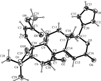
at C-11 or C-12 in ring C. Furthermore, the carbinolic hydrogen appeared as a broad doublet at δ 4.51 (J= 6.7 Hz, 1H), which has been associated to δ 66.4 signal (HSQC), indicating that 11 couples only with one H-12eq. No coupling was observed between H-11 and H-9 and H-12ax. The HMBC spectrum showed a long distance coupling (J3) with C-8 (δ 48.5), confirming that the hydroxylated carbon is C-11. The α stereochemistry of the OH group was determined by X-ray crystallography (Figure 2). The H-12 methylenic hydrogens were observed at δ 1.83 (d, J= 15 Hz, H-12ax) and δ 1.61 (m, H-12eq). HMBC showed long range coupling (J3) between H-12ax
Table 2. 13C NMR spectral dataa for compounds 1-3
C 1 2 3
1 151.6 152.1 79.1
2 123.2 123.1 35.6
3 165.5 165.9 169.1
4 87.6 84.9 80.2
5 49.9 49.8 60.4
6 38.1 38.6 36.3
7 207.3 208.2 206.1
8 48.5 48.6 51.3
9 43.9 42.5 48.1
1 0 53.6 52.3 45.9
1 1 66.4 66.5 18.8
1 2 41.2 40.9 30.1
1 3 36.5 36.3 37.9
1 4 65.1 65.6 65.6
1 5 52.6 52.2 53.8
1 6 166.9 166.6 167.0
1 7 77.8 77.6 77.7
1 8 19.2 19.3 21.3
1 9 182.5 178.2 65.3
2 0 120.1 119.8 119.9
2 1 141.1 141.1 141.1
2 2 109.9 109.8 109.8
2 3 142.9 143.1 143.2
2 8 24.4 24.3 *30.8
2 9 32.4 32.8 *20.7
3 0 16.7 16.2 17.6
3 1 52.3 52.0 _
3 2 _ 170.5 _
3 3 _ 21.2 _
a Chemical shifts in δ from TMS taken in CDCl
3 measured at 50 MHz; * Interchangeable.
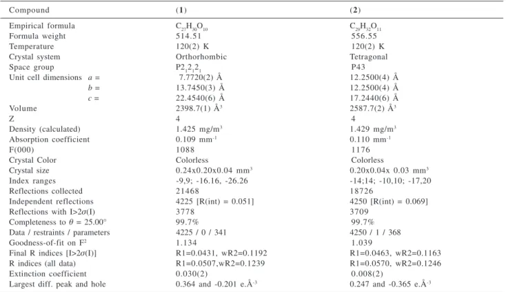
Table 3. Crystal data and structure refinement.
Compound (1) (2)
Empirical formula C27H30O10 C29H32O11
Formula weight 514.51 556.55
Temperature 120(2) K 120(2) K
Crystal system Orthorhombic Tetragonal
Space group P212121 P43
Unit cell dimensions a = 7.7720(2) Å 12.2500(4) Å
b = 13.7450(3) Å 12.2500(4) Å
c = 22.4540(6) Å 17.2440(6) Å
Volume 2398.7(1) Å3 2587.7(2) Å3
Z 4 4
Density (calculated) 1.425 mg/m3 1.429 mg/m3
Absorption coefficient 0.109 mm-1 0.110 mm-1
F(000) 1088 1176
Crystal Color Colorless Colorless
Crystal size 0.24x0.20x0.04 mm3 0.20x0.04x 0.03 mm3
Index ranges -9,9; -16.16, -26.26 -14;14; -10,10; -17,20
Reflections collected 21468 18726
Independent reflections 4225 [R(int) = 0.051] 4250 [R(int) = 0.069]
Reflections with I>2σ(I) 3778 3709
Completeness to θ = 25.00° 99.7% 99.7%
Data / restraints / parameters 4225 / 0 / 341 4250 / 1 / 368
Goodness-of-fit on F2 1.134 1.039
Final R indices [I>2σ(I)] R1=0.0431, wR2=0.1192 R1=0.0463, wR2=0.1163
R indices (all data) R1=0.0507,wR2=0.1239 R1=0.0570, wR2=0.1246
Extinction coefficient 0.030(2) 0.008(2)
Spiranthera odoratissima
with C-9 (δ 43.9), C-14 (δ 65.1), C-17 (δ 77.8) and C-18 (δ 19.2).
Four singlets (3H) relative to the methyl groups, were observed at δ 1.02 (H-18), δ 1.16 (H-30), δ 1.44 (H-29) and
δ 1.49 (H-28). The correct attribution of each methyl group position was carried out by using HMBC and HSQC experiments. The long distance correlation of δ 1.02 (Me-18) with C-12 (δ 41.2), C-13 (δ 36.5), C-14 (δ 65.1), C-17 (δ 77.8), and δ 1.16 (C-30) with C-7 (δ 207.3), C-8 (δ
48.5), C-9 (δ 43.9) and C-14 (δ 65.1), could be easily observed in HMBC spectrum.
The structure presented for compound (1) was definitely
confirmed by single crystal X-ray diffraction (Figure 2).
The second new limonoid (2), was obtained from the
methanol extract of the roots of S. odoratissima, as a white solid presenting a high mp (> 300 oC) and [α]
D25 -20
(CHCl3). The IR spectrum presented absorptions at 1766 (lactone), 1742 (α,β-unsaturated ester) and 1707 (ketone). Compound (2) 1H NMR spectrum presented high
similarity with the one from compound (1), indicating a
limonoid with a β-substituted furan ring, as shown by the two α-furan hydrogen at δ 7.39 (m) and the β-furan hydrogen at d 6.34 (m). An additional signal was observed at δ 2.10 (s, 3H), being attributed to the presence of an extra methyl group in (2).
13C NMR spectrum presented 29 signals, showing that
compound (2) should have two more carbons than the
analogous (1); a quaternary carbon at δ 170.5 and a methyl
group at δ 21.2 were observed by the DEPT experiment indicating the presence of an acetyl group in this compound.
These 1H and 13C NMR data, in association with the
absence of a hydroxyl absorption in IR spectrum led to the conclusion that the hydroxyl group was esterified in (2).
Single crystal X-ray diffraction has confirmed the proposed structure for (2) (Figure 3).
The known compounds were identified through comparison of their spectral data with the ones in the literature, limonin (3),17 dictamnine, skimmianine;18 the β
-indoloquinazoline alkaloids rutaecarpine (4), evodiamine
(5)19 and 1-hydroxyrutaecarpine (6),20 and the coumarin
aurapten.21 The isolated alkaloids and limonoids have been
described as typical metabolites from the Rutaceae.22
Supplementary Information
Crystallographic data (excluding structure factors) for the structures in this paper has been deposited with the
Figure 1. Limonoids isolated from the roots of S. odoratissima.
Figure 2. ORTEP-314 diagram of compound (1), showing the atoms labeling and the 50% probability ellipsoids.
Figure 3. ORTEP-314 diagram of compound (2), showing the atoms labeling and the 50% probability ellipsoids.
Cambridge Crystallographic Data Centre as supplementary publication no CCDC 238939 and 238940. Copies of the data can be obtained, free of charge via www.ccdc.cam.ac.uk/conts/retrieving.html (or from the Cambridge Crystallographic Data Centre, CCDC, 12 Union Road, Cambridge CB2 1EZ, UK; fax: +44 1223 336033; or e-mail: [email protected]).
Acknowledgements
The authors are grateful to Dr. Antônio Gilberto Ferreira from Laboratório de Ressonância Magnética Nuclear (Universidade Federal de São Carlos) for obtaining NMR spectroscopic data. Financial assistance from FAPEMAT, CAPES, CNPq, FAPESP is acknowledged.
References
1. Pirani, J.R.; Estudos Taxonômicos de Rutaceae, Departamento de Biociências, Universidade de São Paulo, Brazil,1999. 2. De- La Cruz, M.G.F.; MSc Dissertation, Instituto de Saúde
Coletiva, Universidade Federal de Mato Grosso, Brazil,1997. 3. Freitas, C.M.D.; Lucchese, A.M.; Silva, F.S.; Velozo, E.D.;
Biochem. Syst. Ecol.2003, 31, 805.
4. Waterman, P.G.; Biochem. Syst. Ecol. 1999, 27, 395. 5. Sartor, C. F. P.; da Silva, M. F. G. F.; Fernandes, J. B.; Vieira, P.
C.; Rodrigues-Filho, E.; Cortez, D.A.G.; Phytochemistry 2003, 63, 185.
6. Biavatti, M. W.; Vieira, P. C.; da Silva, M. F. G. F.; Fernandes, J. B.; Victor, S. R.; Pagnocca, F. C.; Albuquerque, S.; Caracelli, I., Zukerman-Schpector, J.; J. Braz. Chem. Soc. 2002, 13, 66. 7. Biavatti, M. W.; Vieira, P. C.; da Silva, M. F. G. F.; Fernandes,
J.B.; Albuquerque, S.; J. Nat. Prod. 2002, 65, 562.
8. Santos, C. S.; Januário, A.H.; Vieira, P. C.; Fernandes, J. B.; da Silva, M. F. G. F.; Pirani, J.R.; J. Braz. Chem. Soc. 1998, 9, 39.
9. Enraf-Nonius, COLLECT. Nonius BV: Delft, The Netherlands, 1997-2000.
10. Otwinowski, Z.; Minor, W. In Methods in Enzymology; Carter Jr., C. W.; Sweet, R.M., eds.; Academic Press: New York, 1997, vol. 276, p. 307.
11. Sheldrick, G.M.; SHELXS86 - Program for Crystal Structure solution, Institüt für Anorganische Chemie der Universität, Tammanstrasse 4, D-3400 Göttingen, Germany, 1986. 12. Sheldrick, G.M.; SHELXS-97, Program for Crystal Structure
Resolution, University of Göttingen, Germany, 1997. 13. Sheldrick, G.M.; SHELXL-97, Program for Crystal Structures
Analysis, University of Göttingen, Germany, 1997. 14. Farrugia, L. J.; J. Appl. Cryst. 1997, 30, 565.
15. Farrugia, L. J.; WinGX . An Integrate System of Windows Programs for the Solution, Refinement and Analysis of Single
Crystal X-Ray Diffraction Data. Department of Chemistry, University of Glasgow, Scotland, 1997-2003
16. Champagne, D. E.; Koul, O.; Isman, M. B.; Scudder, G. G. E.; Towers, G. H. N.; Phytochemistry 1992, 31, 377.
17. Biavatti, M. W.; Vieira, P.C.; da Silva, M. F. G. F.; Fernandes, J. B.; Albuquerque, S.; Z. Naturforsch.2001, 56c, 570. 18. Facundo, V. A.; da Silveira, A. S. P.; Braz Fo., R.; Pinto, A. C.;
Rezende, C. M.; Quim. Nova2005, 28, 224.
19. Bergman, J.; Bergman, S.;. J. Org. Chem. 1985, 50, 1246. 20. Ayafor, J. F.; Sondengam, B. L.; Ngadjui, B. T.; Phytochemistry
1982, 21, 2733.
21. Agrawal, A.; Siddiqui, I. R.; Singh, J.; Phytochemistry 1989, 28, 1229.
22. Wattanapiromsakul, C.; Forster, P. I.; Waterman, P. G.; Phytochemistry 2003, 64, 609.
Received: September 23, 2004
Published on the web: August 30, 2005