O
RIGINALA
RTICLE Revista Brasileira de FisioterapiaRelationship between the metatarsophalangeal
joint angle and anthropometric measures and
foot posture among older adults
A relação do ângulo da articulação metatarsofalangeana e de medidas
antropométricas com a postura dos pés de idosos
Castro AP, Rebelatto JR, Aurichio TR
Abstract
Objectives: To investigate the relationship between the first metatarsophalangeal joint angle (Ang-I), the age, anthropometric measures
and foot posture of older adults. Methods: The sample was composed of 227 older women with a mean age of 69.6 (±6.8) years and
172 older men with a mean age of 69.4 (±6.7) years. The studied variables were: the width and circumference of the metatarsal heads, the height of the first metatarsal head and the dorsum of the foot, the length of the foot, the Ang-I and fifth metatarsophalangeal joint angles, the arch index and the foot posture index. The measurements were taken with analog instruments. The data were analyzed using
Pearson’s correlation. Results: There was no association between Ang-I and age or arch index, but there were positive associations
between Ang-I and the width and circumference of the metatarsal heads, the foot posture index and the fifth metatarsophalangeal angle. There was a negative association between Ang-I and the height of the dorsum of the foot. Conclusions: Relationships were found between greater Ang-I values and greater widths and circumferences of the forefeet, greater fifth metatarsophalangeal angles and greater pronation of the feet and smaller values for the height of the dorsum of the foot.
Key words: aging; foot; hallux valgus; anthropometry.
Resumo
Objetivos: Verificar a relação entre o ângulo da articulação metatarsofalangeana I (Ang-I) e a idade, as medidas antropométricas e a
postura dos pés de mulheres e homens idosos. Métodos: A amostra foi composta por 227 mulheres idosas, com média de idade de
69,6 anos (±6,8) e 172 homens idosos, com média de idade de 69,4 anos (±6,7). As variáveis estudadas foram: a largura e o perímetro da cabeça dos metatarsos, a altura da cabeça do metatarso I e do dorso do pé, o comprimento do pé, os ângulos articulares Ang-I e metatarsofalangeana V, o índice do arco e o índice postural do pé. As medidas foram tomadas com instrumentos analógicos. Os
dados foram analisados por meio de Correlação de Pearson. Resultados: O Ang-I não apresentou relação com a idade e com o índice
do arco, porém apresentou associação positiva com a largura e o perímetro da cabeça dos metatarsos, com o índice postural do
pé e com o ângulo da articulação metatarsofalangeana V e associação negativa com a altura do dorso do pé. Conclusões: Foram
encontradas relações entre maior Ang-I e maiores largura e perímetro de antepé, maior ângulo da articulação metatarsofalangeana V, pés mais pronados e com menor altura do dorso do pé.
Palavras-chave:envelhecimento; pé; hálux valgo; antropometria.
Received: 15/05/2008 – Revised: 29/07/2008 – Accepted: 26/09/2008
Physical Therapy Department, Universidade Federal de São Carlos (UFSCar), São Carlos (SP), Brazil
Introduction
he feet need to be a irm base for the maintenance of an upright posture, being at the same time elastic and lexible enough to absorb reactive forces from the ground and to gen-erate propulsion. hey also act as sensors of the ground and take part in strategies of body balance1. he morphological,
biomechanical, and functional changes that take place with aging may produce lesions and impairments. An example of that is the hallux valgus.
he hallux valgus consists of a lateral deviation of the hal-lux proximal phalanx on the irst metatarsal head and is char-acterized by a degree greater than 9º between the irst and the second metatarsals, a valgus angle greater than 15º from the irst metatarsophalangeal joint and a lateral subluxation of the sesamoid bones2. It is common for the lexor and extensor
tendons to displace themselves laterally, which makes the hal-lux insuicient. he lateral toes, particularly the second one, are submitted to the laterally displaced hallux and may sufer dorsal or ventral luxations, or lateral displacements. A frequent cause is congenial varus of the irst metatarsal, which makes the forefoot wide. Narrow-pointed shoes and high-heels also contribute to the onset of these deformities. In a study involv-ing 784 older adults, 37.1% had hallux valgus, a condition more commonly found among women3.
he hallux valgus determines signiicant diiculties in the adaptation to shoes, generating balance problems and therefore increasing the risk of falls4. Moreover, this structural change in
the foot implies modiications in its dynamics and punctual overload. Menz and Lord5 observed that older adults who had
hallux valgus had a slower marching speed and a shorter step. In addition to this, the pain on the feet seems to be intimately related to hallux deformities6,7.
Other changes in the feet of older adults include bowing, pes planus, widening of the forefoot, and ungueal problems1,8.
hese morphological and postural changes may be related to the angle deformity of the hallux in older adults. Stemming from this hypothesis, this study aimed to verify the relation-ship between the angle of the irst metatarsophalangeal joint (Ang-I) and age, the anthropometric measures and the posture of the feet in older adults.
Methods
Individuals aged 60 or over, of both sexes, residing in the city of São Carlos, SP, Brazil, were included in this study. We excluded those who had amputated any segment of the lower limbs or who made use of dressings or orthoses that might prevent the measurement instruments from having a direct contact with
the skin. he sample was determined based on the older adult population of the town (20,335, according to the Instituto Brasil-eiro de Geograia e Estatística – Brazilian Institute of Geography and Statistics9) and by means of quotas of age and sex variables.
Hence, it deliberately consisted of 227 older adult women, with a mean age of 69.6 years (±6.8), and 172 older adult men, with a mean age of 69.4 years (±6.7). he selection of the participants was not random, having been made out of convenience, by se-lecting members of the population who lived near the collection points most accessible to the researchers.
he data were collected at Universidade Aberta da Terceira Idade (Open University for Older Adults), the Health-School Unit of Universidade Federal de São Carlos, and two Basic Health Units of the São Carlos Municipality. he participants received the information about the study and signed the con-sent form.
he variables studied were: the anthropometric measures for width, perimeter, heights, and length of the foot described by Manio and Ávila10; Ang-I and ifth metatarsophalangeal
joint angles, as proposed by Norkin and White11; the Arch Index
(AI) described by Cavanagh and Rodgers12 and the Foot Posture
Index (FPI), as described by Redmond, Crosbie and Ouvrier13.
he materials and instruments used were: a pedigraph, a goniometer for the toes with a 1-degree resolution, an analog height gauge with a 1-millimeter resolution, an analog caliper with a 1-millimiter resolution, a iberglass tape measure with a 1-millimeter resolution, alcohol, cotton balls, a marker, and the software AutoCad 2005.
he evaluation began with the participant barefoot and in orthostasis, distributing the weight of the body evenly onto both lower limbs. he foot length and the width of the meta-tarsal heads were measured with the caliper. he foot length is deined as the distance between the most prominent point in the region of the calcaneal tuberosity, up to the most promi-nent point in the anterior region of the tuberosity of the distal phalanx of the big toe, along the longitudinal axis of the foot (heel – 2nd toe). he width of the metatarsal heads is the
dis-tance measured from the most prominent point of the medial region of the tuberosity of the irst metatarsal head up to the most prominent point of the lateral region of the tuberosity of the ifth metatarsal head10.
the irst metatarsal head. he height of the dorsum of the foot is the vertical distance between the foot support plane and the most prominent region of the navicular bone10.
he measurements of the joint angles were taken with a toe goniometer that was placed onto the dorsum of the foot with the axis centered on the metatarsophalangeal joint. he proximal arm of the instrument was aligned with the irst metatarsal and the distal arm with the medial line of the proximal phalanx11. he
degrees that represented valgus of the irst toe and varus of the ifth were considered as positive; negative degrees were those that represented varus of the irst toe and valgus of the ifth one. All measures were taken by the same examiner, who en-sured the instruments exerted the least possible pressure on the skin. Before the measurements took place, the instruments were sanitized with cotton and a 70% alcohol solution, and the anatomical landmarks of the foot were marked so that the measures were always taken from the same place.
With the evaluated patient still in orthostasis, the postural evaluation of the foot was carried out by means of the FPI, already validated for older adults14. his instrument consists
of the sum of six evaluation criteria scored with whole num-bers from -2 to +2 and therefore the test can have a minimum score of -12, indicating maximum supination; and a maximum score of +12, indicating maximum pronation. he criteria are as follows: (1) talar head palpation, considering that the most medially palpable talar head scores positive points, and most laterally, negative points; (2) comparison between the upper and lower curvatures of the lateral malleolus, considering that if the infra-malleolar curvature was more convex than the supra, that would score positive points, otherwise, negative points; (3) evaluation of the position of the calcaneal frontal plane, considering that the calcaneal eversion scores positive points, and its inversion, negative points; (4) evaluation of the talonavicular joint region which, when convex scores positive points, and when concave, negative points; (5) height and con-gruence of the longitudinal arch, which scores positive points when it is low and lattened out and negative points otherwise; and (6) the alignment of the forefoot over the rearfoot (poste-rior view), which scores positive points when there is an abduc-tion of the forefoot over the rearfoot, and negative points when the opposite situation is observed.
he footprints of the right and left foot were taken using the pedigraph, so that the AI – already validated for older adults14 –
could be calculated later. he footprints were taken by the same examiner. he participant was instructed to place one foot next to the pedigraph and place the other foot onto the device, plac-ing the weight of the body evenly on both legs. hey were also instructed to remove the foot that was on the pedigraph irst to ensure that the weight was never placed only on the foot being tested. he same procedure was repeated for the other foot.
he footprints were scanned and converted into images that were later reined by an experienced designer with the aid of the AutoCad 2005 software. he plantar area, except for the digital area, was divided into three equal parts along the longitudinal axis of the foot, and the AI is the ratio between the mid-third area and the total area. Greater values correspond to latter feet, and lower values to high arch feet.
In order to verify the reliability of the AI calculation, the designer calculated the indexes of the right and left feet of 30 older adults three times, and then applied the replicability test, as suggested by Bland and Altman15. he diferences between
the measurements of each subject were smaller than the limit set by the test (replicability=0.122), indicating that it was safe to make only one calculation per participant.
he data were analyzed by means of Pearson’s correlation, and the level of signiicance was set at 5%. As observed by Keenan et al.16, although the data obtained from the FPI are
not continuous, they have the potential to be analyzed by using parametric strategies.
he perimeter, length, and height variables depend on the foot length, and therefore had to be adjusted to this variable so that they could become comparable in the study of individuals with diferent foot sizes. In that sense, the k variable described
by Chouquet-Stringer and Bernard17 was used as the measure
multiplied by 100 and divided by the length of the foot. he present study is in accordance with the ethical stan-dards put forward by Resolution 196/96 of the Conselho Nacio-nal de Saúde (NatioNacio-nal Health Council) and was approved by the Research Ethics Committee of Universidade Federal de São Carlos, under protocol number 241/2006.
Results
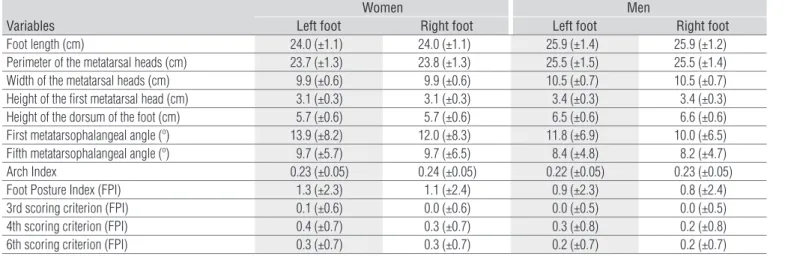
he men had a mean of 11.8º (±6.9) in the left Ang-I, and of 10.0º (±6.5) in the right Ang-I. Among the several variables ana-lyzed by Pearson’s correlation, only the height of the 1st meta-tarsal head/foot length, the AI, and the age did not evidence any association with Ang-I (Table 3). Positive, medium-inten-sity correlations were established between Ang-I and the foot width/foot length ratio, the angle of the ifth metatarsophalan-geal joint, the FPI, and the forefoot alignment (sixth criterion of the FPI). Positive and weak relationships were found between
Ang-I and the foot perimeter, and the 3rd and 4th criteria of the FPI. he height for the dorsum of the foot had a weak negative correlation with Ang-I.
Discussion
Ang-I was weakly associated with age only in the female group, and even so only in relation to the measurements of the
Table 1. Means and standard deviations of the variables studied in the female and male groups.
Women Men
Variables Left foot Right foot Left foot Right foot
Foot length (cm) 24.0 (±1.1) 24.0 (±1.1) 25.9 (±1.4) 25.9 (±1.2)
Perimeter of the metatarsal heads (cm) 23.7 (±1.3) 23.8 (±1.3) 25.5 (±1.5) 25.5 (±1.4)
Width of the metatarsal heads (cm) 9.9 (±0.6) 9.9 (±0.6) 10.5 (±0.7) 10.5 (±0.7)
Height of the first metatarsal head (cm) 3.1 (±0.3) 3.1 (±0.3) 3.4 (±0.3) 3.4 (±0.3)
Height of the dorsum of the foot (cm) 5.7 (±0.6) 5.7 (±0.6) 6.5 (±0.6) 6.6 (±0.6)
First metatarsophalangeal angle (º) 13.9 (±8.2) 12.0 (±8.3) 11.8 (±6.9) 10.0 (±6.5)
Fifth metatarsophalangeal angle (º) 9.7 (±5.7) 9.7 (±6.5) 8.4 (±4.8) 8.2 (±4.7)
Arch Index 0.23 (±0.05) 0.24 (±0.05) 0.22 (±0.05) 0.23 (±0.05)
Foot Posture Index (FPI) 1.3 (±2.3) 1.1 (±2.4) 0.9 (±2.3) 0.8 (±2.4)
3rd scoring criterion (FPI) 0.1 (±0.6) 0.0 (±0.6) 0.0 (±0.5) 0.0 (±0.5)
4th scoring criterion (FPI) 0.4 (±0.7) 0.3 (±0.7) 0.3 (±0.8) 0.2 (±0.8)
6th scoring criterion (FPI) 0.3 (±0.7) 0.3 (±0.7) 0.2 (±0.7) 0.2 (±0.7)
Table 3. Pearson’s correlation between the values for Ang-I and the
other variables studied in the male group.
Men
Variables Correlation
coefficient p-value
Age L 0.050 0.513
R -0.023 0.764
K Width of the metatarsal heads LR 0.3470.331 <0.001<0.001
K Perimeter of the metatarsal heads L 0.229 0.003 R 0.263 0.001
K Height of the first metatarsal head LR -0.0940.023 0.2190.761
K Height of the dorsum of the foot LR -0.171-0.153 0.0250.046
Fifth metatarsophalangeal angle L 0.394 <0.001 R 0.366 <0.001
Arch Index L 0.071 0.352
R 0.025 0.744
Foot Posture Index (FPI) L 0.337 <0.001
R 0.313 <0.001
3rd scoring criterion (FPI) L 0.211 0.005
R 0.184 0.016
4th scoring criterion (FPI) L 0.240 0.001
R 0.212 0.005
6th scoring criterion (FPI) L 0.327 <0.001 R 0.312 <0.001
K=value of the measurement multiplied by 100 and divided by the foot length; L=left; R=right.
Table 2. Pearson’s correlation between the values for Ang-I and the
other variables studied in the female group.
Women
Variables Correlation
coefficient p-value
Age L 0.092 0.165
R 0.133 0.045
K Width of the metatarsal heads LR 0.5600.443 <0.001<0.001
K Perimeter of the metatarsal heads LR 0.4260.320 <0.001<0.001
K Height of the first metatarsal head L 0.005 0.938 R -0.141 0.034
K Height of the dorsum of the foot LR -0.150-0.010 0.0240.880
Fifth metatarsophalangeal angle L 0.471 <0.001 R 0.347 <0.001
Arch Index L 0.049 0.458
R 0.054 0.420
Foot Posture Index (FPI) L 0.075 0.262
R 0.175 0.008
3rd scoring criterion (FPI) L 0.086 0.197
R 0.057 0.395
4th scoring criterion (FPI) L -0.038 0.564
R 0.113 0.090
6th scoring criterion (FPI) L 0.133 0.045
R 0.175 0.008
right foot. Perhaps the limited age span being studied (older adults only) prevented us from verifying that association. Ma-fart18 already reported that the prevalence of the hallux valgus
increases with age, which would justify a possible association. Nevertheless, it is necessary to highlight that the diagnosis of hallux valgus does not depend on a single joint angle, as inves-tigated in this study.
Ang-I maintained a positive correlation with the width and the perimeter of the metatarsal heads (in their proportions with the foot length) and with the angle of the 5th metatar-sophalangeal joint. Lamur et al.19 also noted an association
between the hallux valgus and wider forefeet, perhaps due to the lateral exostosis of the irst metatarsal head, common in hallux valgus cases. he angle of the ifth metatarsophalangeal joint, that may characterize the ifth varus toe, often seems to be a consequence of the hallux valgus, as well as the luxation of the central toes and the periostitis of the second and third metatarsal bones20.
The AI did not have an association with Ang-I. Saragas and Becker21 did not find differences in the incidence of flat
feet among women with and without hallux valgus. Some authors agree that flat feet exert very little influence on the genesis of the hallux valgus due to the great difference between the incidence and the weak coincidence of both deformities22-25.
A positive correlation was found between Ang-I and some of the criteria of the FPI. This finding suggests that older adults who had a greater angle in the first metatar-sophalangeal joint also had more proned feet. Kilmartin and Wallace26, Komeda et al.27 and Nery28 also observed this
association, reinforcing the idea that the valgus of the rear-foot also prones the 1st metatarsal and the hallux, forcing it to lean on its medial face in the propulsion phase of gait, resulting in a valgus force that acts on the hallux29. Besides
the valgus of the rearfoot, the proned foot is accompanied by the internal rotation and the medial displacement of the talus and of the navicular, which are responsible for the re-duction in theheight of the dorsum of the foot, which may explain the negative association found between Ang-I and the foot dorsal height.
he Pearson correlations between the anthropometric vari-ables and Ang-I had diferent results according to the laterality. In the female group, the diferences between the correlation coeicients of the left and right sides were of about 0.1 and in the male group they were even smaller. his inding may be due to the natural variation between the right and left sides. In the cases in which the correlations were weak (correlation coeicient <0.2), they were only signiicant on one of the sides (p≤0.05) because in these cases the coeicient neared the cut-of value for α≤0.05.
he main limitation detected in this study was the fact that the measurements were taken on diferent periods of the day, which might interfere with the volume of the foot, especially in individuals with vascular problems. Moreover, the use of ana-log instruments, less precise than their digital counterparts, may have represented a limitation, even though these are the resources most commonly used in clinical activity.
Based on the results obtained, it is possible to conclude that Ang-I did not show a relationship with age or with the AI, although it did show a positive association with the width and the perimeter of the metatarsal heads, as well as with the FPI and with the angle of the fifth metatarsopha-langeal joint; conversely, it was negatively associated with the height of the dorsum of the foot. Such findings reinforce the hypothesis that the hallux valgus does not occur in iso-lation, but takes part in the morphological changes of the feet of older adults, which may in turn originate pain, dif-ficulty to find adequate to shoes, and gait problems. There-fore, the evaluation of the feet should be part of the older adult patient’s physical therapy treatment, as it will provide a more thorough functional diagnosis, particularly concern-ing disorders affectconcern-ing body balance, as well as those related to pain disabilities.
Acknowledgments
he authors wish to thank the Coordenação de Aper-feiçoamento de Pessoal de Nível Superior (CAPES) for the inancial aid.
1. Petroianu A, Pimenta LA. Pé do idoso. In: Clínica e cirurgia geriátrica. Rio de Janeiro: Guanabara Koogan; 1999. p.503-11.
2. Ignácio H, Chueire AG, Carvalho-Filho G, Nascimento LV, Vasconcelos UMR, Barão GTF. Retrospective study of first metatarsal base osteotomy as a treatment of hallux valgus. Acta Ortop Bras. 2006;14(1): 48-52.
References
3. Dunn JE, Link CL, Felson DT, Crincoli MG, Keysor JJ, McKinlay JB. Prevalence of foot and ankle conditions in a multiethnic community sample of older adults. Am J Epidemiol. 2004;159(5):491-8.
5. Menz HB, Lord SR. Gait instability in older people with hallux valgus. Foot Ankle Int. 2005;26(6):483-9.
6. Benvenuti F, Ferrucci L, Guralnik JM, Gangemi S, Baroni A. Foot pain and disability in older persons: an epidemiologic survey.J Am Geriatr Soc. 1995;43(5):479-84.
7. Roddy E, Zhang W, Doherty M. Prevalence and associations of hallux valgus in a primary care population. Arthritis Rheum. 2008;59(6): 857-62.
8. Carvalho Filho ET, Papaléo Neto M. Anatomia e Fisiologia do Envelhecimento. In: Geriatria: fundamentos, clínica e terapêutica. São Paulo: Atheneu; 1998. p.32-4.
9. Instituto Brasileiro de Geografia e Estatística 2001 [homepage na Internet]. São Carlos: Censo 2000; [acesso em March 27, 2008]. Disponível em: http://www.ibge.gov.br/cidadesat/default.php.
10. Manfio EF, Ávila AIV. Um Estudo dos parâmetros antropométricos do pé feminino brasileiro. Rev Bras Biomecânica. 2003;4 Suppl 1:S39-48.
11. Norkin CC, White DJ. Medida do movimento articular: manual de goniometria. 2ª ed. Porto Alegre: Artmed; 1997.
12. Cavanagh PR, Rodgers MM. The arch index: a useful measure from footprints. J Biomech. 1987;20(5):547-51.
13. Redmond AC, Crosbie J, Ouvrier RA. Development and validation of a novel rating system for scoring standing foot posture: the foot posture index. Clin Biomech (Bristol, Avon). 2006;21(1):89-98.
14. Menz HB, Munteanu SE. Validity of 3 clinical techniques for the measurement of static foot posture in older people. J Orthop Sports Phys Ther. 2005;35(8):479-86.
15. Bland JM, Altman DG. Statistics notes: Measurement error. BMJ. 1996;313(7059):744.
16. Keenan AM, Redmond AC, Horton M, Conaghan PG, Tennant A. The foot posture index: rasch analysis of a novel, foot-specific outcome. Arch Phys Med Rehabil. 2007;88(1):88-93.
17. Chouquet-Stringer J, Bernard JH. Etude statistique sur la mesure des pieds en France. Bull Soc Fr Med Chir Pied. 1972;9:52-70.
18. Mafart B. Hallux valgus in a historical French population: paleopathological study of 605 first metatarsal bones. Joint Bone Spine. 2007;74(2): 166-70.
19. Lamur KS, Huson A, Snijders CJ, Stoeckart R. Geometric data of hallux valgus feet. Foot Ankle Int. 1996;17(9):548-54.
20. Salomão O. Hálux valgo: etiologia e tratamento. Rev Bras Ortop. 2005;40(4):147-52.
21. Saragas NP, Becker PJ. Comparative radiographic analysis of parameters in feet with and without hallux valgus. Foot Ankle Int. 1995;16(3): 139-43.
22. Mann RA, Coughlin MJ. Hallux valgus: etiology, anatomy, treatment and surgical considerations. Clin Orthop Relat Res. 1981;157:31-41.
23. Kilmartin TE, Wallace WA. The significance of pes planus in juvenile hallux valgus. Foot Ankle. 1992;13(2):53-6.
24. Hetherington VJ. Preoperative assessment in hallux valgus. In: Hallux valgus and forefoot surgery. New York: Churchill Livingstone Inc; 1994. p.107-23.
25. Coughlin MJ, Thompson FM. The high price of high-fashion footwear. Instr Course Lect. 1995;44:371-7.
26. Kilmartin TE, Wallace WA. The aetiology of hallux valgus: a critical review of the literature. The Foot. 1993;3(4):157-67.
27. Komeda T, Tanaka Y, Takakura Y, Fujii T, Samoto N, Tamai S. Evaluation of the longitudinal arch of the foot with hallux valgus using a newly developed two-dimensional coordinate system. J Orthop Sci. 2001;6(2): 110-8.
28. Nery CAS. Hálux valgo. Rev Bras Ortop. 2000;36(6):183-200.