Idiopathic popliteal artery pseudoaneurysm: emergency
diagnosis and treatment
Pseudoaneurisma idiopático da artéria poplítea: abordagem
diagnóstico-terapêutica na urgência
Germano da Paz Oliveira1, Ana Terezinha Guillaumon1, Iran Batista de Brito2, Joana Mayra Teixeira Lima2,
Sérgio Clementino Benvindo3, Lina Gomes dos Santos4
Abstract
Pseudoaneurysms or false aneurysms of the popliteal artery are uncommon arterial disorders. hese disorders most commonly result from trauma and iatrogenic lesions following orthopedic procedures. he authors report a rare case of popliteal artery pseudoaneurysm in which etiology was unknown. he authors also demonstrate that Doppler ultrasonography may be suicient for planning vascular surgical procedures and that the open surgical approach is the treatment of choice for cases in which the symptomatic lesion causes local compression.
Keywords: aneurysm, false; popliteal artery; ultrasonography, Doppler; vascular surgical procedures.
Resumo
Pseudoaneurismas ou aneurismas falsos de artéria poplítea são doenças arteriais incomuns. Eles resultam, mais frequentemente, de traumatismos e lesões iatrogênicas após procedimentos ortopédicos. Os autores relatam um raro caso de pseudoaneurisma de artéria poplítea para o qual não foi encontrada etiologia. Demonstram ainda que a ultrassonograia com Doppler pode ser suiciente para o planejamento de procedimentos cirúrgicos vasculares, sendo a abordagem aberta a escolha para casos em que se tenha uma lesão com sintomas compressivos locais.
Palavras-chave: aneurisma falso; artéria poplítea; ultrassonograia; Doppler; procedimentos cirúrgicos vasculares.
1Universidade Estadual de Campinas – UNICAMP, Faculdade de Ciências Médicas – FCM, Campinas, SP, Brasil. 2Universidade Federal do Piauí – UFPI, Teresina, PI, Brasil.
3Centro Universitário UNINOVAFAPI, Teresina, PI, Brasil.
4Universidade Federal do Piauí – UFPI, Centro de Ciências da Saúde – CCS, Teresina, PI, Brasil.
Financial support: None.
Conlicts of interest: No conlicts of interest declared concerning the publication of this article. Submitted: 11.15.13. Accepted: 04.02.14.
INTRODUCTION
By deinition, a “true” aneurysm is a dilatation of an artery that involves all three layers of the artery wall (intima, media and adventitial). In contrast, a pseudoaneurysm – or “false” aneurysm – develops from an injury to the artery wall, followed by formation of a hematoma and its containment by surrounding tissues and by the inlammatory process.1
The artery most frequently associated with pseudoaneurysms is the common femoral, since it is often subject to invasive diagnostic and/or therapeutic procedures.2 Pseudoaneurysms of the
popliteal artery (PPAs) are uncommon and there are a variety of etiologies described in the literature, with associations often described with traumatisms, infections and iatrogenic injuries after orthopedic procedures.3-12 Differential diagnosis is accomplished
by observation of knee luxation and abscesses and tumors in the popliteal fossa.4
The objective of this report is to describe the diagnostic and therapeutic approach taken to a case of an expanding idiopathic pseudoaneurysm affecting the popliteal artery. The project was approved by the local Ethics Committee and the patient described a consent form.
CASE DESCRIPTION
A 45-year-old, black, male agricultural worker presented at the Emergency Department complaining of pain and swelling in the left knee and leg with onset 10 days previously and exacerbation over the previous 12 hours. The patient reported no history of fever, surgery, traumas or any type of intervention to the affected limb. The patient was a smoker (10 pack-years) and had hypertension (under control). He reported being free of any other comorbidities.
Physical examination revealed a voluminous and painful pulsating mass in the left popliteal fossa, with thrill and murmur in the left popliteal cavity (Figure 1). There was infragenicular edema on the left with pitting (2+/4+), without cyanosis, pallor, or abnormal temperatures in the extremities, although distal pulses were dificult to take (probably because of edema). Leg extension movement was also limited, although neurological tests were normal.
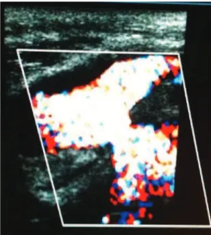
Doppler ultrasonography showed venous system low with normal phasicity and compressibility. However, there was a voluminous hematoma occupying the entire popliteal fossa and distal
thigh, restricting the popliteal vein and exhibiting turbulent low in its interior (Figure 2), with a pedicle communicating with the popliteal artery. The arteries of the leg were patent and showed three-phase low in pulse mode.
With this confirmation of the hypothesis of a pseudoaneurysm of the left popliteal artery in expansion, the patient was prepared for surgical intervention. Via a medial access, a popliteal-popliteal bypass was constructed using the contralateral inverted great saphenous vein and end-to-end anastomoses (Figures 3 and 4).
Material from the pseudoaneurysm capsule and its content were sent for microbiological and histopathological evaluation. The microbiology study ruled out infection. The histological study (Figures 5 and 6) with routine staining (HE), revealed arterial type blood vessel exhibiting luminal dilation but
Figure 1. Voluminous mass in left popliteal fossa.
without signs of atherosclerosis, inlammation or malignity.
During the immediate postoperative period the patient was free from pain and distal pulses were present, which was still the case 1 month later.
DISCUSSION
PPAs are habitually the result of sequelae from injuries to the artery wall.3,4 They rarely occur in
civil settings and the most important element in their genesis is traumas, whether by explosions, penetrating weapons or from iatrogenic causes.3,4
During the Korean war, for example, around 27% of all pseudoaneurysms and 1% of vascular injuries were PPAs and they were responsible for high limb amputation rates due to thromboembolic phenomena.3 In the same study, from 1991,3 Gillespie
and Cantelmo listed explosions and penetrating wounds as the most common etiologic factors, but other traumatic factors can also cause injuries to the popliteal artery, such as orthopedic3,5 and
vascular procedures3,6 and acupuncture3,7 and should
be investigated. Exostosis or osteochondromas of
the distal femur or proximal tibia have also been listed in the literature as pathophysiologic factors in development of PPAs, since they can lacerate the artery wall.8,9
During etiologic investigation, the hypothesis of trauma should be investigated with great insistence and should not be undervalued, even if the trauma happened many years before or there were no signiicant osteomuscular injuries.3,10 If there is no
explanatory history of trauma, however, autoimmune rheumatic causes, such as Behçet’s disease, should be ruled out.3,11 Even rarer causes of PPAs, such as
infection, must also be ruled out.12
In the case described here, the patient denied having suffered any closed or penetrating trauma in the past or having undergone any interventions to the limb. He did not exhibit any sign of toxemia nor did he it the clinical picture of any rheumatologic disease. The results of tests for inlammatory activity (erythrocyte sedimentation rate and C-reactive protein) were normal. Microbiology and pathology test results were normal, without abnormalities in the wall of the vessel that could cause weakening of the
Figure 3. Intraoperative inding.
Figure 4. Popliteal-popliteal graft with inverted great saphenous vein.
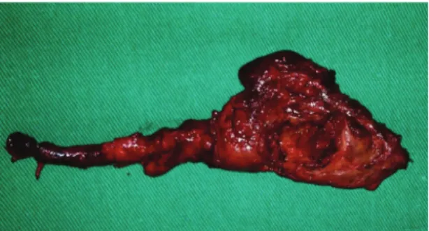
Figure 5. Macroscopic appearance of the popliteal artery pseudoaneurysm.
artery wall and a propensity to rupture in response to minimal trauma.13 There was no evidence of an
inlammatory process in the artery wall or of the presence of giant cells.
Physical examination indings are normally highly suggestive in this condition, with a pulsating mass with palpable thrill, pain and attenuated pulses.14
However, clinical status is not always typical and radiological assessment therefore has an important role to play.15,16 In the case described here, Doppler
ultrasonography confirmed the diagnosis with certainty and provided enough information to plan the surgery that would be used, since it provided the exact location of the injury and his relationship with neighboring structures, in addition to showing the undeniable patency of the distal arteries. This examination was also capable of ruling out deep vein thrombosis. Some authors consider that, although there have been advances in other diagnostic methods, arteriography via puncture with injection of intra-arterial contrast continues to be the gold standard for cases such as the one described here.16
However, there are also reports from well-respected authors17,18 that support the conduct adopted in this
case.
Surgical intervention for popliteal aneurysms and pseudoaneurysms can be performed with open surgery by vein interposition, or using endovascular techniques with exclusion of the aneurysm with covered stents.19 For elective surgery, both methods
appear to be equal in terms of short and medium-term results.20 However, according to
Trinidad-Hernandez et al.,20 the open technique is superior in
emergency situations. Injuries to the popliteal artery, such as the one described here, are in themselves an indication for immediate surgical intervention because of the imminent risk of hemodynamic instability. In the case described here, the injury was very extensive, provoking compressive symptoms such as pain and swelling. Additionally, the possibility of infection as an associated factor cannot be ignored during initial workup. For this reason an endovascular approach was not considered the best option in this case. Finally, the access chosen was the medial approach because of the size of the injury, which extended up to the adductor canal, ruling out the posterior approach.21
The authors’ conclusion is therefore that they were faced with a rare case of idiopathic popliteal artery pseudoaneurysm in which the diagnostic strategy based on ultrasonography defined the surgical intervention chosen.
REFERENCES
1. Tedesco MM, Dalman RL. Arterial aneurysms. In: Cronenwett JL, Johnston KW, editors. Rutherford’s Vascular Surgery. 7th ed. Philadelphia: Elsevier; 2010. p. 559-615.
2. Miyamotto M, Moreira RCR, Erzinger FL, França GJ, Cunha AGP. Pseudo-aneurisma idiopático da artéria poplítea. J Vasc Bras. 2004;3(2):169-71.
3. Gillespie DL, Cantelmo NL. Traumatic popliteal artery pseudoaneurysms: case report and review of the literature. J Trauma. 1991;31(3):412-5. http://dx.doi.org/10.1097/00005373-199103000-00019. PMid:2002532
4. Bel Haj Salah R, Triki W, Gherib SB, Ben Moussa M, Zaouche A. [Traumatic popliteal artery pseudo aneurysm]. Tunis Med. 2011;89(8-9):721-2. PMid:21948669.
5. Szyber P Jr, Skóra J, Rybak W, Pupka A. Iatrogenic pseudoaneurysm of the popliteal artery following corrective tibial osteotomy. Vasa. 2011;40(5):414-7. http://dx.doi.org/10.1024/0301-1526/a000140. PMid:21948786
6. Tsuji Y, Kitano I, Iida O, Kajita S, Sawada K, Nanto S. Popliteal pseudoaneurysm caused by stent fracture. Ann Vasc Surg. 2011;25(6):840.e5-8. http://dx.doi.org/10.1016/j.avsg.2010.12.039. PMid:21620667
7. Kao CL, Chang JP. Pseudoaneurysm of the popliteal artery: a rare sequela of acupuncture. Tex Heart Inst J. 2002;29(2):126-9. PMid:12075870.
8. Pavić P, Vergles D, Sarlija M , Ajduk M , Cupurdija K . Pseudoaneurysm of the popliteal artery in a patient with multiple hereditary exostoses. Ann Vasc Surg. 2011;25(2):268.e1-2. http:// dx.doi.org/10.1016/j.avsg.2010.07.027. PMid:20926234
9. Pellenc Q, Capdevila C, Julia P, Fabiani JN. Ruptured popliteal artery pseudoaneurysm complicating a femoral osteochondroma in a young patient. J Vasc Surg. 2012;55(4):1164-5. http://dx.doi. org/10.1016/j.jvs.2011.01.060. PMid:21459549
10. Ge PS, Ishaque BM, Bonilla J, de Virgilio C. Popliteal artery pseudoaneurysm after isolated hyperextension of the knee. Ann Vasc Surg. 2010;24(7):950.e7-11. http://dx.doi.org/10.1016/j. avsg.2010.01.014. PMid:20471789
11. Koksoy C, Gyedu A, Alacayir I, Bengisun U, Uncu H, Anadol E. Surgical treatment of peripheral aneurysms in patients with Behcet’s disease. Eur J Vasc Endovasc Surg. 2011;42(4):525-30. http://dx.doi.org/10.1016/j.ejvs.2011.05.010. PMid:21641238
12. Ghassani A, Delva JC, Berard X, Deglise S, Ducasse E, Midy D. Stent graft exclusion of a ruptured mycotic popliteal pseudoaneurysm complicating sternoclavicular joint infection. Ann Vasc Surg. 2012;26(5):730.e13-5. http://dx.doi.org/10.1016/j. avsg.2011.09.015. PMid:22664287
13. Erler K, Ozdemir MT, Oguz E, Basbozkurt M. Does false aneurysm behave like a sarcoma? Distal femoral arterial false aneurysm simulated a malign mesenchymal tumor. A case report and review of the literature. Arch Orthop Trauma Surg. 2004;124(1):60-3. http://dx.doi.org/10.1007/s00402-003-0595-8. PMid:14576956
Clin Radiol. 1986;37(6):585-8. http://dx.doi.org/10.1016/S0009-9260(86)80033-2. PMid:3539457
16. Callcut RA, Acher CW, Hoch J, Tefera G, Turnipseed W, Mell MW. Impact of intraoperative arteriography on limb salvage for traumatic popliteal artery injury. J Trauma. 2009;67(2):252-7, discussion 257-8. http://dx.doi.org/10.1097/ TA.0b013e31819ea796. PMid:19667876
17. Proia RR, Walsh DB, Nelson PR, et al. Early results of infragenicular revascularization based solely on duplex arteriography. J Vasc Surg. 2001;33(6):1165-70. http://dx.doi.org/10.1067/ mva.2001.115376. PMid:11389413
18. Ascher E, Hingorani A, Markevich N, Costa T, Kallakuri S, Khanimoy Y. Lower extremity revascularization without preoperative contrast arteriography: experience with duplex ultrasound arterial mapping in 485 cases. Ann Vasc Surg. 2002;16(1):108-14. http://dx.doi.org/10.1007/s10016-001-0130-8. PMid:11904814
19. Pulli R, Dorigo W, Castelli P, et al. A multicentric experience with open surgical repair and endovascular exclusion of popliteal artery aneurysms. Eur J Vasc Endovasc Surg. 2013;45(4):357-63. http://dx.doi.org/10.1016/j.ejvs.2013.01.012. PMid:23391602
20. Trinidad-Hernandez M, Ricotta JJ 2nd, Gloviczki P, et al. Results of elective and emergency endovascular repairs of popliteal artery aneurysms. J Vasc Surg. 2013;57(5):1299-305. http://dx.doi. org/10.1016/j.jvs.2012.10.112. PMid:23375609
21. Zaraca F, Ponzoni A, Stringari C, Ebner JA, Giovannetti R, Ebner H. he posterior approach in the treatment of popliteal artery aneurysm: feasibility and analysis of outcome. Ann Vasc Surg. 2010;24(7):863-70. http://dx.doi.org/10.1016/j.avsg.2010.04.005. PMid:20831987
Correspondence Germano da Paz Oliveira Rua José Olímpio de Melo, 2436/102 – Ilhotas CEP 64014-063 – Teresina (PI), Brasil Fone: +55 (86) 99820901 E-mail: [email protected]
Author information GPO is a Vascular Surgeon; an MSc in Sciences from the School of Medical Sciences of Universidade Estadual de Campinas (UNICAMP). ATG is a Tenured Professor of Peripheral Vascular Diseases at the Department of Surgery, School of Medical Sciences, Universidade Estadual de Campinas (UNICAMP). IBB and JMTL are Medical Students at Universidade Federal do Piauí (UFPI). SCB is a Medical Student at Centro Universitário UNINOVAFAPI. LGS is a Professor at the Department of Pathology of the Center for Health Sciences, Universidade Federal do Piauí (UFPI)
Author contributions Conception and design: GPO, ATG Analysis and interpretation: GPO, ATG, IBB, JMTL, SCB, LGS Data collection: GPO, ATG, IBB, JMTL, SCB, LGS Writing the article: GPO, ATG, IBB, JMTL, SCB, LGS Critical revision of the article: GPO, ATG, LGS Final approval of the article*: GPO, ATG, LGS Statistical analysis: N/A Overall responsibility: GPO Obtained funding: None.