Surgical treatment of false aneurysm of the sciatic artery –
case report and literature review
Tratamento cirúrgico de pseudoaneurisma de artéria isquiática – relato de caso e revisão da literatura
Gustavo Ioshio Handa1, Francisco Eduardo Coral1,2,3, Vinicius Zendrini Buzingnani1, Lucas Marques Mantovani1, Elizana Stella Lopes Rasera1, Denise Krauss1, Maria Carolina S. de Paula4
Introduction
he persistent sciatic artery was described in 1832 by Green1. It is an extension of the internal iliac artery, which
supplies blood to lower limb in the early embryological pe-riod and takes part in the formation of the inferior gluteal, profunda femoral, popliteal and ibular arteries and foot vessels. It rarely persists (0.025 – 0.04%) in the adult, and more than 160 cases have been described in the literature.
Its clinical manifestations are: aneurysm formation (pulsate gluteal mass), acute or chronic limb ischemia and sciatic nerve compression2-4.
Case report
A 43-year-old female patient was admitted with pain in the let gluteal area and a local palpable mass. At physi-cal examination, she presented normal pulses in the lower
Abstract
he persistent sciatic artery is a rare anatomical variation, with few cases described on the literature. It presents clinically as aneurysm formation, pulsate gluteal mass, acute or chronic limb ischemia and sciatic nerve compression. Diagnosis is conirmed by imaging methods: duplex scan, CT angiographt and magnetic resonance angiography. Treatment is indicated in symptomatic cases and when there is aneurysm formation and it is performed by ligation of the sciatic artery or endovascular embolization, associated with limb revascularization in the cases the sciatic artery is the main blood supply to the limb. We report the case of a 43 year-old female patient, ,with a false aneurysm of the sciatic artery conirmed by duplex scan and magnetic resonance angiography who had local pain and sciatic neuropathy due to neural compression. Surgical exploration was performed, with ligation of sciatic artery and thrombus removal. At the 12 months follow up there was signiicant pain relief and she was performing motor physical therapy to recover the neurological functions of the limb.
Keywords: sciatic neuropathy; aneurysm, false; iliac artery.
Resumo
A persistência da artéria isquiática é uma rara variação anatômica, com poucos casos descritos na literatura, manifestando-se por formação de aneurisma, massa pulsátil em glúteo, isquemia aguda ou crônica de membro inferior e compressão de nervo isquiático. O diagnóstico é conirmado com exames de imagem: mapeamento duplex, angiotomograia e angiorressonância magnética. O tratamento é indicado nos casos sintomáticos ou quando há formação de aneurisma, realizado através de ligadura ou embolização por via endovascular, sendo necessário a revascularização do membro nos casos em que a artéria isquiática é a principal responsável pelo suprimento sangüíneo do membro. Apresentamos o caso de uma paciente do sexo feminino, 43 anos, com pseudoaneurisma de artéria isquiática conirmada por mapeamento duplex e angiorressonância magnética, com quadro de neuropatia isquiática por compressão nervosa e dor local. A paciente foi submetida à exploração cirúrgica com ligadura da artéria isquiática e remoção dos trombos. No seguimento de 12 meses, apresentou importante melhora da dor e realizou isioterapia motora para recuperação das funções neurológicas do membro.
Palavras-chave: neuropatia ciática; falso aneurisma; artéria ilíaca.
Study carried out at the Angiology and Vascular Surgery Service, Santa Casa de Misericórdia de Curitiba/Pontifícia Universidade Católica do Paraná (PUCPR) – Curitiba (PR), Brazil.
1 Angiology and Vascular Surgery Service, Santa Casa de Misericórdia de Curitiba/PUCPR – Curitiba (PR), Brazil.
2 Specialist in Vascular Surgery and Endovascular Surgery, Sociedade Brasileira de Angiologia e Cirurgia Vascular – Curitiba (PR), Brazil. 3 Medical residency preceptors in Vascular Surgery, Santa Casa de Misericórdia de Curitiba – Curitiba (PR), Brazil.
4 Nephrology Service, Santa Casa de Misericórdia de Curitiba/PUCPR – Curitiba (PR), Brazil.
limbs and let gluteal palpable mass. She had systemic ar-terial hypertension and chronic renal failure, and she had been in hemodialysis for seven years. Her condition pro-gressed with radiation of the pain to the posterior aspect of the let lower limb, associated with paresthesia, let foot drop and inability to perform dorsal lexion of the foot.
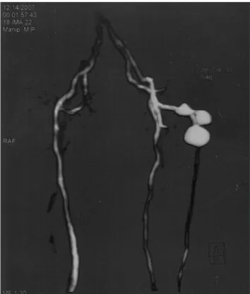
he investigation started with computed tomography and ultrasonography, which showed an expansive mass in the gluteal muscle depth. A duplex scan showed false aneu-rysm of the sciatic artery measuring 46x52 mm transversal-ly and 48x52 mm longitudinaltransversal-ly (Figure 1). Magnetic reso-nance angiography conirmed the presence of incomplete persistent sciatic artery, with false aneurysm of the sciatic artery in the gluteal region (Figures 2, 3 and 4).
Figure 1. Cross-sectional view in a duplex scan, showing the sciatic ar-tery with false aneurysm.
Figure 2. Magnetic resonance angiography showing persistent sciatic artery with false aneurysm.
Figure 3. Magnetic resonance angiography showing persistent sciatic artery with false aneurysm.
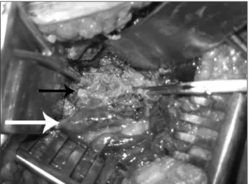
Due to the neurological degradation caused by sciat-ic nerve compression, it was decided to perform surgsciat-ical treatment to decompress the sciatic nerve and relieve the pain and restore the neurological function of the let lower limb. With the patient in the supine position, the procedure consisted of an access to the gluteal region, removal of the thrombi adjacent to the artery, identiication of the sciatic artery and the origin of the false aneurysm, followed by proximal and distal arterial ligation. As the blood supply to the let lower limb was through the femoral artery, revascu-larization was not required (Figures 5 and 6).
In the postoperative period, the patient presented significant pain relief in the lower left limb and gluteal
area and partial recovery of limb sensitivity and motor function. She was discharged from hospital 5 days after the procedure and underwent outpatient physiotherapy sessions for 12 months to fully recover the neurological function of the limb.
Literature review
The sciatic artery supplies axial blood to the em-bryo’s lower limbs and it is an extension of the internal iliac artery that crosses the sciatic foramen and runs be-hind the adductor magnus muscle, becoming eventual-ly the popliteal artery. It usualeventual-ly suffers a process called involution when the embryo reaches about 22 mm and the ilio-femoral system becomes the main blood supply to lower limbs. When the femoral system does not de-velop or the embryonic sciatic artery does not involute, this axial artery becomes the sciatic artery and ensures the blood supply to the limb5.
There are two major categories of persistent sciatic artery: the complete form, which corresponds to 63– 79% of the cases, when the sciatic artery is the dominant blood supply to lower limbs through a direct communi-cation between the internal iliac and popliteal arteries. In most cases, the external iliac and common femoral arteries are complete, but the superficial femoral artery is hypoplastic and communicates with the popliteal ar-tery through collateral arteries. In the incomplete form, the femoral system is dominant and the sciatic artery is hypoplastic and communicates with the femoral system through collateral arteries6,7.
Sultan et al.8 classified the anomalies into five types,
based on the superficial femoral artery: type I is the complete persistent sciatic artery and femoral system that ends up as the popliteal artery; type II corresponds to the complete form associated with aplasia of exter-nal iliac and femoral arteries, with normal superficial femoral and popliteal arteries; type III is the incomplete form with the femoral system in communication with the popliteal and sural arteries; type IV is the incom-plete form with hypoplasia of the sciatic artery and dominant femoral system; and type V corresponds to the incomplete form with hypoplasia of the sciatic and femoral arteries.
The persistent sciatic artery is associated with oth-er congenital anomalies, such as artoth-eriovenous fistula3,
lower extremity hypertrophy9, internal carotid artery
and abdominal aorta aneurysms10, neurofibromatosis,
bone hypertrophy and solitary kidney. In 12–32% of the cases, the persistent sciatic artery is bilateral11,12.
Figure 6. Surgical exploration of false aneurysm with proximal and dis-tal ligation of the artery and removal of thrombi (white arrow). Sciatic nerve exposed (black arrow).
The most frequent complication of this anomaly is aneurysm formation, which occurs in 44–61% of the cases13. The reasons for sciatic artery predisposition to
atheromatous degeneration and aneurysm formation are still not clear.
The clinical presentation is dependent upon the anomaly type and presence of aneurysm. If the femoral artery is hypoplastic or absent, the patient shows no or low femoral pulse and palpable femoral and foot pulses (Cowie’s sign)2,4. The aneurysm thrombosis or
embo-lization causes lower limb ischemia. Forty per cent of the patients are asymptomatic and the sciatic artery is an incidental finding. The most frequent clinical presenta-tion is lower limb ischemia (31.1%), followed by gluteal mass (25.7%). In 25% of the cases of limb ischemia, it is critical, with risk of limb loss. Large aneurysms may cause sciatic nerve compression, with pain, paresthesia and decreased motor function of the affected limb15.
The diagnostic suspicion at the clinical exam should be confirmed with further exams. A duplex scan may show hypoplastic femoral system, the pres-ence of an aberrant artery that fills the popliteal artery and the presence of aneurysm in the gluteal region16.
Computed tomography angiography and magnetic res-onance angiography are useful in the therapeutic plan-ning, as they show whether the aneurysm neck is inside or outside the pelvis, and in the treatment follow-up17.
Arteriography should be used to outline the anatomy, the type of persistence and the distal vessels, which will determine the best surgical method.
The treatment is dependent on the anomaly form, clinical manifestations, concomitant peripheral ob-structive arterial disease and presence or absence of aneurysm. In asymptomatic cases without aneurysm, no intervention is required6, but monitoring should be
made by duplex scan to detect any aneurysm formation. The presence of aneurysm indicates an intervention is required, due to the high risk of thromboembolic com-plications. In incomplete forms, aneurysm can be treat-ed by simple ligation or endovascular embolization2,8.
In complete forms, besides the aneurysm exclusion, the limb requires revascularization, preferably through a femoral-distal bypass. When the aneurysm neck is lo-cated within the pelvis, the access should be transperi-toneal or extra-peritransperi-toneal, increasing the magnitude of the operation and the risk of complications. When the aneurysm is completely outside the pelvis, the posterior transgluteal access is recommended. After aneurysm ex-clusion, a femoral popliteal bypass is made14,18. Fearing
et al.19 and Gabelmann et al.20 successfully performed
endovascular treatment of a sciatic artery aneurysm with the implantation of a stent, recommending this technique to high risk patients, with limited life expec-tancy. The endovascular procedure is not indicated in cases with compression of adjacent anatomical struc-ture, as in the case report.
References
1. Green PH: On a new variety of femoral artery. Lancet. 1832;17(442):730-1.
2. Wilson JS; Bowser AN; Miranda A; et al. Limb ischemia associ-ated with persistent sciatic artery aneurysms – a report of 2 cases. Vasc Endovascular Surg: 2005;39:91-101.
3. Greebe J. Congenital anomalies of the iliofemoral artery. J Cardiovasc Surg (Torino).1977;18(3):317-23.
4. Pirker E, Schmidberger H. Sciatic artery. A rare vessel variant. Fortschr Geb Rontgenrst Nuklearmed. 1972;116(3):434-7.
5. Schutze WP, Garret WV, Smith BL. Persistent sciatic artery: col-lective review and management. Ann Vasc Surg.1993;7(3):303-10.
6. Brantley SK, Rigdom EE, Raju S. Persistent sciatic artery: embry-ology, pathology and treatment. J Vasc Surg. 1993;18(2):242-8.
7. Bower EB, Smullens SN, Parker WW. Clinical aspects of persis-tent sciatic artery: report of two cases and review of literature. Surgery. 1977;81(5):588-95.
8. Sultan SAH, Pacainowski JP, Madhaven P, et al. Endovascular management of rare sciatic artery aneurysm: case report. J Endovasc Ther. 2000;7(5):415-22.
9. Wright FW. Persistent axial or sciatic artery of lower limb in as-sociation with hemihypertrophy. Clin Radiol. 1964;15:291-2.
10. Miyahara T; Miyata T; Shigematsu K; et al. Persistent sciatic ar-tery in a patient with extracranial internal carotid arar-tery aneu-rysm and infrarenal abdominal aortic aneuaneu-rysm. A case report. Int Angiol. 2005;24(4):391-4.
11. Juliá J, Rimbau EM, Gómez F, et al. Arteria ciática persistente bilateral. Rev Angiol. 1995;4:199-205.
12. Bez LG , Costa-Val R, Bastianetto P, et al. Persistência da artéria isquiática: relato de caso. J Vasc Bras. 2006;5(3):233-6.
13. Kritsch D; Hutter HP; Hirschl M; et al. Persistent sciatic artery: an uncommon cause of intermittent claudication. Int Angiol. 2006;25(3):327-9.
14. Cowie TN, McKellar NJ, McLean N, et al. Unilateral congenital absence of the external iliac and femoral arteries. Br J Radiol. 1960;33:520-2.
15. Ikezawa T, Naiki K, Moriura S, et al. Aneurysm of bilateral persis-tent sciatic arteries with ischemic complications: case report and review of the world literature. J Vasc Surg. 1994;20(1):96-103.
16. Erturk SM, Tatli S. Persistent sciatic artery aneurysm. J Vasc Interv Radiol. 2005;16(10):1407-8.
18. Wolf YG, Gibbs BF, Guzzeta VJ, et al. Surgical treatment of aneu-rysm of persistent sciatic artery. J Vasc Surg. 1993;17(1):218-21.
19. Fearing NM, Ammar AD, Hutchinson SA, et al. Endovascular stent graft repair of a persistent sciatic artery aneurysm. Ann Vasc Surg. 2005;19(3):438-41.
20. Gabelmann A, Krämer SC, Wisianowski C, et al. Endovascular interventions on persistent sciatic arteries. J Endovasc her. 2001;8(6):622-8.
Correspondence
Gustavo Ioshio Handa Praça Rui Barbosa, 694 – Centro CEP 80010-030 – Curitiba (PR), Brasil
E-mail: gustavohanda@yahoo.com.br
Author’s contribution
Conception and design: GIH, FEC Analysis and interpretation: GIH, FEC, VZB Data collection: GIH, VZB, ESLR, MCSP Writing the article: GIH, DK, LMM Critical revision of the article: GIH, FEC, LMM, DK Final approval of the article*: GIH, FEC, VZB, ESLR, LMM, DK, MCSP Statistical analysis: GIH Overall responsibility: GIH, FEC Obtained funding: GIH