Biomarkers of inflammation may be of use for identification of
more severe peripheral arterial occlusive disease
Biomarcadores inflamatórios circulantes podem ser úteis para identificar
doença arterial obstrutiva periférica mais grave
Luciana Garofolo1, Sandra Roberta Gouvea Ferreira1, Fausto Miranda Junior1
Abstract
Background: Atherosclerosis is a multifactorial disease with an inlammatory pathophysiological basis. Cytokines released during the atherosclerotic process induce production of C-reactive protein (CRP) in the liver, which is an important marker of inlammation. Objective: We tested whether inlammatory biomarkers were associated with deterioration of peripheral arterial occlusive disease (PAOD) in a population at high cardiovascular risk. Methods: 1,330 subjects ≥30 years of age underwent clinical and laboratory examinations as part of a population-based study of the prevalence of diabetes. PAOD was deined as an ankle-brachial index (ABI) ≤0.90. After application of exclusion criteria, the sample comprised 1,038 subjects. Traditional risk factors, CRP and interleukin 6 (IL-6) were also compared across three ABI categories (≤0.70; 0.71-0.90; ≥0.90). Mean values for these variables were compared by presence/absence of DAOP (Student’s t test) and by ABI categories (ANOVA). Poisson regression and logistic regression models were used to test for associations between risk factors and DAOP and between risk factors and the ABI categories. Pearson’s linear correlation coeicients were calculated for the relationship between CRP and IL-6 levels. Results: Mean age was 56.8±12.9 years, 54% of the sample were women and the prevalence of DAOP was 21.0% (95%CI 18.4-24.1). Individuals with ABI ≤0.70 had higher concentrations of CRP-us (2.1 vs. 1.8) and of IL-6 (1.25 vs. 1.17). Concentrations of CRP and IL-6 were only correlated in patients with DAOP, (p=0.004). Conclusions: he inding that CRP and IL-6 levels were only elevated among people with advanced DAOP may suggest that these biomarkers have a role to play as indicators of more severe disease. Prospective studies are needed to test this hypothesis.
Keywords: atherosclerosis; peripheral arterial occlusive disease; biomarkers.
Resumo
Contexto: Aterosclerose é doença multifatorial, cuja base isiopatológica é um processo inlamatório. Estudos são controversos quanto ao papel dos biomarcadores como fatores de risco. A liberação de citoquinas durante aterogênese promove síntese hepática de proteína C-reativa (PCR), importante marcador inlamatório. Objetivo: Avaliamos se biomarcadores inlamatórios estavam associados à deterioração da doença arterial obstrutiva periférica (DAOP), em população de risco cardiovascular. Métodos: Estudo populacional sobre prevalência de diabetes, em que 1.330 indivíduos com ≥30 anos foram submetidos a exames clínico-laboratoriais. Diagnóstico de DAOP foi feito pelo índice tornozelo-braço (ITB) ≤0,90. Após exclusões, 1.038 indivíduos foram analisados. Fatores de risco tradicionais, PCR e interleucina 6 (IL-6) foram comparados também segundo três categorias de ITB (≤0,70; 0,71-0,90; ≥0,90). Valores médios das variáveis foram comparados segundo presença de DAOP (teste t Student) e categorias do ITB (ANOVA). Utilizou-se modelo de Poisson e regressão logística para avaliar associações da DAOP e categorias do ITB com fatores de risco. Estimou-se coeiciente de correlação linear de Pearson para relação entre os valores de PCR e IL-6. Resultados: A idade média foi 56,8±12,9 anos, 54% mulheres e prevalência de DAOP 21,0% (IC95% 18,4-24,1). Indivíduos com ITB ≤0,70 apresentaram maiores valores de PCR-us (2,1 vs. 1,8) e IL-6 (1,25 vs. 1,17. Apenas em portadores de DAOP, valores de PCR e IL-6 mostraram-se correlacionados (p=0,004). Conclusão: O achado de concentrações mais elevadas de PCR e IL-6 apenas em indivíduos com DAOP avançada pode sugerir um papel destes biomarcadores, indicando doença mais grave. Estudos prospectivos são necessários para testar esta hipótese.
Palavras-chave: aterosclerose; doença arterial periférica obstrutiva; biomarcadores.
1Universidade Federal de São Paulo – UNIFESP, São Paulo, SP, Brazil.
Financial support: FAPESP.
Conlicts of interest: No conlicts of interest declared concerning the publication of this article. Submitted: 03.18.14. Accepted: 06.02.14.
INTRODUCTION
Synthesis of C-reactive protein (CRP) in the liver
is an innate, nonspeciic, immunological defense mechanism1 that activates the complement system
and promotes phagocytosis. To a great extent, synthesis is regulated by interleukin 6 (IL-6), an inlammatory cytokine that is primarily secreted by macrophages and adipocytes.2 Experimental studies
indicate that smooth muscle and endothelial cells of both normal and aneurysmal arteries can also secrete IL-6.3
Ultrasensitive assays are capable of detecting discrete increases in blood CRP concentrations (CRP-us) that characterize subclinical chronic inflammation. With relation to atherosclerotic disease, some authors believe that CRP can also be produced in smooth muscle cells of diseased
coronary arteries,4 but this inding is questionable.5
The several stages of atherogenesis all involve release of cytokines that stimulate CRP production and so determination of its concentration in blood can be used as an inlammatory marker for monitoring cardiovascular risk.6 However, a meta-analysis of the
role of CRP-us in prediction of cardiovascular risk concluded that although it may be promising, to date no adequately-designed studies of suficiently long duration have been conducted that could justify its widespread adoption in clinical practice.7
An initial trigger of formation of fatty streaks is endothelial dysfunction, which can be precipitated by smoking, arterial hypertension, hyperglycemia, hypercholesterolemia, hyperhomocysteinemia and even infections. Initial steps in atherosclerosis include expression of adhesion molecules and transmigration of monocytes into the subendothelial space, where they absorb lipids from foam cells. These cells secrete inlammatory mediators, including IL-6 which acts to reduce de lipoprotein lipase activity, stimulating phagocytosis of lipids further still. There is evidence to indicate that CRP and IL-6 amplify and perpetuate the inlammatory response in atheroma.8
In terms of the different sites affected by atherosclerosis, peripheral arterial occlusive disease (PAOD) is one of the most important conditions because it is becoming ever more prevalent in modern society, as a result of increasing life expectancy. It is estimated that 202 million people had PAOD worldwide in 2010. Over the last ten years, PAOD prevalence in countries with low to medium per capita income increased by 28.7%, and its prevalence in high income countries increased by 13.1%.9 The
growing interest in early diagnosis of PAOD is not
only the result of its increasing prevalence, but also because it is related to diseases in other parts of the body, such as coronary or cerebral conditions.10 In
view of this, identiication of the disease itself and also of indicators of its severity is desirable for the purposes of planning interventions.
While there is evidence that elevated CRP and IL-6 concentrations are indicative of the subclinical inlammatory processes present in atherosclerotic cardiovascular disease, there is no consensus on the etiopathogenic role that these substances play in arterial injury, and this is particularly true of investigations into PAOD.
This research was conducted in order to investigate whether CRP and IL-6 concentrations were related to different classes of PAOD, by means of a population study of Japanese-Brazilian subjects, who are a population group that is known to have high cardiovascular risk.11-13
METHODS
This analysis was conducted with a population of Japanese-Brazilians living in Bauru, SP, Brazil. The sample included both sexes and the minimum age was 30 years. Details of the umbrella study into the prevalence of diabetes and metabolic syndrome have been described elsewhere.11-13 The study was
approved by the Institutional Ethics Committee and free and informed consent forms were signed by all participants. A total of 1,330 people underwent a clinical examination, including anthropometric measurements, arterial blood pressure measurement and calculation of the Doppler ankle-brachial index (ABI). Additionally, blood samples were taken after 12 hours’ fasting. The exclusion criteria were missing clinical or laboratory data preventing analysis (255 participants), ABI >1.40 (one participant) and CRP-us concentration >10 mg/L (36 participants). As a result, 1,038 people were enrolled on the study.
Weight and height were measured with subjects unshod and wearing minimal clothing and the results used to calculate body mass index (BMI). Waist circumference was measured using an inextensible tape measure, at the midpoint between the last loating rib and the iliac crest along a plane parallel to the loor. Hip circumference was measured at the height of the buttocks, passing over the pubic symphysis. The waist-hip ratio was calculated by dividing waist circumference by hip circumference.
recommended by the Japan Society for the Study of Obesity (JASO)14 and the waist-hip ratio cutoff
used was that recommended by the World Health Organization (WHO).15
Arterial blood pressure was measured using an automatic blood pressure meter (Omron HEM-712C, Omron Healthcare, USA), sitting down, after 5 minutes at rest. The igures used for analysis were the arithmetic means of the last two systolic and diastolic pressure readings. Subjects with pressures greater than 140×90 mmHg and those that were on medication for high blood pressure were diagnosed as having arterial hypertension.16
Participants with capillary glycemia greater than 200 mg/dL after 12 hours’ fast were not subjected to the oral glucose tolerance test. Venous blood samples were taken after fasting and 2 hours after intake of 75 grams of glucose. Subjects were allocated to glucose tolerance categories in accordance to American Diabetes Association criteria.17 Therefore,
participants with fasting glycemia <100 mg/dL or post-challenge glycemia <140 mg/dL were considered normoglycemic. Subjects with fasting glycemia greater than or equal to 100 mg/dL, but less than 126 mg/dL, and with glycemia 2 hours after challenge below 140 mg/dL were deined as having abnormal fasting glycemia (AFG). Impaired glucose tolerance (IGT) was defined as fasting glycemia greater than or equal to 100 mg/dL and post-challenge glycemia from 140 to 199 mg/dL. Subjects were diagnosed as diabetic if they exhibited fasting glycemia greater than or equal to 126 mg/dL or post-challenge glycemia greater than or equal to 200 mg/dL, or if they were already on hypoglycemic drugs.
The reference values used to diagnose dyslipidemias were those recommended by the NCEP at the time of enrolment.18 Participants were
considered to have normal lipid proiles if total cholesterol was ≤200 mg/dL, LDL-cholesterol was ≤130 mg/dL, HDL-cholesterol was ≥35 mg/dL for men or ≥45 mg/dL for women and triglycerides were ≤200 mg/dL. Subjects were deined as having a dyslipidemia if any of these variables were outside of these limits.
Uric acid was considered normal up to 6 mg/dL for women or up to 7 mg/dL for men.19
Homocysteine was assayed using high performance liquid chromatography. Homocysteine results were considered normal up to 15 mmol/L.20
Chemiluminescence was used to determine CRP-us and IL-6 concentrations (Immulite, Diagnostic Products Corporation, USA).
Diagnosis of PAOD
Peripheral arterial occlusive disease was diagnosed using an 8 mHz continuous wave Doppler machine by Imbracios®. The ABI was calculated by dividing the pressure at the arteries in the ankle by the greatest pressure measured at the brachial arteries. Indices of ≤0.90 or >1.40 were considered abnormal, as recommended by the TASC II (Transatlantic Society Consensus).21 The sample was also analyzed
after stratiication by ABI into the following three categories: ≤0.70; 0.71 to 0.90; and >0.90.22
Statistical analysis
Variables were expressed as percentages, means and standard deviations. The sample was stratiied by sex or according to PAOD, diagnosed according to ABI results.
Crude analysis was used to identify associations between variables using the chi-square test and prevalence ratios (PR) were estimated for points and for 95% conidence intervals. Mean values for variables were compared according to presence or absence of PAOD or according to ABI categories (≤0.70; 0.71-0.90; >0.90) using Student’s t test or analysis of variance (ANOVA) with Bonferroni’s correction, respectively.
Poisson regression models were used to obtain PR of PAOD by cardiovascular risk factors. The initial model began with all variables that had been associated with PAOD (p<0.150) in the crude analysis and then variables that could be removed without changing the model’s predictive capacity were eliminated one-by-one. A similar procedure was employed to obtain odds ratios in an ordered logistic regression model, by ABI categories (≤0.70; 0.71-0.90; >0.90). Pearson’s linear correlation coeficients were calculated to investigate possible relationships between CRP and IL-6 values. This analysis was supplemented by comparison of mean IL-6 concentrations across CRP-us terciles using ANOVA. Results where P<0.05 were considered signiicant.
Stata 8.0 (Statacorp, 2004, Stata statistical software release 7.0 College Station, TX Stata Corporation) was employed for these analyses.
RESULTS
total cholesterol, LDL-cholesterol, HDL-cholesterol and CRP-us concentrations (Table 1).
Prevalence rates for risk factors stratiied by sex are
shown in Table 2. Prevalence rates were higher among
men for diabetes, smoking, hypertriglyceridemia, hyperuricemia, hyperhomocysteinemia, low HDL-cholesterol and elevated IL-6 concentration, while elevated LDL-cholesterol and CRP-us were more frequent among the women. Anthropometry results showed that elevated BMI was more frequent among the men, while abdominal obesity was more prevalent among the women (Table 2).
The prevalence of PAOD was 21.1% (95%CI 18.4-24.1), with no difference between the sexes (19.2% vs. 22.7%, p>0.05). Since clinical variables for subjects with and without PAOD behaved similarly when broken down by sex, these results are shown for both sexes together. Individuals with PAOD were, on average, older than those free from the disease (60.0 vs. 56.0 years, p<0.001) and had higher results for systolic arterial blood pressure and homocysteine, but not for CRP-us or IL-6 (data not shown in table). Higher prevalence rates of PAOD were therefore observed in the older age range, among hypertense subjects and in those with hyperhomocysteinemia. No differences were detected between strata with and without PAOD in any of the other variables (Table 3).
After adjustment of the PAOD prevalence ratios for the variables analyzed, only smoking and arterial hypertension remained independently associated with the disease (Table 4).
These associations were unchanged by stratiication into three ABI categories (data not shown in tables).
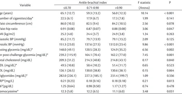
Analyzing individuals with ABI ≤0.70 in isolation, this subset was older, smoked a higher number of cigarettes/day and had higher mean CRP-us and IL-6. Furthermore, these subjects had higher mean systolic pressure, fasting glycemia, 2 hour post-challenge glycemia, triglycerides and homocysteine (Table 5). Eighty-ive percent of these individuals with ABI ≤0.70 were hypertense and 70% had diabetes and high waist-hip ratios (data not shown in tables).
Figures 1 and 2 illustrate the relationships between CRP-us and IL-6 for individuals with and without PAOD, respectively, in the form of scatter plots. Signiicant correlations between these variables were only detected for subjects with PAOD (r=0.24, p=0.010). Stratiication of CRP-us
concentrations into terciles (Figures 3 and 4) only
revealed signiicant differences between mean IL-6 concentrations for individuals with PAOD.
DISCUSSION
In this study of a population of Japanese-Brazilians, a relatively high prevalence of PAOD (21.1%) was expected, considering the unfavorable cardiometabolic profile that has been described previously.11-13,23 Although mean values and
frequencies of risk factors among men were higher, the prevalence of PAOD was similar for both sexes. Similar igures for PAOD have been observed in other populations at high cardiovascular risk. The
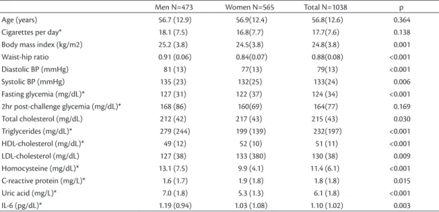
Table 1. Means (SD) of demographic, anthropometric, clinical and biochemical variables, by sex.
Men N=473 Women N=565 Total N=1038 p
Age (years) 56.7 (12.9) 56.9(12.4) 56.8(12.6) 0.364
Cigarettes per day* 18.1 (7.5) 16.8(7.7) 17.7(7.6) 0.138
Body mass index (kg/m2) 25.2 (3.8) 24.5(3.8) 24.8(3.8) 0.001
Waist-hip ratio 0.91 (0.06) 0.84(0.07) 0.88(0.08) <0.001
Diastolic BP (mmHg) 81 (13) 77(13) 79(13) <0.001
Systolic BP (mmHg) 135 (23) 132(25) 133(24) 0.006
Fasting glycemia (mg/dL)* 127 (31) 122 (37) 124 (34) <0.001
2hr post-challenge glycemia (mg/dL)* 168 (86) 160(69) 164(77) 0.169
Total cholesterol (mg/dL) 212 (42) 217 (43) 215 (43) 0.030
Triglycerides (mg/dL)* 279 (244) 199 (139) 232(197) <0.001
HDL-cholesterol (mg/dL)* 49 (12) 52 (10) 51 (11) <0.001
LDL-cholesterol (mg/dL) 127 (38) 133 (380) 130 (38) 0.009
Homocysteine (mg/dL)* 13.1 (7.5) 9.9 (4.1) 11.4 (6.1) <0.001
C-reactive protein (mg/L)* 1.6 (1.7) 1.9 (1.8) 1.8 (1.8) 0.015
Uric acid (mg/L)* 7.0 (1.8) 5.3 (1.3) 6.1 (1.8) <0.001
IL-6 (pg/dL)* 1.19 (0.94) 1.03 (1.08) 1.10 (1.02) 0.003
Table 2. Prevalence rates of cardiovascular risk factors in a Japanese-Brazilian population, by sex.
Male Female
p-value
N % N %
Age ≤60 years 284 60.0 335 59.3 0.806
<60 years 189 40.0 230 40.7
Smoking No 218 46.2 499 88.8 <0.001
Yes (in past) 91 19.3 39 6.9
Yes (current) 163 34.5 24 4.3
Central obesity No 350 74.3 153 27.1 <0.001
Yes 121 25.7 411 72.9
BMI (kg/m2) <23 131 27.8 213 37.7 0.001
23.0 to 24.9 107 22.6 130 23.0
≥25 234 49.6 222 39.3
Arterial hypertension No 250 52.8 317 56.1 0.295
Yes 223 47.2 248 43.9
Glucose tolerance Normal 16 3.4 42 7.5 0.008
AFG 170 35.9 207 36.7
IGT 104 22.0 136 24.1
DM 183 38.7 179 31.7
Hypercholesterolemia No 189 40.0 203 35.9 0.182
Yes 284 60.0 362 64.1
Low HDL No 395 83.5 514 91.0 <0.001
High LDL Yes
No
78 269
16.5 56.9
51 267
9.0 47.3
0.002
Yes 204 43.1 298 52.7
Hypertriglyceridemia No 131 27.7 236 41.8 <0.001
Yes 342 72.3 329 58.2
Homocysteine ≤15 mg/dL 286 77.3 414 92.2 <0.001
>15 mg/dL 84 22.7 35 7.8
Hyperuricemia No 216 45.7 139 24.6 <0.001
Yes 257 54.3 426 75.4
CRP-us (mg/L) 0.00-0.07 184 38.9 197 34.9 0.044
0.08-0.18 163 34.5 177 31.3
0.19-0.99 126 26.6 191 33.8
IL6 (pg/dL) 0.00-0.73 33 25.0 66 42.0 0.005
0.74-1.10 42 31.8 46 29.3
1.10-9.3 57 43.2 45 28.7
US PARTNERS program was designed to study the prevalence of PAOD, among other cardiovascular diseases, inding a 29% prevalence of PAOD among individuals aged >70 years or aged >50 years with comorbidities (diabetes and smoking).24 The
POPADAD study assessed 8,000 diabetic people aged ≥40 years and found a PAOD prevalence of 20.1%.25
In our setting, a multicenter study into the prevalence of PAOD among the general population (n=1,170) of 72 urban centers in Brazil found a prevalence of just 10.5%. It is worth noting that the age range was lower (≥18 years) and the sample was more representative of the Brazilian population because it was not comprised of genetically homogenous
subjects nor of people at high cardiovascular risk.26
Results published previously by our research team have shown that Japanese-Brazilians are a high-risk group, in terms of their igures for obesity, diabetes mellitus, arterial hypertension and dyslipidemia.11-13,23
the signiicant correlation between CRP and IL-6 values was only detected among subjects who did
have PAOD (Figure 2). The signiicant increase in
IL-6 concentrations in CRP terciles was also in line with this inding (Figure 4).
Tzoulaki et al. investigated 1592 smokers aged 55 to 74 years and found a 23% prevalence of asymptomatic PAOD, observing that IL-6 increased in proportion to reductions in ABI over follow-up periods ranging from 5 to 12 years.27
Their indings are compatible with the hypothesis that the proinlammatory state is exacerbated in
proportion to the degree to which the disease worsens. Similarly, we also found a relationship between PAOD severity and inlammatory marker levels. However, it is known that smoking affects IL-6 and CRP levels.28 Both IL-6 and CRP levels
are associated with cumulative tobacco use (years/ packs) and, among ex-smokers, with time free from smoking.29 Signorelli et al. also observed elevated
IL-6 levels in 20 non-smoking patients with a mean ABI of 0.72 (p<0.001).30
The same findings have been described by Danielsson et al. after analyzing five groups of Table 3.Number, percentage and prevalence ratios (95% conidence intervals) for categories of demographic and clinical vari-ables from Japanese-Brazilians, stratiied by presence or absence of peripheral arterial occlusive disease (PAOD).
Has PAOD n=219
Free from PAOD n=819
Total
n=1038 Chi-square PR 95%CI
N % N % N %
Sex Female 128 22.7 437 77.3 565 100 1.80 1
Male 91 19.2 382 80.8 473 100 0.85 0.67-1.08
Age ≤60 years 110 17.8 509 82.2 619 100 10.2 1
>60 years 109 26.0 310 74.0 419 100 1.46 1.16-1.85
Smoking No 150 20.9 567 79.1 717 100 0.39 1
Yes (in past) 38 20.3 149 79.7 187 100 0.97 0.71-1.33
Yes (current) 30 23.1 100 76.9 130 100 1.10 0.78-1.56
High waist-hip ratio No 101 20.1 402 79.9 503 100 0.57 1
Yes 117 22.0 415 78.0 532 100 1.10 0.86-1.39
Body mass index (kg/m2) <23 76 22.1 268 77.9 344 100 3.10 1
23.0 to 24.9 57 24.1 180 75.9 237 100 1.09 0.81-1.47
≥25 85 18.6 371 81.4 456 100 0.84 0.64-1.11
Arterial hypertension No 101 17.8 466 82.2 567 100 8.1 1
Yes 118 25.1 353 74.9 471 100 1.41 1.11-1.78
Glucose tolerance Normal 8 13.8 50 86.2 58 100 4.40 1
AFG 73 19.4 304 80.6 377 100 1.40 0.71-2.76
IGT 59 24.6 181 75.4 240 100 1.78 0.90-3.52
DM 79 21.8 283 78.1 362 100 1.58 0.81-3.10
Hypercholesterolemia No 81 20.7 311 79.3 392 100 0.07 1
Yes 138 21.4 508 78.6 646 100 1.03 0.81-1.32
Low HDL No 190 20.9 719 79.1 909 100 0.17 1
Yes 29 22.5 100 77.5 129 100 1.08 0.72-1.52
High LDL No 112 20.9 424 79.1 536 100 0.03 1
Yes 107 21.3 395 78.7 502 100 1.02 0.81-1.29
Hypertriglyceridemia No 76 20.7 291 79.3 367 100 0.05 1
Yes 143 21.3 528 78.7 671 100 1.03 0.80-1.32
CRP-us# (mg/L) 0.00-0.07 72 18.9 309 81.1 381 100 0.38 1
0.08-0.18 62 18.2 279 81.8 341 100 0.96 0.71-1.31
0.19-0.99 64 20.1 255 79.9 319 100 1.06 0.78-1.44
Homocysteine (mg/dL) ≤15 140 20.0 560 80.0 700 100 5.36 1
IL-6#(pg/dL) >15
0.00-0.73 0.74-1.10 1.10-9.3
35 35 41 44
29.4 35.4 46.6 43.1
84 64 47 58
70.6 64.6 53.4 56.9
119 99 88 102
100 100 100 100
2.59 1.47
1 1.32 1.22
1.07-2.02 0.93-1.87 0.86-1.73
people: patients with intermittent claudication and critical ischemia, both with and without diabetes, and a control group. An analysis adjusted for multiple factors only detected a relationship between inflammatory markers (IL-6 and CRP-us) and
critical ischemia. These authors concluded that the relationship is dependent on the severity of PAOD, irrespective of the presence of other risk factors.31
The relationship between inlammatory markers and PAOD severity has also been investigated in the
Table 4.Prevalence ratios (PR) for PAOD and respective 95% conidence intervals (95%CI) in a population of Japanese-Brazilians, by variables selected for models (initial and inal models).
Variable Initial model Adjusted model
PR 95%CI PR 95%CI
Sex Female 1 1
Male 0.66 0.42-1.02 0.66 0.44-1.01
Age ≤60 years 1 1
>60 years 0.98 0.71-1.36 0.94 0.69-1.23
Smoking No 1 1
Yes (past) 1.45 0.91-2.31 1.44 0.92-2.33
Yes (current) 2.14 1.32-3.50 2.16 1.34-3.48
Arterial hypertension No 1 1
Yes 1.61 1.12-2.31 1.56 1.12-2.22
Glucose tolerance Normal 1
AFG 0.67 0.38-1.21 0.71 0.40-1.32
IGT 0.76 0.42-1.38 0.65 0.43-1.44
DM 0.66 0.37-1.17 0.65 0.39-1.23
Homocysteine (mg/dL) ≤15 1 1
>15 1.25 0.88-1.77 1.26 0.89-1.78
CRP-us# (mg/L) 0.00-0.07 1 1
0.08-0.18 0.62 0.35-1.10 0.77 0.52-1.14
IL-6# (pg/dL) 0.19-0.99
0.00-0.73 0.74-1.10
0.72 1 1.26
0.40-1.30 0.70-2.26
0.97 1 0.88
0.66-1.41 0.76-1.05
1.10-9.3 1.09 0.60-1.98 0.91 0.88-1.18
#Figures log-transformed for analysis.
Table 5.Means and standard deviations (SD) for demographic, anthropometric and clinical variables from a Japanese-Brazilian population, by ankle-brachial index categories.
Variable Ankle-brachial index F statistic
(Anova)
P
≤0.70 0.71-0.90 >0.90
Age (years) 65.1 (12.7) 59.3 (13.2) 56.0 (12.3) 10.14 < 0.001
Number of cigarettes/day* 22.5 (6.1) 17.9 (6.7) 17.3 (7.8) 1.99 0.141
Waist circumference (cm) 86.0 (10.3) 82.5 (9.4) 84.2 (10.5) 2.56 0.078
Waist-hip ratio 0.91 (0.08) 0.87 (0.07) 0.88 (0.08) 3.06 0.047
BMI (kg/m2) 25.2 (4.8) 24.4 (3.7) 24.9 (3.8) 1.34 0.263
Diastolic BP (mmHg) 85.2 (11.7) 79.7 (13.9) 79.1 (13.2) 2.09 0.125
Systolic BP (mmHg) 151.3 (23.0) 137.6 (27.5) 1312.0 (23.4) 9.86 < 0.001
Fasting glycemia (mg/dL)* 148.0 (49.1) 120.5 (28.3) 124.9 (35.2) 6.56 0.002
2hr post-challenge glycemia (mg/dL)* 239.2 (115.9) 164.1 (70.4) 162.2 (77.0) 7.45 0.001
Total cholesterol (mg/dL) 209.3 (31.2) 214.3 (40.8) 214.8 (43.1) 0.17 0.840
HDL (mg/dL)* 49.5 (10.8) 50.4 (10.2) 51.4 (11.7) 0.65 0.523
LDL (mg/dL) 126.1 (26.5) 130.0 (38.8) 130.6 (38.1) 0.15 0.864
Triglycerides (mg/dL)* 282.0 (226.1) 227.3 (185.1) 231.4 (199.7) 1.09 0.336
CRP*(mg/L) 0.21 (0.23) 0.18 (0.16) 0.18 (0.18) 0.21 0.813
IL6*(pg/dL) 1.25 (0.64) 0.98 (0.50) 1.17 (1.27) 0.74 0.478
Homocysteine* 12.3 (5.8) 12.2 (6.5) 11.1 (6.0) 3.48 0.031
carotid territory. No association was detected between IL-6 or CRP-us and subclinical atherosclerosis assessed in 1,111 individuals with carotid myointimal thickening,32 which is similar to our indings since we
also failed to detect a relationship with inlammatory markers in patients with PAOD in the initial phases. However, the study did detect a positive relationship between carotid stenosis (≥25%) and CRP-us. These authors also failed to detect any relationship between the marker and myointimal hyperplasia of the carotid, suggesting that CRP-us is a marker of more advanced disease.33
The same indings have been observed in the coronary territory. A case-control study of 926 men aged 50 to 59 found that among subjects who suffered infarction or sudden death over a 5-year follow-up period had had elevated IL6 and CRP levels at the start of the study. Patients who only exhibited angina during this period had had normal levels of these markers at the study outset. Additionally, the relative
risk of infarction and death increased in line with increases in IL6 levels.34
Another prospective study, analyzing the association between IL-6 and mortality in 718 patients with coronary disease, found that serum IL-6 concentrations of patients who died from coronary disease during a 2.3-year follow-up period were signiicantly higher at the study outset. Interleukin 6 levels were associated with cardiovascular mortality, conferring a relative risk of 2.04 (95%CI 1.34-3.68). These indings support the hypothesis that a subclinical chronic inlammatory process plays a role in determining the outcome of atherosclerotic disease and is a sign of patients with poor prognosis.35
Biasucci et al. have published further evidence in support of this theory, showing that IL-6 levels increased after admission in patients with unstable angina who went on to develop complications.36
Our indings are in agreement with the literature reviewed. Both IL-6 and CRP-us concentrations Figure 1. Correlation between CRP and IL-6 levels in people
without PAOD. r=0.04 (p=0.624).
Figure 2. Correlation between CRP and IL-6 levels in people with PAOD. r=0.24 (p=0.010).
Figure 3. IL-6 levels by CRP terciles for people without PAOD. (p=0.658).
appear to relect the intensity of occult inlammation in the atherosclerotic plaque and may be capable of determining its vulnerability to rupture and, consequently, of indicating disease severity.37
Although the cross-sectional design has inherent limitations that prevent conclusions on cause-effect relationships, the strengths of our study lie in its populational characteristic and in the uniformity of the population investigated.
In summary, our data suggest the existence of a relationship between atherosclerosis of peripheral arteries and inlammatory markers and show that these markers can indicate disease severity. Patients with advanced PAOD may have few or even no symptoms if they engage in little or no activity, as is the case with bedridden patients. The same may be true of atherosclerotic disease in the cerebral and coronary territories. The ability to detect these patients with more advanced forms of the disease would be of great value and the hypothesis merits testing in prospective studies, since this information would facilitate the tasks of mapping those patients with least favorable atherosclerotic disease prognoses and of planning more aggressive treatments.
REFERENCES
1. Ablij H, Meinders A. C-reactive protein: history and revival. Eur J Intern Med. 2002;13(7):412-22. http://dx.doi.org/10.1016/S0953-6205(02)00132-2. PMid:12384129
2. Ridker PM, Rifai N, Stampfer MJ, Hennekens CH. Plasma concentration of interleukin-6 and the risk of future myocardial infarction among apparently healthy men. Circulation. 2000;101(15):1767-72. http://dx .doi.org/10.1161/01. CIR.101.15.1767. PMid:10769275
3. Pearson TA, Mensah GA, Alexander RW, et al, and the Centers for Disease Control and Prevention, and the American Heart Association. Markers of inflammation and cardiovascular disease: application to clinical and public health practice: A statement for healthcare professionals from the Centers for Disease Control and Prevention and the American Heart Association. Circulation. 2003;107(3):499-511. http://dx.doi.org/10.1161/01. CIR.0000052939.59093.45. PMid:12551878
4. Calabró P, Willerson JT, Yeh ET. Inflammatory cytokines stimulated C-reactive protein production by human coronary artery smooth muscle cells. Circulation. 2003;108(16):1930-2. http://dx.doi. org/10.1161/01.CIR.0000096055.62724.C5. PMid:14530191
5. Sun H, Koike T, Ichikawa T, et al. C-reactive protein in atherosclerotic lesions: its origin and pathophysiological significance. Am J Pathol. 2005;167(4):1139-48. http://dx.doi. org/10.1016/S0002-9440(10)61202-3. PMid:16192648
6. Yudkin JS, Kumari M, Humphries SE, Mohamed-Ali V. Inflammation, obesity, stress and coronary heart disease: is interleukin-6 the link? Atherosclerosis. 2000;148(2):209-14. http:// dx.doi.org/10.1016/S0021-9150(99)00463-3. PMid:10657556
7. Goff DC Jr, Lloyd-Jones DM, Bennett G, et al. 2013 ACC/AHA guideline on the assessment of cardiovascular risk: a report of the American College of Cardiology/American Heart Association
Task Force on Practice Guidelines. Circulation. 2014;129(25, Suppl 2):S49-73. http://dx.doi.org/10.1161/01.cir.0000437741.48606.98. PMid:24222018.
8. Pearson TA, Mensah GA, Alexander RW, et al, and the Centers for Disease Control and Prevention, and the American Heart Association. Markers of inflammation and cardiovascular disease: application to clinical and public health practice: A statement for healthcare professionals from the Centers for Disease Control and Prevention and the American Heart Association. Circulation. 2003;107(3):499-511. http://dx.doi.org/10.1161/01. CIR.0000052939.59093.45. PMid:12551878
9. Fowers FGR, Rudan D, Rudan I, et al. Comparison of global estimates of prevalence and risk factors for peripheral artery disease in 2000 and 2010: a systematic review and analysis. Lancet. 2013;38(9901):1329-40. http://dx.doi.org/10.1016/ S0140-6736(13)61249-0.
10. Criqui MH, Langer RD, Fronek A, et al. Mortality over a period of 10 years in patients with peripheral arterial disease. N Engl J Med. 1992;326(6):381-6. http://dx.doi.org/10.1056/ NEJM199202063260605. PMid:1729621
11. Lerario DD, Gimeno SG, Franco LJ, Iunes M, Ferreira SRG. [Weight excess and abdominal fat in the metabolic syndrome among Japanese-Brazilians]. Rev Saude Publica. 2002;36(1):4-11. http:// dx.doi.org/10.1590/S0034-89102002000100002. PMid:11887223
12. Gimeno SG, Ferreira SR, Franco LJ, Hirai AT, Matsumura L, Moisés RS. Prevalence and 7-year incidence of Type II diabetes mellitus in a Japanese-Brazilian population: an alarming public health problem. Diabetologia. 2002;45(12):1635-8. http://dx.doi. org/10.1007/s00125-002-0963-x. PMid:12488952
13. Siqueira AFA, Franco LJ, Gimeno SGA, et al, and the JBDSG. Macrovascular disease in a Japanese-Brazilian population of high prevalence of metabolic syndrome: associations with classical and non-classical risk factors. Atherosclerosis. 2007;195(1):160-6. http://dx.doi.org/10.1016/j.atherosclerosis.2002007;195(1):160-6.09.012. PMid:17064712
14. Examination Committee of Criteria for ‘Obesity Disease’ in Japan, Japan Society for the Study of Obesity (JASO). New criteria for ‘obesity disease’ in Japan. Circ J. 2002;66(11):987-92.
15. International Diabetes Institute, Regional Office of the Western Pacific Region – WPRO, World Health Organization – WHO, International Association for the Study of Obesity – IASO, International Obesity Task Force. The Asia-Pacific perspective:
redefining obesity and its treatment. Melbourne: Health
Communications Australia Pty; 2000. www.wpro.who.int/ nutrition. Acessado: 10/01/2014.
16. U.S. Department of Health and Human Services. The Seventh
Report of the Joint National Committee on Prevention Detection, Evaluation, and Treatment of High Blood Pressure. Bethesda: NIH Publications; 2004. (NIH Publication, no. 04-5230). www.nhlbi.nih. gov/guidelines/hypertension/jnc7full.pdf. Acessado: 10/01/2014.
17. American Diabetes Association. Standards of medical care in diabetes—2011. Diabetes Care. 2011;34(Suppl. 1):S11-S61.
18. Expert Panel on Detection, Evaluation, and Treatment of High Blood Cholesterol in Adults. Executive Summary of The Third Report of The National Cholesterol Education Program (NCEP) Expert Panel on Detection, Evaluation, and Treatment of High Blood Cholesterol In Adults (Adult Treatment Panel III). JAMA. 2001;285(19):2486-97. http://dx.doi.org/10.1001/ jama.285.19.2486. PMid:11368702
and gout. N Engl J Med. 2005;353(23):2450-61. http://dx.doi. org/10.1056/NEJMoa050373. PMid:16339094
20. Garofolo L, Barros N Jr, Miranda F Jr, D’Almeida V, Cardien LC, Ferreira SR. Association of increased levels of homocysteine and peripheral arterial disease in a Japanese-Brazilian population. Eur J Vasc Endovasc Surg. 2007;34(1):23-8. http://dx.doi.org/10.1016/j. ejvs.2007.02.008. PMid:17482486
21. Norgren L, Hiatt WR, Dormandy JA, Nehler MR, Harris KA, Fowkes FG, and the TASC II Working Group. Inter-Society Consensus for the Management of Peripheral Arterial Disease (TASC II). J Vasc Surg. 2007;45(1, Suppl S):S5-67. http://dx.doi.org/10.1016/j. jvs.2006.12.037. PMid:17223489
22. McDermott MM, Green D, Greenland P, et al. Relation of levels of hemostatic factors and inflammatory markers to the ankle brachial index. Am J Cardiol. 2003;92(2):194-9. http://dx.doi. org/10.1016/S0002-9149(03)00537-X. PMid:12860223
23. Ferreira SRG, Almeida-Pittito B, and the Japanese-Brazilian Diabetes Study Group. Reflexão sobre a imigração japonesa no Brasil sob o ângulo da adiposidade corporal. Arq Bras Endocrinol Metabol. 2009;53(2):175-82. http://dx.doi.org/10.1590/S0004-27302009000200009. PMid:19466210
24. Hirsch AT, Criqui MH, Treat-Jacobson D. Peripheral arterial disease detection, awareness, and treatment in primary care. JAMA. 2001;286(11):1317-24. PMid:11560536
25. Elhadd T, Robb R, Jung R, Stonebrigde P, Belch J. Pilot study of prevalence of asymptomatic peripheral arterial occlusive disease in patients with diabetes attending a hospital clinic. Pract Diab Int. 1999;16(6):163-6. http://dx.doi.org/10.1002/pdi.1960160605.
26. Makdisse M, Pereira AC, Brasil DP, et al, and the Hearts of Brazil Study and Peripheral Arterial Disease Committee of the Brazilian Society of Cardiology/Funcor. Prevalence and risk factors associated with peripheral arterial disease in the Hearts of Brazil Project. Arq Bras Cardiol. 2008;91(6):370-82. http://dx.doi. org/10.1590/S0066-782X2008001800008. PMid:19142364
27. Tzoulaki I, Murray GD, Lee AJ, Rumley A, Lowe GD, Fowkes FG. C-reactive protein, interleukin-6, and soluble adhesion molecules as predictors of progressive peripheral atherosclerosis in the general population: Edinburgh Artery Study. Circulation. 2005;112(7):976-83. http://dx.doi.org/10.1161/CIRCULATIONAHA.104.513085. PMid:16087797
28. Deswal A, Petersen NJ, Feldman AM, Young JB, White BG, Mann DL. Cytokines and cytokine receptors in advanced heart failure: an analysis of the cytokine database from the Vesnarinone trial (VEST). Circulation. 2001;103(16):2055-9. http://dx.doi. org/10.1161/01.CIR.103.16.2055. PMid:11319194
29. Sunyer J, Forastiere F, Pekkanen J, et al, and the AIRGENE Study Group. Interaction between smoking and the interleukin-6 gene affects systemic levels of inflammatory biomarkers. Nicotine Tob Res. 2009;11(11):1347-53. http://dx.doi.org/10.1093/ntr/ntp144. PMid:19828434
30. Signorelli SS, Mazzarino MC, Di Pino L, et al. High circulating levels of cytokines (IL-6 and TNFalpha), adhesion molecules (VCAM-1 and ICAM-1) and selectins in patients with peripheral arterial disease at rest and after a treadmill test. Vasc Med. 2003;8(1):15-9. http://dx.doi.org/10.1191/1358863x03vm466oa. PMid:12866607
31. Danielsson P, Truedsson L, Eriksson K-F, Norgren L. Inflammatory markers and IL-6 polymorphism in peripheral arterial disease with
and without diabetes mellitus. Vasc Med. 2005;10(3):191-8. http:// dx.doi.org/10.1191/1358863x05vm617oa. PMid:16235772
32. Chapman CM, Beilby JP, McQuillan BM, Thompson PL, Hung J. Monocyte count, but not C-reactive protein or interleukin-6, is an independent risk marker for subclinical carotid atherosclerosis. Stroke. 2004;35(7):1619-24. http://dx.doi.org/10.1161/01. STR.0000130857.19423.ad. PMid:15155967
33. Wang TJ, Nam BH, Wilson PW, et al. Association of C-reactive protein with carotid atherosclerosis in men and women: the Framingham Heart Study. Arterioscler Thromb Vasc Biol. 2002;22(10):1662-7. http://dx.doi.org/10.1161/01. ATV.0000034543.78801.69. PMid:12377746
34. Luc G, Bard JM, Juhan-Vague I, et al, and the PRIME Study Group. C-reactive protein, interleukin-6, and fibrinogen as predictors of coronary heart disease: the PRIME Study. Arterioscler Thromb Vasc Biol. 2003;23(7):1255-61. http://dx.doi.org/10.1161/01. ATV.0000079512.66448.1D. PMid:12775578
35. Su D, Li Z, Li X, et al. Association between serum interleukin-6 concentration and mortality in patients with coronary artery disease. Mediators Inflamm. 2013;2013:726178. http://dx.doi. org/10.1155/2013/726178. PMid:23818744
36. Biasucci LM, Liuzzo G, Fantuzzi G, et al. Increasing levels of interleukin (IL)-1Ra and IL-6 during the first 2 days of hospitalization in unstable angina are associated with increased risk of in-hospital coronary events. Circulation. 1999;99(16):2079-84. http://dx.doi.org/10.1161/01.CIR.99.16.2079. PMid:10217645
37. Ikeda U. Inf lammation and coronar y arter y disease. Curr Vasc Pharmacol. 2003;1(1):65-70. http://dx .doi. org/10.2174/1570161033386727. PMid:15320854
Correspondence
Luciana Garofolo Rua Dr. Rafael de Barros, 497 CEP 04003-040 – São Paulo (SP), Brazil Tel.: +55 (11) 999039505 / +55 (11) 38873271 Fax: +55 (11) 38870868 E-mail: [email protected]
Author information
LG is a PhD in Interdisciplinary Surgical Science from Universidade Federal de São Paulo (UNIFESP), São Paulo, Brazil. SRGF is a professor at the School of Public Sciences, Universidade de São Paulo (USP), São Paulo, Brazil. FMJ is a full professor of Vascular Surgery, Department of Surgery, Universidade Federal de São Paulo (UNIFESP), São Paulo, Brazil.
Author contributions
Conception and design: LG, SRGF Analysis and interpretation: LG, SRGF Data collection: LG, SRGF Writing the article: LG, SRGF Critical revision of the article: SRFG, FMJ Final approval of the article*: LG, SRGF, FMJ Statistical analysis: LG, SRGF Overall responsibility: LG Obtained funding: FAPESP.