*Correspondence: Alceu Afonso Jordão Júnior. Curso de Nutrição e Metabo-lismo, Faculdade de Medicina de Ribeirão Preto – USP, Av. Bandeirantes 3900, 14049-900 - Ribeirão Preto – SP, Brasil. E-mail: [email protected]
A
rti
Pharmaceutical Sciences vol. 45, n. 4, oct./dec., 2009
Effect of methionine load on homocysteine levels,
lipid peroxidation and DNA damage in rats receiving ethanol
Alceu Afonso Jordao Júnior*, Fernanda Aparecida Domenici, Renata Cristina Lataro,
Guilherme Vannucchi Portari, Helio Vannucchi
Laboratory of Nutrition and Metabolism,
Faculty of Medicine of Ribeirão Preto, University of São Paulo
Changes in the metabolism of methionine can cause hyperhomocysteinemia, inducing a triad of atherosclerosis, hypertension, and increased oxidative stress. The generation of free radicals and oxidative damage to DNA is important in the liver damage caused by ethanol. In this study, the effect of methionine overload associated or otherwise with acute administration of ethanol on homocysteine values, damage to DNA, lipoperoxidation and vitamin E was evaluated. Thirty rats were divided into 3 groups: Group Ethanol 24 hours (EG24), Group Methionine 24 hours (MG24), and Group Methionine and Ethanol 24 hours (MEG24). TBARS, vitamin E, GS and, homocysteine values were determined and the Comet assay was carried out. Increased GSH, vitamin E and homocysteine levels were observed for MEG24, and increased TBARS were observed in EG24. The Comet assay showed an increase in DNA damage in EG24 and DNA protection in MEG24. The administration of ethanol decreased antioxidant levels and increased TBARS, indicating the occurrence of oxidative stress with possible DNA damage. The combination of methionine and ethanol had a protective effect against the ethanol-induced damage, but increased the levels of homocysteine.
Uniterms: Methionine. Homocysteine. Free Radicals. Antioxidants. Ethanol/induced damage.
Alterações no metabolismo da metionina podem ocasionar hiper-homocisteinemia, quadro indutivo de aterosclerose, hipertensão e aumento do estresse oxidativo. A geração de radicais livres e dano oxidativo ao DNA são importantes na injúria hepática provocada pelo etanol. Neste estudo avaliaram-se os efeitos da sobrecarga de metionina associada ou não à administração aguda de etanol sobre valores de homocisteína, dano ao DNA, lipoperoxidação e vitamina E. Foram utilizados 30 ratos Wistar distribuídos em 3 Grupos: Grupo Etanol 24 horas (GE24), Grupo Metionina 24 horas (GM24) e Grupo Metionina e Etanol 24 horas (GME24). Realizaram-se determinações hepáticas de SRATB, vitamina E, GSH, homocisteína e Teste do Cometa e determinações plasmáticas de GSH e homocisteína. Valores aumentados de GSH, vitamina E e homocisteína foram observados para o GME24, e de SRATB no GE24. O Teste do Cometa mostrou aumento do dano ao DNA no GE24 e proteção ao DNA no GME24. A administração de etanol diminuiu os níveis de antioxidantes e aumentou o de SRATB, indicando ocorrência de estresse oxidativo, podendo ocasionar dano ao DNA. A presença da metionina associada com o etanol agiu como protetora contra os danos do etanol, mas aumentou os níveis de homocisteína.
Unitermos: Metionina. Homocisteína. Radicais Livres. Antioxidantes. Etanol/danos.
INTRODUCTION
Methionine-rich diets can lead to hyperhomocys-teinemia, a condition that induces the development of atherosclerosis and hypertension in rats and humans and
glutathio-ne (GSH), with a strong involvement in the process of free radical formation and lipid peroxidation (Vannucchi et al., 1998; Jordão Jr et al., 1998).
Changes in methionine metabolism may result in high HCY levels (hyperhomocysteinemia), relecting the deiciency of one or all of the three vitamins (folic acid, vitamin B6 or B12) involved in the process (El-Saleh et al., 2004). The association of hyperhomocysteinemia in patients on hemodialysis may also lead to increased lipid peroxidation and changes in antioxidant levels, a condi-tion that can be partially corrected by the administracondi-tion of folate (Chiarello et al., 2003).
Experimental studies have suggested that HCY may have a pro-oxidant effect on the presence of me-tals such as iron and copper (Ventura et al., 2000). One of the classical ways to study the effects of hyperho-mocysteinemia is to induce this condition by means of an oral methionine load test (100 mg/kg body weight), which can be applied to both animals and humans. This hyperhomocysteinemia mode due to oral methionine load leads to increased plasma oxidation and also to a reduction of antioxidants, whereas normal HCY levels have not demonstrated signiicant changes in antioxidant metabolism (Ventura et al., 2000).
Abnormal methionine metabolism occurs in ani-mals subjected to ethanol intake and also in cirrhotic patients. The consequences of this metabolism may in-clude a reduction of S-adenosylmethionine and of hepatic GSH, changes in methylation processes and a reduction of homocysteine catabolism resulting in hyperhomocys-teinemia (Lee et al., 2004). An acute dose of ethanol (3 g/kg body weight) induces signiicant changes in the metabolism of sulfurated amino acids in the liver, redu-cing the availability of cysteine for the synthesis of glu-tathione and also increasing the irreversible catabolism to taurine via hypotaurine (Jung et al., 2003).
The generation of free radicals is an important step in the liver damage provoked by ethanol. Acute or chronic ethanol administration can lead to increased lipid peroxidation via the exacerbated production of reactive oxygen species and/or a reduction of antioxidants such as vitamin E (Jordão Jr et al., 2004).
A study on lipid peroxidation caused by ethanol clearly showed that the maintenance of normal vitamin E levels is essential for antioxidant production. Vitamin E-deicient animals showed increased malondialdehyde (MDA) levels even without receiving ethanol. The same study demonstrated that the effects of ethanol are obser-ved in a time-dependent manner, i.e., the results crucially depend on the time when the sample is collected (Jordão Jr et al., 2004).
After a single dose of ethanol (5 g/kg body weight), urinary acetaldehyde excretion increases by about 5.8 times during a period of 6-12 hours, while formaldehyde increases only after 24 hours and lipid peroxidation me-asured by MDA concentration increases after 18 hours, thus representing an event secondary to the increase in acetaldehyde levels (Moser et al., 1993).
The above data show that there is a relationship between methionine overload and HCY levels, lipid peroxidation and oxidative DNA damage in rats and that ethanol intake can also inluence the metabolism of sulfu-rated amino acids, correlations that would be interesting to study. Thus, the objective of the present study was to assess the effect of a single methionine overload on HCY levels, on the possible oxidative damage to DNA, on the rate of lipoperoxidation and on vitamin E levels after acute ethanol administration in rats.
MATERIAL AND METHODS
The experiment was conducted in the Animal Fa-cilities of the Department of Internal Medicine, Faculty of Medicine of Ribeirão Preto, University of São Paulo (USP). Thirty newly weaned male Wistar rats weighing approximately 60 g provided by the Central Animal House of FMRP-USP were used. The animals were di-vided into 3 groups:
Twenty-four hour Ethanol Group (n = 10): rats re-ceiving ethanol and a normal diet and sacriiced 24 hours after the period of acute ethanol intake.
Twenty-four hour Methionine Group (n = 10): rats receiving ethanol and a normal diet and sacriiced 24 hours after methionine overload.
Twenty-four hour Methionine plus Ethanol Group (n = 10): rats receiving ethanol and a normal diet and sacriiced 24 hours after the period of acute ethanol intake and methionine overload.
Diets were prepared according to AIN-93 recom-mendations and offered to the rats in the animal house for one week for adaptation. Both the diets and water were available ad libitum.
The animals were weighed at the time of sacriice. The animals of the Ethanol and Methionine plus Ethanol groups received by gavage an ethanol solution at the concentration of 5 g/kg body weight.
plasma determination of HCY and GSH. The liver was then removed and weighed and immediately placed in liquid nitrogen (–196 °C) for later determination of thiobarbituric acid reactive substances (TBARS), GSH and α-tocopherol.
Lipid peroxidation in the liver was measured by TBARS determination (Buege and Austi, 1978), GSH was determined by the method of Sedlack and Lindsay (1968), and hepatic vitamin E (α-tocopherol) was
de-termined by HPLC (Arnaud et al., 1991). Protein deter-mination, used to express the hepatic values of TBARS and GSH, was carried out by the method of Lowry et al. (1951).
Plasma and liver HCY was determined by HPLC with fluorimetric detection. This method is based on compound derivatization with o-ophthaldehyde in the presence of 2-mercaptoethanol, and separation through a C18 column with isocratic elution using 17% metha-nol and 0.04 M sodium phosphate buffer, pH 7.0, 0.002 Na2EDTA and luorimetric detection at 340 nm excitation and 450 nm emission (Tcherkas, Denisenko, 2001).
Oxidative DNA damage to the hepatocytes was determined by the Comet test (single cell gel assay) ac-cording to Rojas and coworkers (1999). After staining with ethidium bromide, the slides were examined at 250 to 400x magniication under a luorescent micros-cope equipped with excitation filters and a barrier of 515-560 nm and 590 nm, respectively. Approximately 50 cells selected at random were analyzed according to comet size and classiied into ive distinct classes, with the absence of damage being scored as 0 and maximum dama-ge as 4. Thus, the total score for 50 cells could randama-ge from 0 (no damage) to 200 (maximum damage for all cells).
Data are reported as means ± SD. Groups were compared by analysis of variance (ANOVA) and by the Tukey test, with the level of signiicance set at p<0.05.
RESULTS AND DISCUSSION
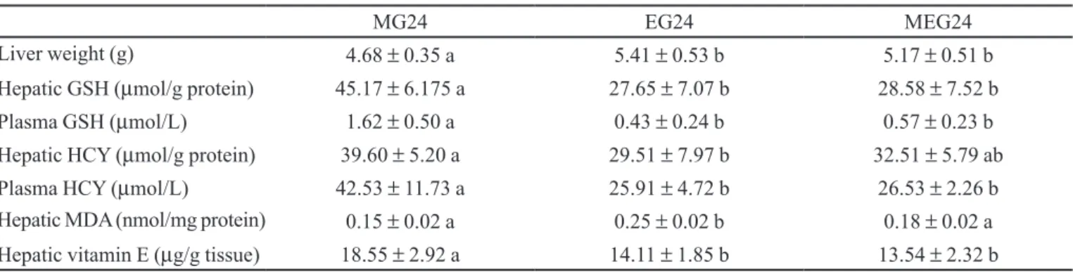
The data presented in Table I initially indicate that ethanol administration increases liver weight over a 24-hour period, probably due to a greater deposition of hepatic fat, leading the animal to a state of alcoholic steatosis.
Ethanol causes steatosis by altering several steps in the hepatic processing of fatty acids, including their plasma uptake, their use as energy substrates, and their release as triglycerides (Maher, 2002).
Currently, it is of vital importance to study the role not only of ethanol but also of other nutritional compo-nents such as methionine and HCY in hepatic steatosis, in view of the great interest in the study of steatosis of non-alcoholic origin (Cortez-Pinto et al., 2006).
It should be pointed out that one of the most fre-quently used models of induction of steatosis and of non-alcoholic steatohepatitis is the offer of a choline- and methionine-free diet to the animals (Kirsch et al., 2003).
Table I also lists the values of antioxidant metabo-lism, with data on the hepatic and plasma concentration of reduced GSH, HCY and the hepatic concentrations of MDA and vitamin E.
The present results principally indicate a reduction of plasma and hepatic reduced GSH levels in animals receiving ethanol, but a preservation of these values in animals receiving methionine in combination with ethanol. The group receiving methionine alone showed increased GSH values, indicating that methionine administration can spare GSH in situations of oxidative stress or can even increase GSH in a natural manner, in the absence of acute stress such as that induced by ethanol administration.
These GSH data initially reflect the depletion of this antioxidant which is considered to play a key role in drug detoxiication, being affected by ethanol. GSH levels decreased over a 24 hour period (Table I), showing that
TABLE I - Biochemical data for the animals that received methionine and/or ethanol and were sacriiced 24 hours later
MG24 EG24 MEG24
Liver weight (g) 4.68 ± 0.35 a 5.41 ± 0.53 b 5.17 ± 0.51 b
Hepatic GSH (µmol/g protein) 45.17 ± 6.175 a 27.65 ± 7.07 b 28.58 ± 7.52 b
Plasma GSH (µmol/L) 1.62 ± 0.50 a 0.43 ± 0.24 b 0.57 ± 0.23 b
Hepatic HCY (µmol/g protein) 39.60 ± 5.20 a 29.51 ± 7.97 b 32.51 ± 5.79 ab
Plasma HCY (µmol/L) 42.53 ± 11.73 a 25.91 ± 4.72 b 26.53 ± 2.26 b
Hepatic MDA (nmol/mg protein) 0.15 ± 0.02 a 0.25 ± 0.02 b 0.18 ± 0.02 a
Hepatic vitamin E (µg/g tissue) 18.55 ± 2.92 a 14.11 ± 1.85 b 13.54 ± 2.32 b
during this period there is not yet complete GSH recovery in the presence of acutely administered ethanol.
GSH plays an important role in the antioxidant de-fense system, in the metabolism of various nutrients and in the regulation of cellular events including DNA damage, gene expression and apoptosis where its deiciency contri-butes to oxidative stress (Wu et al., 2004). However, these effects of GSH on the mechanisms of lipid and protein oxi-dation need to be better elucidated (Biswas et al., 2006).
As expected, a signiicant increase in HCY levels was demonstrated only in animals receiving methionine, while an intermediate value was observed in the group re-ceiving methionine plus ethanol, and ethanol administered alone had no effect of HCY levels.
A recently published study described the inluence of various factors such as ethanol intake and polymor-phisms of the enzyme methylene tetrahydrofolate re-ductase (MTHFR) on total HCY values, indicating that many of these effects can be modulated by nutritional status in terms of folic acid (Chiuve et al., 2005). Folate deiciency may also be directly responsible for the incre-ased activity of the enzyme glycine n-methyltransferase, which regulates the levels of S-adenosylmethionine and S-adenosylhomocysteine (Uthus et al., 2006).
Ethanol administration can result in DNA hypome-thylation and in greater lipid deposition in the liver and also alters the hepatic metabolism of methionine, leading to changes in the S-adenosylmethionine-dependent trans-methylation process (Schalinske et al., 2005).
The present results show that the administration of methionine in combination with ethanol was able to pre-serve several important parameters such as GSH levels.
On the other hand, lipoperoxidation indirectly me-asured by the TBARS test indicated that hepatic TBARS levels were higher only in the group receiving ethanol, with the addition of methionine to the acute ethanol dose preventing the elevation of this biochemical parameter of lipoperoxidation.
The levels of hepatic vitamin E, the main liposoluble antioxidant, were higher in the group receiving methionine only (Table I), being reduced in the groups that received ethanol or ethanol plus methionine.
The reduction of vitamin E levels probably de-monstrates the utilization of this vitamin in the process of detoxiication and recovery of the animal after ethanol consumption. We suggest that further studies prolonging the time in a similar experiment may determine whether vitamin E is recycled and recovered in the subsequent period. An experiment using different vitamin E levels (normal, deicient and supplemented) in the presence of acute ethanol intake, with the animals being sacriiced
at different time points, clearly showed that vitamin E is reduced up to 8 hours after ethanol intake but that these hepatic levels are restored within a period of 24 hours, except in animals who already have vitamin E deiciency (Jordão Jr et al., 2004).
Table II presents the data regarding oxidative DNA damage determined by the Comet test. The data clearly demonstrate the increased DNA damage induced by acute ethanol administration and DNA protection when ethanol is administered together with methionine, a fact that may be explained by protection against the increase in lipid peroxidation in the ethanol plus methionine group. This shows the existence of a relation between TBARS levels and DNA damage, with consumption of hepatic antioxi-dants occurring in both groups receiving ethanol.
The DNA changes observed by the comet test are known to depend on the ethanol amount administered and also on the form of administration (chronic or acute), with higher ethanol doses leading to greater oxidative DNA damage (Lamarche et al., 2003).
Pretreatment with vitamin C (400 mg/kg/day) or vitamin E (100 mg/kg/day) for 5 days before acute etha-nol administration (5 g/kg) inhibits the generation of the hydroxyethyl radical by 30 and 50%, respectively, and prevents oxidative DNA damage, which is high in groups receiving no antioxidants (Navasumrit et al., 2000).
The results demonstrate that acute ethanol adminis-tration reduces antioxidant levels (vitamin E and GSH) and increases TBARS levels, events that indicate the occurrence of oxidative stress in the liver of these animals possibly leading to oxidative DNA damage. Methionine administered in combination with ethanol prevented the increase in TBARS, probably with antioxidant (vitamin E and GSH) consumption and this lack of TBARS increase was relected in the preservation of DNA from oxidative damage.
TABLE II - DNA damage (Comet test) in the animals that
received methionine and/or ethanol and were sacriiced 24 hours later. Data are reported as mean + SD DNA damage (Comet test) for animals that received Methionine and/or Ethanol and were sacriiced 24 hours later
Groups DNA Damage
MG24 141.38 ± 11.98a
EG24 172.63 ± 18.94b
MEG24 144.75 ± 8.60a
Two distinct, although interlinked processes may be present, i.e., an unquestionable increase of oxidative damage in the presence of acute ethanol intake and the preservation of some of these parameters in the presence of also acute administration of methionine, with the con-sequent expected harmful occurrence of increased HCY levels.
REFERENCES
ARNAUD, J.; FORTIS, I.; BLACHIER, S.; KIA, D.; FAVIER, A. Simultaneous determination of retinol, alpha-tocopherol and beta-carotene in serum by isocratic high-performance liquid chromatography. J. Chromatogr., v.572, n.1/2, p.103-116, 1991.
BISWAS, S.; CHIDA, A. S.; RAHMAN, I. Redox modiications of protein-thiols: emerging roles in cell signaling. Biochem. Pharmacol., v.71, n.5, p.551-564, 2006.
BUEEG, J. A.; AUST, S. D. Microsomal lipid peroxidation.
Methods Enzymol., v.52, p.302-310, 1978.
CHIARELLO, P. G.; VANNUCCHI, M. T.; MOYSES NETO, M.; VANNUCCHI, H. Hyperhomocysteinemia and oxidative stress in hemodialysis: effects of supplementation with folic acid. Int. J. Vitam. Nutr. Res., v.73, n.6, p. 431-438, 2003.
CHIUVE, S. E.; GIOVANNUCCI, E. L.; HANKINSON, S. E.; HUNTER, D. J.; STAMPFER, M. J.; WILLETT, W. C.; RIMM, E. B. Alcohol intake and methylenetetrahydrofolate reductase polymorphism modify the relation of folate intake to plasma homocysteine. Am. J. Clin. Nutr., v.82, n.1, p.155-162, 2005.
CORTEZ-PINTO, H.; DE MOURA, M. C.; DAY, C. P. Non-alcoholic steatohepatitis: from cell biology to clinical practice. J. Hepatol., v.44, n.1, p.197-208, 2006.
EL-SALEH, S. C.; AL-SAGAIR, O. A.; AL-KHALAF, M. I. Thymoquinone and Nigella sativa oil protection against
methionine-induced hyperhomocysteinemia in rats. Int. J.
Cardiol., v.93, n.1, p.19-23, 2004.
JORDAO JR, A. A.; CHIARELLO, P. G.; ARANTES, M. R.; MEIRELLES, M. S.; VANNUCCHI, H. Effect of an acute dose of ethanol on lipid peroxidation in rats: action of vitamin E. Food Chem. Toxicol., v.42, n.3, p.459-464, 2004.
JORDÃO JR., A. A; CHIARELLO, P. G.; BERNARDES, M. M.; VANNUCCHI, H. Peroxidação lipídica e etanol: papel
da glutationa reduzida e da vitamina E. Medicina, v.31,
p.434-449, 1998.
JUNG, Y. S.; KWAK, H. E.; CHOI, K. H.; KIM, Y. C. Effect of acute ethanol administration on S-amino acid metabolism: increased utilization of cysteine for synthesis of taurine rather than glutathione. Adv. Exp. Med. Biol., v.526, p.245-52, 2003.
KIRSCH, R.; CLARKSON, V.; SHEPHARD, E. G.; MARAIS, D. A.; JAFFER, M. A.; WOODBURNE, V. E.; KIRSCH, R. E.; HALL PDE, L. Rodent nutritional model of non-alcoholic steatohepatitis: species, strain and sex difference studies. J. Gastroenterol. Hepatol., v.18, n.11, p.1272-1282, 2003.
LAMARCHE, F.; GONTHIER, B.; SIGNORINI, N.; EYSSERIC, H.; BARRET, L. Acute exposure of cultured neurones to ethanol results in reversible DNA single-strand breaks; whereas chronic exposure causes loss of cell viability. Alcohol Alcohol, v.38, n.6, p.550-8, 2003.LEE, T. D.; SADDA, M. R.; MENDLER, M. H.; BOTTIGLIERI, T.; KANEL, G.; MATO, J. M.; LU, S. C. Abnormal hepatic methionine and glutathione metabolism in patients with
alcoholic hepatitis. Alcohol Clin. Exp. Res., v.28, n.1,
p.173-181, 2004.
LOWRY, O. H.; ROSEBROUGH, N. J.; FARR, A. L.; RANDALL, R. J. Protein measurement with the Folin phenol reagent. J. Biol. Chem., v.193, n.1, p.265-275, 1951.
MAHER, J. J. Alcoholic steatosis and steatohepatitis. Semin.
Gastrointest. Dis., v.13, n.1, p.31-9, 2002.
MOSER, J.; BAGCHI, D.; AKUBUE, P. I.; STOHS, S. J. Excretion of malondialdehyde, formaldehyde, acetaldehyde and acetone in the urine of rats following acute and chronic administration of ethanol. Alcohol Alcohol, v.28, n.3, p.287-295, 1993.
NAVASUMRIT, P.; WARD, T. H.; DODD, N. J.; O’CONNOR, P. J. Ethanol-induced free radicals and hepatic DNA strand breaks are prevented in vivo by antioxidants: effects of
acute and chronic ethanol exposure. Carcinogenesis, v.21,
ROBIN, S.; COURDEROT-MASUYER, C.; NICOD, L.; JACQUESON, A.; RICHERT, L.; BERTHELOT, A. Opposite effect of methionine-supplemented diet, a model of hyperhomocysteinemia, on plasma and liver antioxidant status in normotensive and spontaneously hypertensive rats.
J. Nutr. Biochem., v.15, n.2, p.80-89, 2004.
ROJAS, E.; LOPEZ, M. C.; VALVERDE, M. Single cell gel
electrophoresis assay: methodology and applications. J.
Chromatogr. B Biomed. Sci. Appl., v.722, n.1/2, p.225-254,1999.
SCHALINSKE, K. L.; NIEMAN, K. M. Disruption of methyl group metabolism by ethanol. Nutr. Rev., v.63, n.11, p.387-391, 2005.
SEDLAK, J.; LINDSAY, R. H. Estimation of total, protein-bound, and nonprotein sulfhydryl groups in tissue with
Ellman’s reagent. Anal. Biochem., v.25, n.1, p.192-205,
1968.
TCHERKAS, Y. V.; DENISENKO, A. D. Simultaneous determination of several amino acids, including homocysteine, cysteine and glutamic acid, in human plasma by isocratic reversed-phase high-performance liquid
chromatography with luorimetric detection. J. Chromatogr.
A, v.913, n.1-2, p.309-313, 2001.
UTHUS, E. O.; ROSS, S. A.; DAVIS, C. D. Differential effects of dietary selenium (Se) and folate on methyl metabolism in liver and colon of rats. Biol. Trace Elem. Res., v.109, n.3, p.201-214, 2006.
VANNUCCHI, H.; MOREIRA, E. A. M.; CUNHA, D. F.; JUNQUEIRA-FRANCO, M. V. M.; BERNARDES, M. M.; JORDÃO JR., A.A. Papel dos nutrientes na peroxidação
lipídica e no sistema de defesa antioxidante. Medicina,
v.31, p.31-44, 1998.
VENTURA, P.; PANINI, R.; VERLATO, C.; SCARPETTA, G.; SALVIOLI, G. Peroxidation indices and total antioxidant capacity in plasma during hyperhomocysteinemia induced
by methionine oral loading. Metabolism, v.49, n.2,
p.225-228, 2000.
WU, G.; FANG, Y. Z.; YANG, S.; LUPTON, J. R.; TURNER, N. D. Glutathione metabolism and its implications for health.
J. Nutr., v.134, n.3, p.489-492, 2004.
Received for publication on 13th August 2008.