Rhinometric evaluation of nasal cavity
geometry and its relation to the upper
arch transverse distance
Abstract: The objective of this study was to evaluate children’s respira-tory patterns in the mixed dentition, by means of acoustic rhinometry, and its relation to the upper arch width development. Fifty patients were examined, 25 females and 25 males with mean age of eight years and seven months. All of them were submitted to acoustic rhinometry and upper and lower arch impressions to obtain plaster models. The upper arch analysis was accomplished by measuring the interdental transverse distance of the upper teeth, deciduous canines (measurement 1), decidu-ous irst molars (measurement 2), decidudecidu-ous second molars (measure-ment 3) and the irst molars (measure(measure-ment 4). The results showed that an increased left nasal cavity area in females means an increased interdental distance of the deciduous irst molars and deciduous second molars and an increased interdental distance of the deciduous canines, deciduous irst and second molars in males. It was concluded that there is a correla-tion between the nasal cavity area and the upper arch transverse distance in the anterior and mid maxillary regions for both genders.
Descriptors: Nasal cavity; Dental arch; Acoustic rhinometry; Dental models; Respiration.
João Batista Paiva(a) Adriana Silva Alves(b) Annelise Nazareth Cunha Ribeiro(b)
José Rino Neto(a)
Solange Mongeli de Fantini(c)
(a) Associate Professor; (c)PhD, Professor
– Department of Orthodontics, School of Dentistry, University of São Paulo, São Paulo, SP, Brazil.
(b) Dental Clinician, São Paulo, SP, Brazil.
Corresponding author:
João Batista de Paiva Av Prof Lineu Prestes, 2227 Cidade Universitária
CEP 05508-000 - São Paulo - SP E-mail: [email protected]
Braz Oral Res. 2009 Oct-Dec;23(4):424-31 425
Introduction
When nasal breathing is compromised and oral breathing becomes predominant, the airlow and pressure in the oral and nasal cavities are modi-ied, causing alterations in the development of these structures. Among the most common results of these alterations is the development of a long and narrow upper arch.1,2
It is said that the predominantly nasal respirato-ry pattern contributes to the craniofacial structures balanced growth.3-5 Shanker et al.5 (2004) wrote that oral breathing individuals have often been as-sociated with many unfavorable characteristics in craniofacial development. In this study, it was con-cluded that the upper dental arch width among the nasal breathing group was signiicantly bigger than in the oral breathing group.
Other authors,6,7 however, reported that there is no signiicant association between craniofacial mor-phology and the respiratory pattern, and that ad-ditional and well controlled studies should still be done.
The respiratory pattern has been diagnosed in part by clinical observations, according to the litera-ture. The acoustic rhinometry, introduced by Hil-berg et al., has been described as a reliable method to analyze the respiratory pattern.2,8-12
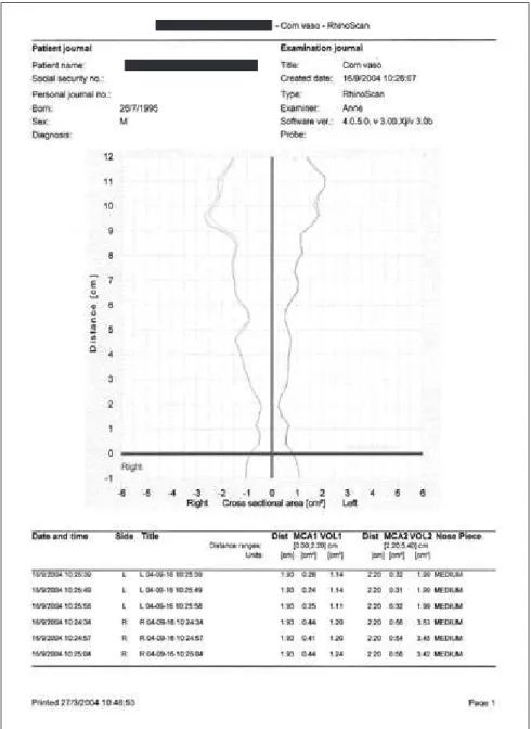
The clinical value of this exam is associated with its ability of measuring reliably the geometry of the nasal cavity. The exam is presented in a Cartesian graph, in which it is possible to accurately localize the transversal areas from 0 to 54 mm in depth.8-12 The graph shows the minimum transversal area val-ues observed from the nostril toward the nasophar-ynx. The readings describe the nasal respiratory way capacity, that is, the observed degree of nasal obstruction. The method is based on analysis of the sound waves relected inside the nasal cavity, taking into account the sound wave incidence and relec-tion properties.
The objective of this study was to evaluate the re-spiratory pattern in children in the mixed dentition by means of acoustic rhinometry and its relation to the upper arch width development.
Materials and Methods
Fifty white children, 25 females and 25 males with a mean age of eight years and seven months, were examined. These patients were chosen random-ly from a sample of enrolled ones to be submitted to orthodontic treatment at the School of Dentistry, University of São Paulo, without previous evaluations of their respiratory pattern or their upper arch shape. The requirements for the sample selection were not having undergone orthodontic treatment and or sur-gery, palatine and/or pharynx tonsillectomy.
In the present study, acoustic rhinometry exams were performed using the RhinoScan (Rhinomet-rics A/S - Assens, Funen, Denmark) and upper plas-ter models.
The acoustic rhinometry exam is made in a stat-ic, rapid, and non invasive manner, without nasal low. A probe is utilized, coupled to the nostrils’ in-dividual adapters to transmit and receive the sound from the electronic source inside the nasal cavity. The narrowest nasal cavity area is measured and standardized as the minimal cross-sectional area (MCA), and the relation of this narrowness with the respective localization throughout the nasal cavity is established. Normally, these area values are given in two moments known as: MCA1 and MCA2. In this study only MCA1 values were used.
The exams were done with the patient correctly and comfortably positioned on the chair, leaning the head, avoiding any possible head delection or extension. At the beginning of the examination, the patient was asked to interrupt his or her respiration so that the sound waves penetrated the nasal cavity and allowed the calibration that would generate the graph. The exam was repeated until producing three green curves for each nostril. For this study, the av-erage of these three readings was used.
Two exams were performed: the irst being with-out a vasoconstrictor, and the second exam, approx-imately 15 minutes after the use of a vasoconstrictor (nasal decongestant – oxymetazoline hydrochloride 0.25 mg/ml), applied after the irst exam.
In this study, even though the acoustic rhinom-etry exam was performed with and without a va-soconstrictor, only the average values found for the nasal cavity minimum transversal area (MCA1) without the vasoconstrictor were used, thus aiming to analyze the patient in his or her normal daily life. Before beginning the examination, no evident nasal inlammation was detected.
Impressions of the upper and lower dental arches were made for each patient with Orthoprint algi-nate (Zhermack – Badia Polesine, Rovigo, Italy)
us-Figure 1 - Acoustic Rhinometry exam.
Braz Oral Res. 2009 Oct-Dec;23(4):424-31 427
ing the proportions indicated by the manufacturer. Immediately afterwards, orthodontic plaster models were made using a ratio of 30 ml of water to 100 g of powder according to the manufacturer’s instruc-tions.
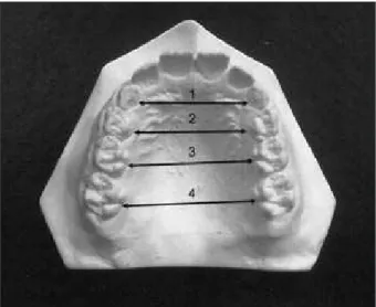
The study model analysis of the upper arch was accomplished by measuring the interdental distance of the deciduous canines (measurement 1), decidu-ous irst molars (measurement 2) and decidudecidu-ous second molars (measurement 3). For these measure-ments, the intersection between the center of the lin-gual surface and the marginal gum tissue was used. For the transverse distance measurement between the upper irst molars, the lingual groove and the marginal gum tissue point of intersection was used (measurement 4) as in Figure 3.
For the method’s error performance, 20 plas-ter models were randomly chosen. The same steps were repeated with them to obtain the transversal measurement values. With the obtained values, the Dahlberg (1940) formula was applied to calculate the method’s error.
The calibrations were made using a Mitutoyo digital caliper (Suzano, São Paulo, SP, Brazil) (Fig-ure 4). The same examiner meas(Fig-ured the plaster models.
To verify if there was a statistical difference be-tween genders in the measurements, the t-Student test was applied at the 5% signiicance level.
Results
The acoustic rhinometry exam and the measure-ments taken of the plaster models, whose values pre-sented a normal distribution, were compared by the Pearson’s parametric test of linear correlation at the 5% signiicance level. The Pearson’s correlation test results for the two genders, female and male, are represented in Tables 1 and 2, respectively.
As seen in Table 1, the interdental distances of the irst deciduous molars (r = 0.401 and p = 0.047) and second deciduous molars (r = 0.406 and p = 0.044) in females without vasoconstrictor use were directly correlated with the rhinometric measurements of the left nasal cavity area (p < 0.05).
As shown in Table 2, the interdental distances of the deciduous canines (r = 0.400 and p = 0.047), deciduous irst molars (r = 0.493 and p = 0.012) and deciduous second molars (r = 0.435 and p = 0.030) were directly and signiicantly correlated (p < 0.05) with the left nasal cavity area in males.
As shown in Table 3, the average interdental distance between the permanent irst molars was statistically higher in males (p = 0.036). The other measurements did not present average differences in relation to gender (p > 0.05).
Discussion
The relevance of nasal respiratory obstruction associated with orthodontics has remained a
con-Figure 4 - Transversal measurements made with the Mitu-toyo digital caliper in the orthodontic study models.
troversy for over a century. There are many studies that try to deine a correlation between these two factors.2,9-12
The greatest challenge in establishing a rela-tion between the respiratory pattern and the facial growth is to quantify the degree of this relation. Is the genetic factor the primary or secondary
determi-nant? This question remains unanswered for more than a century. In the 1980s, Harvold et al.11 (1981) performed a study altering the respiratory pattern from nasal to oral in monkeys, using silicone to close the air passage through the nostrils, and found an alteration in the growth pattern and occlusion in re-lation to the control group. But, it should be empha-Table 1 - Pearson’s correlation results for females.
Measurement Interdental distance of deciduous canines
Interdental distance of deciduous first molars
Interdental distance of deciduous second molars
Interdental distance of permanent first molars
Right W/V area Correlation 0.324 0.307 0.349 0.367
P 0.114 0.135 0.087 0.071
Left W/V area Correlation 0.311 0.401 0.406 0.185
P 0.131 0.047 0.044 0.376
W/V: without vasoconstrictor.
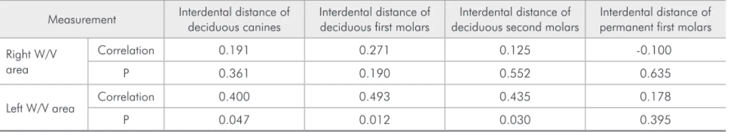
Table 2 - Pearson’s correlation results for males.
Measurement Interdental distance of deciduous canines
Interdental distance of deciduous first molars
Interdental distance of deciduous second molars
Interdental distance of permanent first molars Right W/V
area
Correlation 0.191 0.271 0.125 -0.100
P 0.361 0.190 0.552 0.635
Left W/V area Correlation 0.400 0.493 0.435 0.178
P 0.047 0.012 0.030 0.395
W/V: without vasoconstrictor.
Table 3 - Description and results of the t-Student test comparing the measurements between genders.
Measurement Gender Average Standard deviation N Value of t df p-value Interdental distance of
deciduous canines
Feminine 24.67 mm 1.81 25
-1.93 48 0.059 Masculine 25.90 mm 2.63 25
Interdental distance of deciduous first molars
Feminine 26.42 mm 2.42 25
-1.45 48 0.154 Masculine 27.39 mm 2.31 25
Interdental distance of deciduous second molars
Feminine 30.36 mm 3.12 25
-1.69 48 0.097 Masculine 31.64 mm 2.15 25
Interdental distance of permanent first molars
Feminine 34.07 mm 2.43 25
-2.16 48 0.036 Masculine 35.50 mm 2.26 25
Right W/V area Feminine 0.28 cm
2 0.10 25
0.51 48 0.610 Masculine 0.27 cm2 0.13 25
Left W/V area Feminine 0.29 cm
2 0.10 25
-0.39 48 0.701 Masculine 0.30 cm2 0.11 25
Braz Oral Res. 2009 Oct-Dec;23(4):424-31 429
sized that humans breathe predominantly through the nose or predominantly through the oral cavity, but not exclusively by either of them. Consequently, the indings of Harvold et al.11 (1981) can not be an-alyzed in a simple manner, having in mind that pre-dominantly oral breathing patients are found, and they do not present vertical growth predominance.
Nowadays however, there is no undisputed meth-od for registering the degree of nasal obstruction. Nonetheless, acoustic rhinometry has demonstrated to be an adequate method for the nasal area evalua-tion, presenting reproducible, reliable, and objective data.12
These indings are consistent with those of
Çak-mak et al.8 (2003), who report that acoustic
rhinom-etry exam results and computerized tomogram re-sults were similar, thus validating the method.
Many other authors, among them Roithmann et al.13 (1995), Zancanella, Lima12 (2004), Grymer10 (2000), Hilberg, Pedersen3 (2000), Tomkinson, Eccles14 (1995), Çakmak et al.8 (2003), performed studies aiming to determine MCA minimum values in order to be able to distinguish between a normal nasal cavity from an obstructed one. However, most of them were accomplished with adult patients.
Hinton et al.15 (1987) and Warren et al.16 (1988) afirmed that when the transversal section of the na-sal cavity, in adults, is smaller than 0.4 cm², there is a decrease of the air volume that passes through the nasal cavity, compromising the respiratory capac-ity and harming the respiration qualcapac-ity. An almost linear relation was found between the nasal cavity transversal area decrease and the nasal air volume decrease in this type of patient.
Believing that the nasal transversal area interferes with the respiratory pattern, and that the smaller transversal area would be in the anterior region of the nasal cavity,3 acoustic rhinometry was proposed to analyze this region, and compare it with the up-per arch transversal dimensions, in an attempt to ind a correlation between these factors.
The nasal geometry in children with nantly nasal respiration and children with predomi-nantly oral respiration was already reported by Za-vras et al.2 (1994). The authors found statistically signiicant differences in the total volume of the
na-sal cavity, where the oral breathers presented small-er values in relation to the nasal breathsmall-ers. Howev-er, differences were not observed in relation to the nasal cavity transversal area. In this same study, it was observed that the minimum transversal area in the nasal cavity was 0.28 cm² in males and 0.29 cm² in females. These values were smaller than the ones found in the study performed by Vig, Zajac17 (1993), where the minimum transversal area average was 30 mm² in males and 32 mm² in females.
The value differences between the two studies may be associated, partially, with the studied popu-lation, knowing that in the study conducted by Za-vras et al.2 (1994), the age samples varied from 6 to 10 years, and in that conducted by Vig, Zajac17 (1993), between 5 and 12 years. According to
War-ren et al.18 (1990), the air passage increases
approxi-mately 0.032 cm² per year. This statement was made by the authors led by the study in which it was found that a transversal area average of 0.21 ± 0.05 cm² at 6 years of age increased to 0.46 ± 0.15 cm² at 14 years of age.
In the present study, where the mean age was eight years and seven months, the transversal area average was 0.28 cm² in the right nasal cavity and 0.29 cm² in the left nasal cavity in females, and 0.27 cm² in the right nasal cavity and 0.30 cm² in the left nasal cavity in males.
Analyzing the correlation between the nasal ge-ometry and the upper arch, as shown in Table 1, females presented a much larger left nasal cavity; in addition, the interdental distances of the decidu-ous irst molars (r = 0.401 and p = 0.047) and of the deciduous second molars (r = 0.406 and p = 0.044) were also larger in females.
As for the measurements of males, Table 2 shows that there is a correlation between the interdental distances of the deciduous canines (r = 0.400 and p = 0.047), of the deciduous irst molars (r = 0.493 and p = 0.012) and of the deciduous second molars (r = 0.435 and p = 0.030), with the left nasal cavity. Therefore, an increased left nasal cavity area means an increased interdental distance of the deciduous canines, deciduous irst molars and deciduous sec-ond molars.
that results were not uniform between the relation of the nasal cavity minimum transversal area and the upper dental arch transverse distance.
In the permanent irst molar area, there was no correlation between the nasal area and the upper dental arch transverse distance. However, an in-creased upper arch transverse distance was observed in males in relation to females, as can be observed in Table 3.
Analyzing these correlations, it can be seen that there was no uniformity of results between the left nasal cavity minimum transversal area and the up-per dental arches transverse distance.
There was no correlation between the right na-sal cavity minimum transverna-sal area and the upper arch transverse distances. Even though studies in the literature do not mention the division between the right and left nasal cavity, this division was used in this research. It is interesting to observe that if the nasal cavity minimum transverse distance had been studied utilizing the average between the left
and right nasal cavity, there would have been no difference of this value for gender. The nasal cavity area in females would be the same in males, that is, 0.285 cm².
Knowing that the facial structure is determined in the irst decade of life, a longitudinal study dur-ing the growth phase may answer today’s persistent questions in the near future, using more objective exams for measuring the nasal cavity, among them, acoustic rhinometry.
Conclusion
There was correlation between the left nasal cav-ity transversal area and the upper arch interdental distance of deciduous irst molar and deciduous second molar for both genders. For the interdental distance of deciduous canines, there was correlation only in males. There was no correlation between the right nasal cavity minimum transversal area and the upper arch width.
References
1. McNamara JA. Influence of respiratory pattern on craniofa-cial growth. Angle Orthod. 1981;51(4):269-300.
2. Zavras Al, White GE, Rich A, Jackson AC. Acoustic rhinom-etry in the evaluation of children with nasal or oral respiration. J Clin Pediatr Dent. 1994 Spring;18(3):203-10.
3. Hilberg O, Pedersen OF. Acoustic rhinometry: recommen-dations for technical specifications and standard operating procedures. Rhinol Suppl. 2000;16:3-17.
4. Principato JJ, Kerrigan JP, Wolf P. Pediatric nasal resistance and lower anterior vertical face height. Otolaryngol Head Neck Surg. 1986;95(2):226-9.
5. Shanker S, Fields HW, Beck FM, Vig PS, Vig KWL. A longitu-dinal assessment of upper respiratory function and dentofacial morphology in 8- to 12-year-old children. Semin Orthod. 2004;10(1):45-53.
6. Kluemper GT, Vig PS, Vig KW. Nasorespiratory characteristics and craniofacial morphology. Eur J Orthod. 1995;17(6):491-5.
7. Vig KWL. Nasal obstruction and facial growth: the strength of evidence for clinical assumptions. Am J Orthod Dentofac Orthop. 1998;113(6):603-11.
8. Çakmak O, Coskun M, Çelik H, Buyuklu F, Ozluoglu LN. Value of acoustic rhinometry for measuring nasal valve area. Laryngoscope. 2003;113(2):295-302.
9. Carlini D. Rinometria acústica na avaliação de pacientes entre 7 e 13 anos de idade com obstrução nasal por rinite crônica hi-pertrófica não infecciosa [Dissertação de Mestrado]. São Paulo: Universidade Federal de São Paulo; 1999.
10. Grymer LF. Clinical applications of acoustic rhinometry. Rhi-nol Suppl. 2000;16:35-43.
11. Harvold EP, Tomer BS, Vargervik K, Chierici G. Primate ex-periments on oral respiration. Am J Orthod. 1981,79(4):359-72.
12. Zancanella E, Lima WTA. Uso da rinometria acústica como mé-todo diagnóstico. Rev Bras Otorrinolaringol. 2004;70(4):500-3.
13. Roithmann R, Cole P, Chapnik J, Shpirer I, Hoffstein V, Zamel N. Acoustic rhinometry in the evaluation of nasal obstruction. Laryngoscope. 1995;105(3 Pt 1):275-81.
14. Tomkinson A, Eccles R. External facial dimensions and mini-mum nasal cross-sectional area. Clin Otolaryngol Allied Sci. 1995;20(6):557-60.
15. Hinton VA, Warren DW, Hairfield WM, Seaton D. The re-lationship between nasal cross-sectional area and nasal air volume in normal and nasally impaired adults. Am J Orthod Dentofac Orthop. 1987;92(4):294-8.
Braz Oral Res. 2009 Oct-Dec;23(4):424-31 431
breathing. Am J Orthod Dentofacial Orthop. 1988;93(4):289-93.
17. Vig PS, Zajac DJ. Age and Gender effects on nasal respira-tory function in normal subjects. Cleft Palate Craniofac J. 1993;30(3):279-84.