MASTICATORY MUSCLE ELECTRICAL ACTIVITY PATTERN
IN OBESE AND EUTROPHIC CHILDREN
Padrão de atividade elétrica dos músculos mastigatórios
em crianças obesas e eutróficas
Talita Cristina Favero(1); Ana Maria Toniolo da Silva(2), Leris Bonfanti Haeffner(3),
Angela Ruviaro Busanello-Stella(4), Eliane Correa(5)
(1) Universidade Federal de Santa Maria (UFSM), Santa
Maria, RS, Brasil.
(2) Departamento de Fonoaudiologia - Universidade Federal
de Santa Maria (UFSM), Santa Maria, RS, Brasil.
(3) Departamento de Pediatria - Universidade Federal de
Santa Maria (UFSM), Santa Maria, RS, Brasil.
(4) Departamento de Fonoaudiologia – Universidade Federal
de Santa Catarina (UFSC), Florianópolis, SC, Brasil.
(5) Departamento de Fisioterapia - Universidade Federal de
Santa Maria (UFSM), Santa Maria, RS, Brasil.
Conlict of interest: non-existent
program. Changes in lifestyle and eating habits, increased inactivity and consumption of high energy density foods can explain this scenario 2.
Obesity, among nutritional disorders, generates the greatest amount of musculoskeletal problems. Critical periods of progressive obesity onset occur on the irst 12 months of life, in kindergarten and during puberty. Progressive obesity is associated with hyperplastic obesity, making it diicult to control body weight in adulthood. This implies the impor
-tance of further studies on the obese population with excessive weight gain and its contribution to changes in the performance of the stomatognathic system (SS) 3.
Changes in the morphology, tone, and posture of the structures of SS, which may occur due to excess weight, directly interfere with its functioning. The imbalance of the stomatognathic system can afect the postural system as a whole, just as postural
INTRODUCTION
Obesity is a chronic, complex disease of multi
-factorial etiology that determines several complica
-tions in childhood and adulthood 1. The increasing
number of obese people in the world indicates a strong environmental contribution to genetic
ABSTRACT
Purpose: to compare the masticatory muscle electrical activity pattern of obese and eutrophic chil
-dren during muscle on-of timing using surface electromyography. Methods: a total of 32 children from
6 to 12 years of age were divided into two equal groups - 16 obese and 16 eutrophic children - and assessed. The variables studied included the electromyography activity of the muscles of mastication (anterior temporalis, masseter and orbicularis oris) during rest, maximum voluntary contraction, mas
-tication (regular and directed), and swallowing. For statistical analysis, the median, and the irst and third quartiles were found and the Wilcoxon test was used, considering signiicance level of p < 0.05.
Results: obese children showed similarities in muscle activation compared with eutrophic children
during maximum voluntary contraction and rest. However, for dynamic activities - regular and directed mastication and swallowing - obese children had lower muscle activation medians than eutrophic chil
-dren in most proposed situations, both in the activation period (on) and in the inactivation period (of), with signiicant statistical diference (p < .05). Conclusion: obese children, probably due to excessive
facial adiposity, present changes in the conditioning of the masticatory muscles, which are relected in the performance of the stomatognathic system.
KEYWORDS: Nutritional Status; Child; Stomatognathic System; Electromyography; Mastication;
The following were used as inclusion criteria: both genders, aged from 6 to 12 years, and having
a parent or guardian sign the FPIC. Individuals
were excluded if they presented signs of neuro
-logical impairment, absence of third molar tooth (for occlusal stability), changes in dental anatomy due to restorations or trauma, and if they had a history of orthodontic treatment, orofacial motricity phono
-audiological therapy, facial or bariatric surgery, and craniofacial malformations.
The anamnesis was conducted individually with caregivers who agreed to participate in the study, to gather data on children proile, complaints, family history, medical complications, motor devel
-opment and diiculties, health and respiratory issues, sleeping and treatments; eating facts from breastfeeding to current habits, as well as chewing, swallowing, oral habits, and information on commu
-nication, speech, hearing, voice and education.
Children weight, height and nutritional status
were measured throughout the stages of evalu
-ation, by using a 100g precision G- Tech ® digital scale, and a measuring tape ixed on wall with no footer, to then determine their BMI. Anthropometric variables were transformed into z-scores, in accor
-dance with the growth curves of the World Health Organization 11. Children whose BMI z-score was ≥
-2 were considered eutrophic, and < +1 and +2 ≥ were considered obese.
ENT examination was intended to evaluate and diagnose children respiratory pattern and its probable etiology. Children underwent clinical examination, as well as cephalometry, when further investigation was required to classify respiratory behavior (nose-breather, habitual mouth-breather or obstructive mouth-breather). Tonsils were classiied according to the degree of obstruction on the level of the oropharynx. Tonsil obstruction of up to 25 % of the oropharynx was rated as Grade I; 25-50 % obstruction as Grade II; 50-75 % obstruction as Grade III; and greater than 75 % as Grade VI 12.
The same criteria used to assess the degree of obstruction produced by tonsillar hypertrophy were used to evaluate obstruction caused by increased
adenoid tissue.
The dental evaluation aimed to observe the type of dentition and occlusion of children through clinical examination. The type of molar ratio was considered for occlusion, in accordance with the classiication of Angle (1899)13, as well as transverse relationship.
The following was also observed: midline, conigu
-ration of the hard palate, presence of overjet, overbite, and teeth conservation, in order to meet the study criteria for inclusion or exclusion8.
All children underwent electromyographic evalu
-ation of the right anterior temporalis (RT), left anterior changes may negatively impact the stomatognathic
system 4; 5.
Excess weight in young populations and its implications on the SS has been rarely addressed in Phonoaudiology 6. It is well known that muscle
dysfunction may act on the facial and postural growth and development of the individual. Thus, stomatognathic system components can sometimes act as agents of structural changes, and sometimes be the targets of such changes 7.
With the purpose of assisting on the evaluation and diagnosis of these patients, electromyography (EMG) comes as a possibility to objectively analyze muscle electrical activity and, it has also been studied in Phonoaudiology in recent years. The use of a device that captures and expands muscle action potential is useful for patient diagnosis, since it relects their neuromuscular system condition 8; 9.
Portney (1993)10 states that as an assessment
procedure, clinical EMG involves the inding and recording of muscle iber electrical potential, providing important data for patient diagnosis and determining rehabilitation goals for patients with muscle disorders, such temporomandibular dysfunctions. Based on that, and because there are only few researches on this matter, the aim of this study was to compare the masticatory muscle electrical activity pattern of obese and eutrophic children during muscle on-of timing using surface electromyography (EMG).
METHODS
This research project has been previously submitted to evaluation and was approved by the Ethics Committee of the institution of origin under protocol number 01120243000-10.
This was a cross-sectional, analytical observa
-tional and quantitative ield study developed at the Orofacial Motricity Lab of the Phonoaudiological Assistance Service from UFSM from May 2012 to March 2013.
The procedures for children screening in this study were: anamnesis, ENT and dental evaluation, and surface EMG assessment. A total of 230 1st to 6th graders from a public middle school located in Santa Maria - RS, Brazil, were submitted to the initial screening process. Of these, 32 completed all phases of research evaluation, 20 female and 12
male.
We used the following verbal command: “... press it, press it, press it...”. For the orbicularis muscle group, in the second phase of the evaluation, children were asked to pressed one lip against the other, also for a period of 5 seconds, while the evaluator used the same verbal command.
Habitual mastication - To perform this test, children were instructed to chew the usual way a piece of French bread (2 x 2cm) and tell the examiner with a hand signal (positive) whenever they swallowed it 9. For this test, we used the
following verbal command: “eat this bread the same way you usually do at home.”
Directed mastication - Children were initially told to chew a Trident gum (produced by Warner-Lambert e Com. Ltda - Adams Division) for an average of 20 seconds to obtain a uniform consistency before records. This gum was chosen because it is easily handled, also for being popular, and for being well accepted among children and widely used in related research. Next, for the electromyographic collections, children were requested to determine their favorite chewing side and after that, they were asked to use that side only for chewing up to 20 seconds 14.
Swallowing liquid - In this test, children were asked to suck 10ml of water (measured with a syringe) from the glass through a straw and hold it in their mouths, with sealed teeth and lips until they received an order to swallow. Five sips were regis
-tered for each of the three collections.
In the EMG signal collection, double electrodes containing gel were used, as well as disposable silver circular adhesive - silver chloride (Ag / AgCl), with a diameter of 10 mm (Hal Indústria e Comércio Ltda.) with a 20mm distance between electrodes center to center. For skin oil removal, before attaching the electrodes, 70% alcohol was used to facilitate both the ixation of the electrode and the transmission of electrical activity 20. To examine the masseter
muscle, electrodes were placed bilaterally between the lower border of the zygomatic arch and the angle of the jaw 21; and to examine the temporal muscle
they were placed on its anterior part, perpendicular to the zygomatic arch above and behind the frontal process of the zygomatic arch 22.
To examine the orbicularis muscle, an electrode was positioned on the upper lip, just above its edge, perpendicular to the ilter, and another on the lower lip, just below its edge. For the three muscles, electrode positioning followed the longitudinal direction of its ibers. A ground electrode was also ixed on the glabellar region, to avoid interference of electromagnetic currents 16.
The equipment used for the electromyographic tests were the Electromyograph Miotool produced temporalis (LT), right masseter (RM), left masseter
(LM), upper orbicularis (UO), and lower orbicularis (LO), through tests that could show their behaviors.
Electromyography was preceded by skin imped
-anciometry. This procedure was conducted to ensure greater safety in the collection and greater reliability of the electromyography results, since there could be interference in the passage of electric current due to facial adiposity. SK-100 ICEL-KAISE was used to determine impedance. When impedance averaged less than or equal to 10 (+ / -1.8) Ω, the electromyographic examination was then performed 14.
The assessment of muscle activity was performed through bilateral electromyographic recordings of the masseter and temporal muscles in the conditions of rest, maximum voluntary contraction, habitual masti
-cation, directed masti-cation, and swallowing. For the orbicular muscles, the same tests were performed, except for directed mastication, once it did not apply to this muscle group. The proposed tests strictly followed an electromyographic miofunctional evalu
-ation protocol developed for the purposes of this research and based on the literature 15; 8; 16.
Children were comfortably seated in a chair when evaluated. Their trunk was straight upward, the soles of their feet rested against the loor (or on a wooden box, if too little to reach the loor) and head positioned according to the Frankfurt horizontal plane, parallel to the ground. The posture of the children was monitored throughout the evaluation.
For each of the tests, three collections were performed. Before acquiring the EMG recordings, children were previously trained to ensure the consis
-tency of results. All movements were monitored by the researcher and, if any inappropriate movement was noted, the collection was stopped and started over. To avoid possible muscle fatigue, children were instructed to remain at rest for a period of 2 minutes between records.
The following are tests conducted in the electro
-myographic evaluation:
Rest - In this test, the child was instructed to
remain seated in the usual position of rest, with lips and jaw relaxed and upright torso. This position lasted 20 seconds in the electromyographic recording 14.
We applied the following verbal command: “ ... relax, look forward and stay in this position ... “.
Maximum voluntary contraction (MVC) - a
Parailm ® (Parailm M Laboratory Film) measuring 3 cm long, 1cm wide and folded into ive equal
parts17;18;19 was positioned bilaterally on the occlusal
surfaces of posterior teeth, and children were told to bilaterally and simultaneously contract their masti
that values higher than this mean were classiied as activation period (on) and the values below as inactivation period (of) 23.
Subsequently, values were normalized by simple rule of three and the results were expressed in percentage. For MVC and rest testing 100 % was considered peak muscle activation during MVC. For other tests (dynamic activities) values were normalized based on the mean muscle activation during MVC 24; 25.
Data were placed on tables using Microsoft Excel 2007 and then statistically analyzed using Stata version 10.0. The Shapiro- Wilk test was used to verify the normality of the variables. Median, irst and third quartiles were calculated by Wilcoxon test to detect diferences between groups. For all tests, a signiicance level of 5 % (p < 0,05) was adopted.
RESULTS
A total of 32 children were included in the study, 20 female and 12 male, aged between 6 and 12 years. Regarding nutritional status, 16 were classiied as eutrophic and 16 as obese, with a statistically signif
-icant diference between groups (p < 0.001). Of all children, 50 % were classiied as mouth-breathers, and the remaining as nose-breathers, with no statis
-tical diference between groups (p = 0.480).
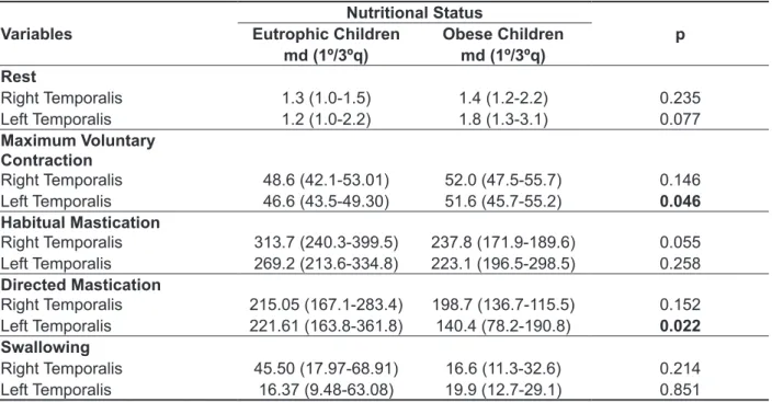
Table 1 presents the temporal muscle electrical activity medians for MVC and rest situations, and activation period (on) for situations of habitual and directed mastication, and swallowing in relation to nutritional status. It was observed that obese and eutrophic children had similar percentages of activation when muscles were at rest. Statistically signiicant diference was noted between groups (p = 0.046) for the left temporal muscle during MVC. For the other situations, obese subjects showed lower activation percentage than the control group, with statistically signiicant diference (p < 0.022) between groups during directed mastication activity for the left temporal muscle.
by Miotec Equipamentos Biomédicos Ltda. and belonging to the Laboratory of Orofacial Motricity from the Department of Phonoaudiology/UFSM.
To capture the electromyographic signal, an acquisition system with 4 channels was used. For data acquisition, the Miograph ® software was used, they were scanned by an A / D (analog - digital) conversion board with 14 bit resolution and signals with sampling frequency of 2000 samples / second / channel, bandpass cut ilter from 20-500 Hz, with an ampliication gain of 1000 times and common rejection mode of 110 dB, installed on a Itautec SA laptop with an Intel Pentium processor and Windows 7 Pro. Equipment calibration followed standard speciications by manufacturers.
It should be noted that the computer was using its own battery, there was no connection to the power grid, and the loor was covered with Pavilex. During data collection, the researcher and the patient remained in the location; all electronic devices that could possibly generate an electromagnetic ield, as well as light sources, were turned of.
The clipping of the rest and MVC test records was made considering 5 sequential seconds of the best collection (best electromyographic signal, lower occurrence of electrical interference or variation based on FFT (Fast Fourier Transform) curve analysis). As for the dynamic activities (habitual and directed mastication, and swallowing) the clipping of three sequential cycles of the best collection was
made.
Table 1 – Temporal muscle electrical activity medians for maximum voluntary contraction and rest situations and activation (on) period of dynamic activities in relation to nutritional status, with normalized and percentage measured data.
Variables
Nutritional Status
p Eutrophic Children Obese Children
md (1º/3ºq) md (1º/3ºq)
Rest
Right Temporalis 1.3 (1.0-1.5) 1.4 (1.2-2.2) 0.235
Left Temporalis 1.2 (1.0-2.2) 1.8 (1.3-3.1) 0.077
Maximum Voluntary Contraction
Right Temporalis 48.6 (42.1-53.01) 52.0 (47.5-55.7) 0.146
Left Temporalis 46.6 (43.5-49.30) 51.6 (45.7-55.2) 0.046 Habitual Mastication
Right Temporalis 313.7 (240.3-399.5) 237.8 (171.9-189.6) 0.055
Left Temporalis 269.2 (213.6-334.8) 223.1 (196.5-298.5) 0.258
Directed Mastication
Right Temporalis 215.05 (167.1-283.4) 198.7 (136.7-115.5) 0.152
Left Temporalis 221.61 (163.8-361.8) 140.4 (78.2-190.8) 0.022 Swallowing
Right Temporalis 45.50 (17.97-68.91) 16.6 (11.3-32.6) 0.214
Left Temporalis 16.37 (9.48-63.08) 19.9 (12.7-29.1) 0.851
Md (1st - 3rdQ)= median (irst and third quartiles); *Wilcoxon Test.
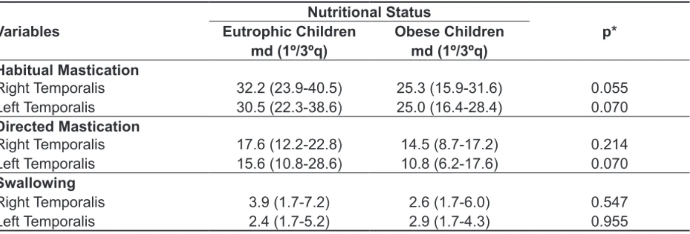
Table 2 presents the temporal muscle electrical activity medians in relation to nutritional status in situations tested during muscle inactivation (of). Similar percentage of muscle activity was found between groups for all situations tested, with no statistically signiicant diference.
Table 3 presents the masseter muscle electrical activity medians for MVC and rest situations, and
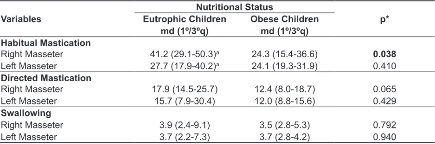
muscle, which reported superiority of activation for the eutrophic group, with statistically signiicant diference (p = 0.038). In this analysis, there was also signiicant diference in muscle activation (left and right) among members of the eutrophic group (p = 0.012) during habitual mastication.
Table 4 presents the masseter muscle electrical activity medians in relation to nutritional status in situations tested during muscle inactivation (of). It was observed that the percentages of activation were similar in all situations tested, except for the activity of habitual mastication for the right masseter
Table 2 – Temporal muscle electrical activity medians in relation to nutritional status in situations
tested during the period of muscle inactivation (of), with normalized and percentage measured data.
Variables
Nutritional Status
p* Eutrophic Children Obese Children
md (1º/3ºq) md (1º/3ºq)
Habitual Mastication
Right Temporalis 32.2 (23.9-40.5) 25.3 (15.9-31.6) 0.055
Left Temporalis 30.5 (22.3-38.6) 25.0 (16.4-28.4) 0.070
Directed Mastication
Right Temporalis 17.6 (12.2-22.8) 14.5 (8.7-17.2) 0.214
Left Temporalis 15.6 (10.8-28.6) 10.8 (6.2-17.6) 0.070
Swallowing
Right Temporalis 3.9 (1.7-7.2) 2.6 (1.7-6.0) 0.547
Left Temporalis 2.4 (1.7-5.2) 2.9 (1.7-4.3) 0.955
Md (1st - 3rdQ)= median (irst and third quartiles); *Wilcoxon Test.
Table 3 – Masseter muscle electrical activity medians for maximum voluntary contraction and rest situations and activation (on) period of dynamic activities in relation to nutritional status, with normalized and percentage measured data.
Variables
Nutritional Status
p* Eutrophic Children Obese Children
md (1º/3ºq) md (1º/3ºq)
Rest
Right Masseter 1.1 (0.8-1.5) 1.5 (2.0-1.1) 0.181
Left Masseter 1.3 (0.9-1.6) 1.4 (1.1-1.9) 0.258
Maximum Voluntary Contraction
Right Masseter 46.8 (36.6-53.3) 47.8 (42.7-50.6) 0.792
Left Masseter 47.3 (45.2-50.3) 48.9 (44.7-52.6) 0.510
Habitual Mastication
Right Masseter 378.9 (272.1-495.6) 211.5 (152.5-267.9) 0.013
Left Masseter 269.6 (170.9-405.2) 224.6 (173.0-274.1) 0.291
Directed Mastication
Right Masseter 236.4 (168.9-318.8) 155.7 (103.3-228.9) 0.097
Left Masseter 169.7 (93.3/398.3) 134.6 (51.1-177.8) 0.228
Swallowing
Right Masseter 22.7 (3.5-55.3) 22.3 (14.9-31.9) 0.547
Left Masseter 22.4 (13.3-56.3) 21.7 (16.7-38.9) 0.851
muscle during habitual mastication (p = 0.008). There were diferences in activation for upper and lower orbicular muscles among the members of the eutrophic group during MVC, with activation percentage of 45.4 % for upper orbicular and 43.3 % for lower orbicular (p = 0.03). Both groups showed signiicant diferences in the activation of the orbicu
-laris muscle among their members for the activities of habitual mastication and swallowing, with higher percentages of activation in the lower orbicularis. Table 5 presents upper and lower orbicular
muscle electrical activity medians to the situations of MVC and rest and activation period (on) of habitual mastication and swallowing in relation to nutritional status. There was similarity in muscle activation between groups during rest and MVC. As for the activities of habitual mastication and swallowing, obese children had lower activation percentage median than eutrophic children, with statistically signiicant diferences for the lower orbicularis
Table 4 – Masseter muscle electrical activity medians in relation to nutritional status in situations
tested during the period of muscle inactivation (of), with normalized and percentage measured data.
Variables
Nutritional Status
p* Eutrophic Children Obese Children
md (1º/3ºq) md (1º/3ºq)
Habitual Mastication
Right Masseter 41.2 (29.1-50.3)a 24.3 (15.4-36.6) 0.038
Left Masseter 27.7 (17.9-40.2)a 24.1 (19.3-31.9) 0.410
Directed Mastication
Right Masseter 17.9 (14.5-25.7) 12.4 (8.0-18.7) 0.065
Left Masseter 15.7 (7.9-30.4) 12.0 (8.8-15.6) 0.429
Swallowing
Right Masseter 3.9 (2.4-9.1) 3.5 (2.8-5.3) 0.792
Left Masseter 3.7 (2.2-7.3) 3.7 (2.8-4.2) 0.940
Md (1st - 3rdQ)= median (irst and third quartiles); *Wilcoxon Test. Similar letters present signiicant statistical diference: a- 0.012.
Table 5 – Orbicularis oris muscle electrical activity medians for maximum voluntary contraction and rest situations and activation (on) period of dynamic activities in relation to nutritional status, with normalized and percentage measured data.
Variables
Nutritional Status
p*
Eutrophic Children Obese Children
md (1º/3ºq) md (1º/3ºq)
Rest
Superior Orbicularis 2.0 (1.3-3.5) 2.0 (1.2-2.2) 0.547
Inferior Orbicularis 1.9 (1.1-3.8) 2.0 (0.9-3.1) 0.638
Maximum Voluntary Contraction
Superior Orbicularis 45.4 (43.4-47.6)a 46.2 (43.4-48.5) 0.509 Inferior Orbicularis 43.1 (37.7-45.5)a 45.5 (42.4-49.2) 0.097
Habitual Mastication
Superior Orbicularis 221.5 (143.9-320.4)b 190.7 (146.6-212.5)d 0.274
Inferior Orbicularis 414.9 (275.0-565.6)b 260.1 (179.1-337.7)d 0.008
Swallowing
Superior Orbicularis 124.5 (90.9-156.7)c 112.8 (81.2-120.7)e 0.386
Inferior Orbicularis 202.0 (117.0-231.6)c 175.3 (123.4-221.1)e 0.706
Md (1st - 3rdQ)= median (irst and third quartiles); *Wilcoxon Test. Similar letters present signiicant statistical diference: a- 0.03;
between the upper and lower orbicular muscles during habitual mastication and swallowing when comparing groups. However, when analyzing the values within each group, both obese and eutrophic children had greater activation percentage for lower orbicularis in all proposed situations, with statisti
-cally signiicant diference (p < .05). Table 6 presents the orbicularis oris muscle
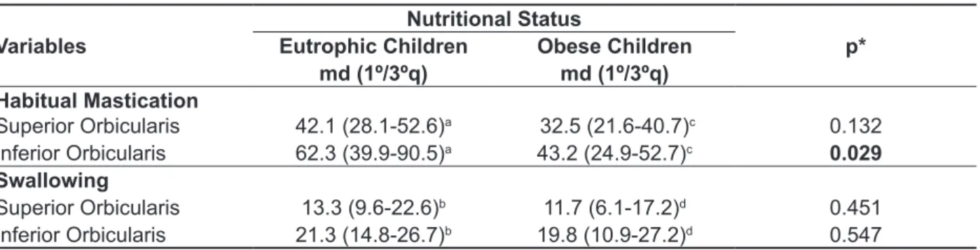
electrical activity medians in relation to nutritional status in situations tested during muscle inactivation (of). It was observed that the obese had lower percentage of muscle activity than eutrophic for lower orbicularis during habitual mastication, with statistically signiicant diference (p = 0.029). There was no statistical diference in muscle activation
Table 6 – Orbicularis oris muscle electrical activity medians in relation to nutritional status in situations
tested during the period of muscle inactivation (of), with normalized and percentage measured data.
Variables
Nutritional Status
p* Eutrophic Children Obese Children
md (1º/3ºq) md (1º/3ºq)
Habitual Mastication
Superior Orbicularis 42.1 (28.1-52.6)a 32.5 (21.6-40.7)c 0.132
Inferior Orbicularis 62.3 (39.9-90.5)a 43.2 (24.9-52.7)c 0.029
Swallowing
Superior Orbicularis 13.3 (9.6-22.6)b 11.7 (6.1-17.2)d 0.451
Inferior Orbicularis 21.3 (14.8-26.7)b 19.8 (10.9-27.2)d 0.547
Md (1st - 3rdQ)= median (irst and third quartiles); *Wilcoxon Test. Similar letters present signiicant statistical diference: a- p=0.003;
b- p=0.010; c- p=0.010; d- p=0.023.
DISCUSSION
Studies involving evaluations of obese stomato
-gnathic system are scarce, which makes this study relevant, although it is diicult to compare its results.
Regarding the EMG data of the anterior temporal muscle during activation (on), it was observed that obese children had similar activation percentage to eutrophic children, when muscles were at rest and MVC; whereas for dynamic situations, obese had lower activation percentage than that observed in the control group. The literature concerning these data shows that the discrepancy of electrical activity and the statistical diference found in some activities involving the temporalis muscle can be correlated to the pattern of lateral chewing preference and altered head posture present in some children26;27;28.
With respect to data on the masseter muscle during activation (on), also both at rest and MVC, obese showed similar activation percentage to eutrophic, which was not conirmed during the dynamic activities proposed, where eutrophic obtained important superiority of muscle activity. In this regard, and given the importance of the masseter muscle exercises during the masticatory process, the literature states that the obese individual may have problems related to chewing because they do not have an oral musculature strengthened by
having dental changes or by decreasing the masti
-catory speed 29.
It is known that this possible muscle decon
-ditioning of obese children, here translated by the inferiority of electrical activity compared to eutrophic children, is probably due to the preference for fast food, whose consistency is characterized by being more shredded, cooked and soft, usually composed of carbohydrates that increase satiety. Lieberman
et al (2004) 30 have showed that consumption of
processed foods has decreased facial growth of the mandibular and maxillary arches in humans in response to decreased occlusal force and masti
-catory required for grinding food.
In relation to the orbicularis oris muscle, it is known that patients with incompetent lips cannot seal their lips naturally and efortlessly; a condition that favors tooth protrusion by reduced labial pressure on them, generating a facial imbalance. The absence of lip contact causes muscle imbalance that can afect various functions, such as breathing, swallowing,
phonation, and the harmonious growth and
devel-opment of the face 31; 32; 33.
muscle activation on the on period, the greater was the value for the inactivation period (of). The high muscle activity, with values above the rest event, observed in the periods between activation cycles (of periods) reveals the absence of a complete muscle relaxation after contraction .
These indings are consistent with the study of Basmajian and De Luca (1985)39, who state that
during the complete rest period the muscle does not lose its tone, even when neuromuscular activity is nil. Thus, the results obtained in this study are considered to agree with the indings from the literature, as the subjects did not reach a state of complete muscle relaxation both between the cycles of activation (of period) and during rest periods. Furthermore, the fact that, even in the period of muscle inactivation (of), the obese children have presented lower values of muscle activity than those of eutrophic children conirms their inferiority of muscle conditioning compared to their eutrophic
peers.
CONCLUSION
From the analysis of the results obtained in this study, it was possible to conclude that obese individuals have similar muscle activation compared to eutrophic individuals during activities of MVC and rest. However, for most dynamic activities - habitual and directed mastication and swallowing - obese children had lower muscle activation medians than eutrophic children, both in the activation period (on) and in the inactivation period (of), for all muscle
groups studied.
Thus, these indings support the hypothesis that obese children, probably due to excessive facial adiposity, present changes in the conditioning of the masticatory muscles, which are relected in the performance of the stomatognathic system.
diferences for lower orbicularis in habitual masti
-cation (p = 0.008), this diference was not conirmed for the upper orbicularis (p = 0.274) in this same activity. Some authors claim that the change in the usual position of the lips is a sign of hypotonicity 34
or hypofunction of the orbicularis oris muscle, especially during mastication 35.
Studies report that both segments of the orbicularis oris muscle function as separate and independent entities. The default behavior of the upper and lower segments of the orbicularis oris, assessed at youth presenting normal occlusion, shows absence of signiicant electromyographic activity in this muscle during mastication and swallowing, as well as in the resting state. The medial
and lateral regions, upper and lower segments,
can function as independent bodies among themselves, even though they constitute the same muscle 36-38; 16.
Regarding the data on the proposed dynamic activities during the period of inactivation (of) of the muscle groups covered in this study, it was observed, for the temporal muscle, a lower percentage of muscle activity in obese compared to eutrophic children in the analyzed situations. The same occurred for the masseter muscles, for which the medians of action percentage were also higher for the eutrophic group . Repeatedly, in the analysis of the values obtained for the orbicular muscles, a superiority of muscle activity in eutrophic was noted, however, for this muscle group, it was observed that there was still a signiicant diference of activity between muscle units, with greater activation for the lower orbicularis compared to the upper orbicularis, for both groups during the situations tested.
REFERENCES
1. Ebbeling CB, Pawlak DB, Ludwig DS. Childhood obesity: public-health crisis, common sense cure. Lancet.2002;360:473-8.
2. Sociedade Brasileira de Pediatria, (2008). Departamento de Nutrologia. Obesidade na Infância e Adolescência – Manual de Orientação. URL: http://www.sbp.com.br/show_item2.cfm?id_ categoria=89&id_detalhe=2740&tipo_detalhe=s 3. Machado PG, Mezzomo CL. A relação da postura corporal, da respiração oral e do estado nutricional em crianças – uma revisão de literatura. Rev CEFAC.2011;13(6):1109-18.
4. Motonaga SM, Berte LC, Anselmo-Lima WT. Respiração bucal: causas e alterações no sistema estomatognático. RevBrasOtorrinolaringol. 2000; [Acesso em: 26 de fevereiro de 2015] 66(4):373-9. Disponível em: http://www.rborl.org.br/conteudo/ acervo/print_acervo.asp?id=2482
5. Bianchini EMG. Disfunções da articulação temporomandibular: relações com a articulação da fala [dissertação]. São Paulo (SP): Faculdade de Fonoaudiologia, Pontifícia Universidade de São Paulo; 1998.
6. Fernandes AR, Casonatto J, Christofaro DGD, Ronque VER, Oliveira AR. Risco para o excesso de peso entre adolescentes de diferentes classes econômicas. Rev AssocMedBras. 2008;54(4):334-8. 7. Marchesan IQ. Protocolo de avaliação miofuncional orofacial. In: Krakauer LH, Di
Francesco RC, Marchesan IQ. (Org.). Respiração oral. São José dos Campos: Pulso, 2003. P. 55-79. 8. Krob CL. Efeito do exercitador facial em crianças respiradoras orais: avaliação eletromiográica. [Dissertação] Santa Maria (RS): Universidade Federal de Santa Maria; 2008.
9. Figueiredo AB. Avaliação fonoaudiológica clínica e eletromiográica da motricidade orofacial do obeso: estudo comparativo. [Dissertação]. São Paulo (SP): Universidade de São Paulo; 2010. 10. Portney L. Eletromiograia e testes de velocidade de condução nervosa. In: O`Sullivan SB, Schmitz TJ editores. Fisioterapia: Avaliação e Tratamento. São Paulo: Manole, 1993. P. 183-217.
11. Onis MDE, Onyango AW, Borghi E, Siyam A, Nishida C, Siekmann J.Development of a WHO growth reference for school-aged children and adolescents. Bulletin of the World Health Organization. 2007;85:660-7.
12. Hiyama S, Ono T, Ishiwata Y, Kuroda T,Ohyama K. Efects of experimental nasal obstruction on human masseter and suprahyoid muscle activities
during sleep. Angle Orthodontist. 2003;73:151-7.
13. Angle EH. Classiication of Malocclusion. Dental Cosmos. 1899;41(3):248-64.
14. Berlese DB, Copetti F, Weimmann ARM, Fontana PF, Haeffner LSB. Atividade dos músculos masseter e temporal em relação às características miofuncionais das funções de mastigação e deglutição em obesos. DistúrbComun.2012;24(2):215-21.
RESUMO
Objetivo: comparar o padrão de atividade elétrica dos músculos mastigatórios de crianças obesas
e eutróicas durante os períodos de ativação (on) e inativação (of) muscular por meio da eletromio
-graia de superfície. Métodos: foram avaliadas 32 crianças, entre 6 e 12 anos de idade, divididas em
dois grupos iguais – 16 obesas e 16 eutróicas. As variáveis estudadas incluíram a atividade eletro
-miográica da musculatura mastigatória (músculo temporal anterior, masseter e orbicular da boca) durante as atividades de repouso, contração voluntária máxima, mastigação (habitual e direcionada) e deglutição. Para a análise estatística calculou-se a mediana, primeiro e terceiro quartis e utilizou-se
o teste de Wilcoxon, considerando nível de signiicância de p<0,05. Resultados: os obesos apre
-sentaram semelhanças de ativação muscular em relação aos eutróicos durante as atividades de contração voluntária máxima e repouso. Porém, para as atividades dinâmicas – mastigação habitual, mastigação direcionada e deglutição - os obesos apresentaram medianas de ativação muscular infe
-riores aos eutróicosna maioria das situações propostas, tanto no período de ativação (on) quanto
no período de inativação (of), com diferença estatística signiicante (p<0,05).Conclusão:crianças
obesas, provavelmente em função do excesso de adiposidade facial, apresentam alterações no con
-dicionamento da musculatura mastigatória, que se reletem durante a realização das funções do sistema estomatognático.
DESCRITORES:Estado Nutricional; Criança; Sistema Estomatognático; Eletromiograia; Mastigação;
de um protocolo de EENM. Rev. bras. isioter.
2009;13(5):422-9.
25. Jardini RSR. Avaliação eletromiográica do músculo bucinador lácido usando o Exercitador Facial. Pró-Fono R Atual Cient.2002;4:331-42. 26. Felício CM. Fonoaudiologia aplicada a casos odontológicos. São Paulo: Pancast, 1999.
27. Corrêa ER, Marchiori SC, Silva AMT. Electromyographics muscle EMG activity in mouth and nasal breathing children. J Craniomandibular Pract. 2004;22:145-50.
28. Oncins MC, Freire RMA, Marchesan IQ. Mastigação: análise pela eletromiograia e eletrognatograia. Seu uso na clínica fonoaudiológica. Distúrbios da Comunicação. 2006;18(2):155-65. 29. Onucleo (2013). Obesidade e Fonoaudiologia. URL:http://www.onucleo.com/index.php/ fono/315-obesidade-fonoaudiologia
30. LiebermanDE,Krovitz GE, Yates FW, Devlin M, Claire MS. Efects of food processing on masticatory strain and craniofacial growth in a retrognathic face. J Hum Evol.2004;46:655-77.
31. Lowe AA, Takada K. Associations between anterior temporal, masseter, and orbicularis oris muscle activity and craniofacial morphology in children. Am J Orthod. 1984;86(4):319-30.
32. Camargo MCF, Azevedo JRO,Briso MLG. Dispositivo indutor de vedamento labial - DIVEL.
Jornal Brasileira de Ortodontia e Ortopedia
Facial.2001;6(33):256-61.
33. Jung MH, YangWS,Nahm DS. Efects of upper lip closing force on craniofacial structures. Am J OrthodDentofacialOrthop. 2003;123(1):58-63.
34. Cattoni DM, Fernandes FDM,
FrancescoRCD,Latorre MRDO. Características do sistema estomatognático de crianças respiradoras orais: enfoque antroposcópico. Pro Fono R Atual
Cient.2007;19(4):347-51.
35. Junqueira P. Avaliação e Diagnóstico Fonoaudiológico em Motricidade Oral. In: Ferreira LP,Bei-Lopes DM, Limongi SCO,Pupo AC, Furkim AM, Chiari BM, Bianchini EM, Ramos SM. Tratado de Fonoaudiologia. Roca; 2004. P. 230-53.
36. Nieberg LG. An electromyographic and cephalometric radiographic investigation of the orofacial muscular complex.Am J Orthod. 1960;46(8):627-8.
37. Essenfelder LRC, Vitti M. Análise eletromiográica dos músculos orbicularisoris em jovens portadores de oclusão normal. Ortodontia.1997;10(3):180-91. 38. Zilli AS. Estudo eletromiográico dos músculos orbiculares da boca, segmentos superior e inferior (região medial), em jovens com maloclusão Classe I de Angle [dissertação]. Piracicaba (SP):
Universidade Estadual de Campinas; 1994.
39. Basmajian JV, De Luca CJ. Muscles alive: their functions revealed by electromyography. Baltimore: Williams & Wilkins, 1985.
15. Ferla A. Padrão de atividade elétrica dos músculos temporal anterior e masseter em crianças respiradoras bucais e em crianças respiradoras nasais. [Dissertação]. Santa Maria
(RS):Universidade Federal de Santa Maria; 2004.
16. Siqueira VCV, SousaMA,BérzinF,Casarini CAS. Análise eletromiográica do músculo orbicular da boca em jovens com Classe II, 1ª divisão, e jovens com oclusão normal. Dental Press.2011;16(5):54-61. 17. Berretin-Felix G, Genaro KF, Trindade IEK, Trindade Júnior AS. Masticatory function in temporomandibular dysfunction patients: electromyographic evaluation. J. Appl. Oral Sci.
2005;13:360-5.
18. Biasotto-Gonzalez DA, Bérzin F. Electromyographic study of patientswithmasticatory muscles disorders, physiotherapeutic treatment (massage). Braz J Oral Sci. 2004;3:516-21.
19. Biasotto DA. Estudo eletromiográico dos músculos do sistema estomatognático durante a mastigação de diferentes materiais. [Dissertação]. Piracicaba (SP): Universidade Estadual de
Campinas; 2000.
20. Goiato MC, Garcia AR, Santos DM. Electromyographic evaluation of masseter and anterior temporalis muscles in resting position and during maximum tooth clenching of edentulous patients before and after new complete dentures. Acta OdontolLatinoam. 2007;20(2):67-72.
21. Rahal A, Goi-Gomez MVS. Avaliação eletromiográica do músculo masseter em pessoas com paralisia facial periférica de longa duração. Rev Cefac.2007;9(2):207-12.
22. Santos MTBR, Biasotto-Gonzalez DA, Bérzin F. Avaliação eletromiográica dos músculos temporal aterior e masseter em pacientes com sequela de acidente vascular encefálico isquêmico. Pesqui Bras Odontopediatria Clin Integr. 2004;4(1):15-8. 23. Briesemeister M, Schmidt KC, Ries LGK. Changes in masticatory muscle activity in children with cerebral palsy.J Electromyography Kinesiol.2013;23:260-6.
24. Faller L, Nogueira Neto GN, Button VLSN, Nohama P. Avaliação da fadiga muscular pela mecanomiograia durante a aplicação
Received on: December 19, 2014 Accepted on: March 30, 2015
Mailing address:
Talita Cristina Favero
Universidade Federal de Santa Maria
Av. Roraima, 1000. Centro de Ciências da Saúde - CCS Campus Universitário - Prédio 26
Santa Maria – RS – Brasil CEP: 97015-560