Angle Class I malocclusion and agenesis
of lateral incisors*
Fernanda Catharino Menezes Franco**
Orthodontic planning for patients with agenesis of lateral incisors should include extremely relevant esthetic and functional considerations so that a satisfactory clinical result is achieved. Both space closure and space opening or maintenance have advantages and disadvantages that should be evaluated according to the patient’s individual characteristics. Some of the important factors that affect planning are the skeletal pattern, the type of malocclusion and the color and shape of canines. This study reports on the treatment of a patient with Class I malocclusion and agenesis of lateral incisors, decreased overjet and overbite, and a tendency to open bite and crossbite. The clinical approach included palatal expansion followed by space closure using extraoral anchorage. This case was presented to the Committee of the Brazilian Board of Orthodontics and Facial Orthopedics (BBO) in the Free Case category as part of the requisites to obtain the BBO Diploma.
Abstract
Keywords: Angle Class I. Tooth agenesis. Corrective orthodontics.
** MSc in Orthodontics, Federal University of Rio de Janeiro, Rio de Janeiro, Brazil. Diplomate of the Brazilian Board of Orthodontics and Facial Orthopedics. Professor, School of Medicine and Public Health of Bahia, Salvador, Brazil. Professor, Graduate Program, Specialization Course of Orthodontics, Federal University of Bahia (UFBA), Salvador, Brazil.
introduction
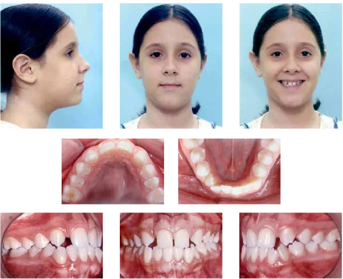
A Caucasian girl aged 10 years and 4 months complained about her teeth esthetics because of the diastemas in the maxillary incisor re-gion, according to herself and her parents. She reported good health except for occasional episodes of allergic rhinitis. The examination of family history showed cases of mandibular prognathism, and the family expressed their concern about the possible genetic transmis-sion of this facial dysplasia.
diAGnoSiS
Facial examination showed a mesocephalic pattern, no evident asymmetries, a straight pro-file, increased nasolabial angle and discrete upper lip retrusion. Analysis of the smile showed broad buccal corridors, a result of transverse deficiency in the maxilla, as well as a low smile line with re-duced exposure of anterior teeth, a greatly rele-vant aspect in orthodontic treatment considering the patient’s age. Intraoral examination showed low risk of caries and healthy periodontal tissues.
* Case Report, category 5, approved by the Brazilian Board of Orthodontics and Dentofacial Orthopedics. (BBO). How to cite this article: Franco FCM. Angle Class I malocclusion and
agen-esis of lateral incisors. Dental Press J Orthod. 2011 July-Aug;16(4):137-47.
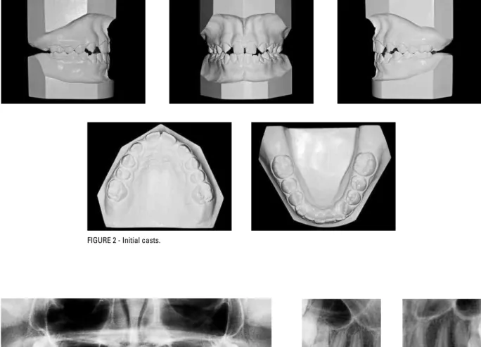
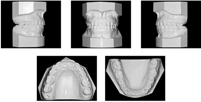
The patient presented Class I molar and ca-nine occlusion, missing maxillary lateral incisors, reduced overbite and overjet, which indicated a tendency to open bite and crossbite in the an-terior region. Additionally, the upper arch was triangular and atresic and there were spaces be-tween the anterior teeth. The lower arch was also narrow and followed the shape of the upper arch, with marked lingual inclination and crowding in the anterior region (Figs 1 and 2).
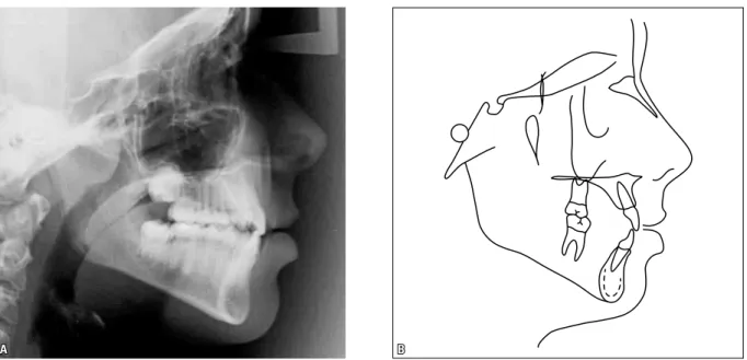
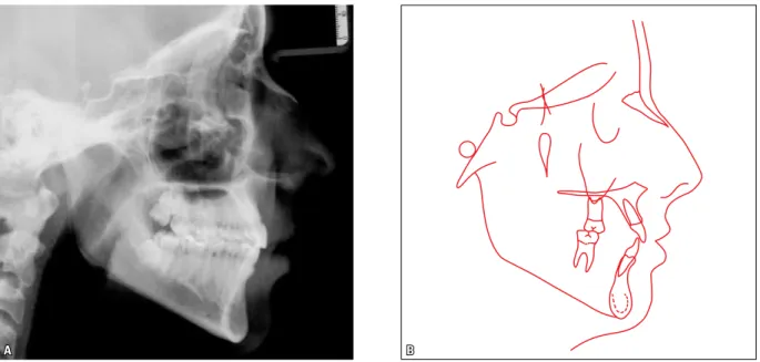
The examination of periapical and panoram-ic radiographs confirmed agenesis of maxillary lateral incisors and showed that the roots of sec-ond molars were in an advanced stage of forma-tion and that third molar germs were missing (Fig 3). Cephalometric analysis (Fig 4, Table 1) revealed a Class I (ANB = 2º) skeletal pattern, predominance of vertical growth (SN-GoGn = 40º) and lingual inclination of mandibular inci-sors (IMPA = 88º).
A B FIGURE 2 - Initial casts.
A B
trEAtMEnt oBJEctiVES
Considering that the deficiency in the anterior region of the maxilla was one of the aspects that most affected the patient’s smile esthetics, the main objective of the treatment was to improve arch shape. In addition to the esthetic benefits, palatal expansion would favor the axial adjust-ment of mandibular teeth and improve crowd-ing in that region. We decided to close the spaces that corresponded to the missing maxillary lat-eral incisors by moving the canines and poste-rior teeth mesially. Another treatment objective was to restore the correct levels of overbite and overjet to achieve a better anterior positioning of the maxilla, control the tendency towards man-dibular prognathism and, therefore, establish the harmony of facial and smile esthetics.
trEAtMEnt PLAn
A comprehensive treatment was planned and included fixed orthodontic apliances in the maxil-lary and mandibular arches after palatal expansion
FIGURE 4 - Initial lateral cephalometric radiograph (A) and cephalometric tracing (B).
using a Hyrax expander associated with maxil-lary protraction using a facial mask. The purpose of the planned treatment was to increase the transverse dimension and to move the maxilla anteriorly to manage the tendency to crossbite and to define proper overjet.
First, the palatal appliance would be installed. On the second week of activation, the extra-oral reverse traction (ERT) appliance would be placed. After expansion, during the six-month retention for the maxillary arch, the ERT would be kept in use, the fixed orthodontic appliance for the mandibular arch would be placed and mandibular alignment and leveling would begin. After the expander was removed, the maxillary fixed orthodontic appliance would be installed. To avoid the tendency towards anterior cross-bite, space closure in the maxillary arch would use extraoral anchorage.
and torques according to esthetic and functional needs in cases of agenesis of lateral incisors. A removable wraparound retainer would by pre-scribed for the maxillary arch, and for the man-dibular arch, a fixed retainer with a 0.028-in stainless steel intercanine wire.
trEAtMEnt ProGrESSion
The Hyrax palatal expander was placed, and directions were given for two daily activations (1/4 of a turn of the screw every 12 hours) for three weeks. The patient experienced intense pain five days later, and instructions were given to reduce activations to once a day. The ERT appliance was placed on the second week; the initial force was 350 g on each side and progres-sively increased up to 500 g, which was kept after expansion. Traction direction was adjusted according to the center of resistance of the max-illary bone to avoid unwanted maxmax-illary rota-tions. Instructions were given to wear the ERT appliance for at least 14 hours per day. Because of the reduction in the number of activations, expansion lasted four weeks; significant opening of the interincisal diastema was achieved, and overjet increased 3 mm.
After active expansion, a fixed orthodontic appliance with a 0.022x0.028-in slot was placed in the mandibular arch for its alignment and lev-eling. Palatal expansion allowed uprighting of the mandibular teeth, which had a lingual inclination in the beginning of the treatment. This move-ment promoted a gain in arch perimeter and re-solved crowding in the anterior region.
The fixed orthodontic appliance was placed in the maxillary arch six months after retention and when radiographs confirmed suture ossifica-tion. After incisor alignment and leveling, the an-terior spaces were closed as a result of the mesial movement of canines, which occupied the space corresponding to the missing lateral incisors. To avoid occlusal interferences, canines were re-shaped, their torques were individualized, and
their anatomic shape was that of the missing lateral incisors. The remaining spaces were closed by moving the posterior teeth mesially by using the ERT appliance as anchorage. This movement began with the first molar and fol-lowed, subsequently, up to the second molar. During finishing, the gingival margins of ante-rior teeth were adjusted to meet esthetic re-quirements in this area. The archwires for the final stage were made to have individualized torques for the canines in the sites of the miss-ing teeth and for the premolars that replaced the canines. Posterior teeth were positioned to promote posterior guidance by mandibular ex-cursion. Results were maintained by using an upper arch wraparound removable retainer and a fixed intercanine retainer for the lower arch.
rESuLtS
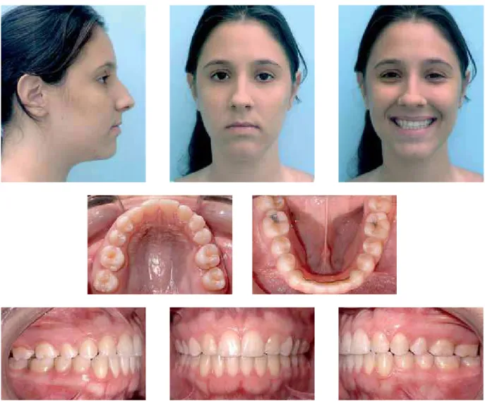
The trend towards anterior crossbite was con-trolled by the action of the ERT appliance. Even during space closure by movement of the posteri-or teeth mesially, there was no direct application of force on the anterior teeth, which preserved their position and avoided unwanted effects on occlusion and profile. The effects of reverse trac-tion mechanics were confirmed by cephalometry, which showed the maintenance of the maxillary position both in absolute values (initial and final
SNA equal to 76º) and in total superimposition (Figs 8 and 9).
Final occlusion had a good relationship be-tween arches and a Class II molar relationship, and normal occlusion of mandibular canines and maxillary first premolars, as well as nor-mal overbite and overjet. These results ensure functional balance and mandibular excursion without interferences, as well as healthy peri-odontal tissues.
A B FIGURE 6 - Final casts.
A B
A B
FIGURE 8 - Final lateral cephalometric radiograph (A) and cephalometric tracing (B).
MEASUREMENTS Normal A B A/B DIFFERENCE
Skeletal Pattern
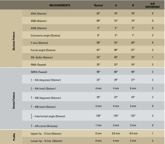
SNA(Steiner) 82° 76° 76° 0
SNB(Steiner) 80° 74° 74° 0
ANB(Steiner) 2° 2° 2° 0
Convexity angle(Downs) 0° 3° 1° 2
Y axis(Downs) 59° 52° 60° 8
Facial angle(Downs) 87° 90° 87° 3
SN–GoGn(Steiner) 32° 40° 39° 1
FMA(Tweed) 25° 22° 24° 2
Dental Pattern
IMPA(Tweed) 90° 88° 90° 2
–
1 – NA (degrees)(Steiner) 22° 29° 27° 2
–
1 – NA (mm)(Steiner) 4 mm 4 mm 6 mm 2
–
1 – NB (degrees)(Steiner) 25° 22° 24° 2
–
1 – NB (mm)(Steiner) 4 mm 4 mm 4 mm 0
– 1
1 – Interincisal angle(Downs) 130° 135° 132° 3
–
1 – APo (mm)(Ricketts) 1 mm 3 mm 3 mm 0
Proile
Upper lip – S line(Steiner) 0 mm 0.5 mm -0.5 mm 1
Lower lip – S line (Steiner) 0 mm 4 mm 2 mm 2
TABLE 1 - Summary of cephalometric measurements.
FinAL conSidErAtionS
Orthodontic treatment of lateral incisors agen-esis has been widely discussed and documented in the literature. The main treatment options are space closure or the maintenance of that space for future implant placement.1,5,7,9,10 In planning and treatment, orthodontic space closure may be ei-ther indicated or contraindicated depending on the
primarily based on the fact that teeth change posi-tions as the individual grows. That growth is not followed by osseointegrated implants, which, moreover, have effects that are similar to that of ankylosed teeth.4,6,8 In addition to that, in mid-dle- and long-term studies about the position of implants in the anterior region of the max-illa, there was progressive loss of marginal bone support in the buccal surface, gingival retractions and exposure of the implant margin.2,7,11,12 The patient and her guardians also received explana-tions about the implicaexplana-tions of the increase of treatment time and the greater need for collabo-ration. Another factor that played a role in treat-ment choice was the anatomy of the patient’s maxillary canines, which were light colored and not very large. These factors favor reshaping and the positioning of these teeth as lateral incisors. To change the shape of the canines, a combina-tion of abrasion and composite resin restoracombina-tion was used. The palatal surface was also adjusted to avoid premature contacts with maxillary lat-eral incisors, which ensured the correct axial
positioning of the teeth and the establishment of occlusion without interference.4 Moreover, in treatment finishing, canine torque was corrected to be similar to lateral incisor torque, and ideal torques for the first and second premolars were included.4,10 The esthetic result might have been better if the first premolar had been intruded to obtain a greater gingival margin height and to ensure a longer premolar crown, whose shape re-sembled that of the canines more closely.4 How-ever, the restorations of the first premolars were rejected by the patient and her guardians after the evaluation of cost and benefits and long-term restoration results.
contact address
Fernanda Catharino Menezes Franco Av. ACM, 585, sl. 1307 – Itaigara CEP: 41.850-000 – Salvador/BA, Brazil E-mail: f.catharino@uol.com.br rEFErEncES
Submitted: May 27, 2011 Revised and accepted: July 5, 2011 1. Al-Anezi SA. Orthodontic treatment for a patient with
hypodontia involving the maxillary lateral incisors. Am J Orthod Dentofacial Orthop. 2011;139(5):690-7. 2. Chang M, Wennström JL, Odman P, Andersson B.
Implant supported single-tooth replacements compared to contralateral natural teeth. Crown and soft tissue dimensions. Clin Oral Implants Res. 1999;10(3):185-94. 3. Cozzani M, Lombardo L, Gracco A. Class III malocclusion
with missing maxillary lateral incisors. Am J Orthod Dentofacial Orthop. 2011;139(3):388-96.
4. Iseri H, Solow B. Continued eruption of maxillary incisors and irst molars in girls from 9 to 25 years, studied by the implant method. Eur J Orthod. 1996;18(3):245-56. 5. Kokich V. Maxillary lateral incisor implants: planning with
the aid of orthodontics. J Oral Maxillofac Surg. 2004;62(9 Suppl 2):48-56.
6. Oesterle LJ, Cronin RJ. Adult growth, aging, and the single-tooth implant. Int J Oral Maxillofac Implants. 2000;15(2):252-60.
7. Robertsson S, Mohlin B. The congenitally missing upper lateral incisor. A retrospective study of orthodontic space closure versus restorative treatment. Eur J Orthod. 2000;22(6):697-710.
8. Rosa M, Zachrisson BU. Integrating esthetic dentistry and space closure in patients with missing maxillary lateral incisors. J Clin Orthod. 2001;35(4):221-34.
9. Schwaninger B, Shaye R. Management of cases with upper incisors missing. Am J Orthod. 1977;71(4):396-405. 10. Suguino R, Furquim LZ. Uma abordagem estética e funcional
do tratamento ortodôntico em pacientes com agenesias de incisivos laterais superiores. Rev Dental Press Ortod Ortop Facial. 2002;8(6):119-57.
11. Thilander B, Odman J, Lekholm U. Orthodontic aspects of the use of oral implants in adolescents: a 10-year follow-up study. Eur J Orthod. 2001;23(6):715-31.