Agreement among orthodontists
regarding facial pattern diagnosis
Sílvia Augusta Braga Reis*, Jorge Abrão**, Cristiane Aparecida Assis Claro***, Renata Ferraz Fornazari****, Leopoldino Capelozza Filho*****
Objective: To assess agreement among orthodontists trained in facial pattern diagnosis
through the morphological evaluation of the face. Methods: Facial photographs were taken in front and side views, as well as photographs of the smiles of 105 individuals randomly se-lected among patients seeking orthodontic treatment. The photographs were sent to ortho-dontists trained in facial pattern classification. Intra-rater agreement, agreement between raters and the Gold Standard, as well as inter-rater agreement were assessed using the Kappa index. Results: Intra-rater agreement was almost perfect, with Kappa index reaching 0.85. Agreement between raters and the Gold Standard was moderate (Kappa = 0.48), higher for Pattern I (Kappa = 0.62) and lower for the Short Face Pattern (Kappa = 0.33). Agreement between raters was significant (Kappa = 0.61) and even higher than agreement with the
Gold Standard for all patterns. Conclusions: The criteria used by raters to determine the
facial pattern were the same in the first and second evaluation. Agreement between raters and the Gold Standard was moderate, with raters exhibiting greater agreement between them than with the Gold Standard.
Abstract
Keywords: Orthodontic diagnosis. Facial analysis. Facial pattern.
* PhD in Orthodontics, University of São Paulo (USP - São Paulo, Brazil). Master in Orthodontics, Methodist University, São Paulo. Specialist
in Orthodontics Prois-USP, Bauru.
** Full Professor, School of Dentistry, University of São Paulo. *** PhD in Orthodontics, USP - São Paulo.
**** Specialist in Orthodontics, Prois-USP, Bauru.
***** PhD and Professor at the University of São Paulo – Bauru. Coordinator of the Specialization Course in Orthodontics, Prois–USP, Bauru.
» The authors report no commercial, proprietary, or inancial interest in the
products or companies described in this article. How to cite this article: Reis SAB, Abrão J, Claro CAA, Fornazari RF,
intROduCtiOn
Disagreement on the diagnosis of orthodontic problems is still very prevalent among orthodon-tists10 despite efforts to standardize the
classifica-tion of malocclusions.
Initially, Angle3 classified malocclusions
ac-cording to the mesiodistal relationship of first mo-lars. After studying the quality of cases finished by
North American orthodontists, Andrews2 noted a
lack of standardization among different practitio-ners and designed the six keys to a normal occlu-sion, which would serve as a guide to diagnose and assess the quality of finished cases.
These classifications were based, however, only on dental relationships and would eventu-ally prove insufficient to define the diagnosis of sagittal and vertical facial discrepancies.
In an attempt to create a diagnosis method supported by evidence-based treatment plans, Ackerman and Proffit1 developed a classification
system based on five key malocclusion character-istics: Facial profile, alignment, vertical, horizontal and sagittal deviations. However, little agreement was reached among professionals who teach this classification, and consequently on the treatment strategy indicated by each professional.10
Driven by the same concern, to organize a di-agnostic method supported by protocols and ca-pable of providing specific predictions, Capelozza Filho6 developed a classification system for
orth-odontic problems based on facial morphology. According to this classification, the morphologi-cal analysis of the face is the main diagnostic tool for facial pattern determination.
Organizing orthodontic diagnosis according to facial patterns allows orthodontists to treat mal-occlusions based on the location of skeletal dis-crepancies if present, or the etiology of the mal-occlusion, establishing treatment protocols that are tailored specifically to each pattern in each age group, with short-term protocols and predict-able long-term prospects by taking into account discrepancy severity.
The benefit of this new approach to orthodon-tics begins, however, with appropriate facial pat-tern diagnosis.
Considering the limitation of facial measures
in expressing form or normality,14 pattern
clas-sification should be performed by means of an evaluation of facial morphology in front and side views.
Individuals can be classified as Pattern I, II, III, Long Face or Short Face. Pattern I consists of a normal face. Whenever the malocclusion is pres-ent it is just a dpres-ental malocclusion not associated with any sagittal or vertical skeletal discrepancy. Patterns II and III are characterized, respectively, by a positive and negative sagittal discrepancy be-tween maxilla and mandible. The Long Face and Short Face patterns feature vertical discrepancies. In patients with skeletal errors, malocclusions usually result from these discrepancies.
The use of concepts that have been long estab-lished in the orthodontic literature, applied under this new perspective, imparts greater predictabil-ity to orthodontic treatment and assurance to professionals who learn to use such concepts, in addition to patient security and comfort.
The aim of this study was to evaluate agree-ment among orthodontists trained on facial pat-tern determination through the morphological evaluation of the face.
MAteRiAl And MethOds
The study sample consisted of 105 adults of both genders, aged at least 18 years, selected from the records of offices belonging to specialists in orthodontics. The authors randomly selected pa-tients with the five following facial patterns: Pat-tern I, PatPat-tern II, PatPat-tern III, Long Face PatPat-tern and Short Face Pattern. Individuals who were still in the growth period and those with craniofacial syndromes were excluded from the sample.
Standardized facial photographs in front and side views, as well as photographs of each pa-tient’s smile, were taken from their initial orth-odontic records for use in the study. The photo-graphs were inserted in a PowerPoint presenta-tion, with the photographs of the smile of each patient and of their face in front and side views on the same slide.
The rater sample consisted of 20 orthodontists trained in facial pattern classification. Most were teachers who taught this classification at different institutions.
Each rater received by mail a CD containing a presentation with 105 slides, each comprising the photographs in front and side views, along with the photograph of the smile of the same patient.
Raters were instructed to classify the facial pat-tern of each patient according to the following clas-sification: (1) Pattern I, (2) Pattern II, (3) Pattern III, (4) Long Face Pattern, and (5) Short Face Pattern.
The sample was also assessed by the author
who developed the classification method,6 whose
evaluation was set as the Gold Standard.
Thirty-five patients were randomly selected and subjected to the same classification with at least one week interval between the two evalu-ations with the purpose of assessing intra-rater agreement in terms of profile morphology.
Kappa coefficient, which evaluates agreement between two non-parametric variables, was used to analyze evaluation error.
Determining the value that defines a good cor-relation is inherent in each study, but in general, the following classification is acceptable:8
» If Kappa < 0.0 = Poor agreement;
» If 0.00 < Kappa < 0.20 = Slight agreement; » If 0.21 < Kappa < 0.40 = Fair agreement; » If 0.41 < Kappa < 0.60 = Moderate agreement; » If 0.61 < Kappa < 0.80 = Substantial agreement; » If 0.81 < Kappa < 1.0 = Almost perfect agreement.
Kappa coefficient was used in the total sample and in the different patterns in order to assess agree-ment between raters, as well as between raters and the Gold Standard.
Results
Eighteen of the 20 raters performed sample classification. As regards intra-rater assessment, the mean percentage of correct classifications found between the first and second evaluations was 84%. Kappa coefficient was 0.8 and showed substantial agreement between the first and second evaluation (Table 1). However, while most raters exhibited higher than 80% correct classifications between the two evaluations, two of them showed significantly lower agreement (60%, with Kappa = 0.5 - Moderate agreement) and were excluded from the sample, since these raters either did not master the method or did not implement it with due attention. The study was therefore conducted with 16 raters and the Gold Standard. Intra-rater agreement among the 16 raters was almost perfect, with Kappa index reaching 0.85 (Table 1).
Kappa coefficient was used in the total sam-ple and in the different patterns in order to as-sess agreement between raters and the Gold Standard. The percentage of agreement was 59% with Kappa = 0.48 (moderate agreement).
TABLE 1 - Intra-rater agreement between first and second evaluations with 18 and 16 raters.
TABLE 2 - Agreement between raters and the Gold Standard.
p(a) = percentage of correct classification, p(e) = percentage of error. p(a) = percentage of correct classifications, p(e) = percentage of error.
Number of
raters p(a) p(e) Kappa Index
18 raters 84% 16% 0.8
16 raters 88% 12% 0.85
Pattern (Gold Standard)n p(a) p(e) Kappa Index
I 27 70% 30% 0.62
II 37 56.5% 43.5% 0.46
III 17 53% 47% 0.41
Long Face 14 59% 41% 0.49
Short face 10 47% 53% 0.33
TABLE 3 - Assessment of inter-rater agreement.
p(a) = percentage of correct classifications, p(e) = percentage of error.
For the different patterns the percentage of agree-ment with the Gold Standard was: 70% for Pattern I (Kappa = 0.62), 56.5% for Pattern II (Kappa = 0.46) and 53% for Pattern III (Kappa = 0.41), 59% for the Long Face Pattern (Kappa = 0.49) and 47% for the Short Face Pattern (Kappa = 0.33) (Table 2).
Kappa index was also employed to assess agree-ment between raters. A 72% percentage of correct classifications was found for the total sample, with Kappa index = 0.65 (substantial agreement). For Pattern I this percentage was 77% (Kappa = 0.71); for Pattern II, 76% (Kappa = 0.7); for Pattern III, 69% (Kappa = 0.61), for the Long Face Pattern, 71% (Kappa = 0.64) and for the Short Face Pat-tern, 61% (Kappa = 0.51) (Table 3).
disCussiOn
Different diagnoses by different orthodontists for the same patient result in different treatment plans and outcomes which are not always
com-patible.9 Lack of agreement among professionals
instills great uncertainty in patients, tarnishing the scientific credibility of orthodontics, a dental specialty whose literature is replete with studies that allow evidence-based practice.
Diagnoses based on occlusal classification and cephalometric landmarks lead to results that of-ten fail to meet the esthetic expectations of
pa-tients.11 Above and beyond seeking an ideal
oc-clusal relationship one should seek the best pos-sible esthetics.15 To this end, diagnoses should be
based primarily on the evaluation of facial mor-phology in front and lateral views, and evaluation of the smile, complemented by an assessment of the occlusion, whose discrepancy is often a con-sequence of skeletal error. Radiographs are also important complementary exams but 75% of orthodontic diagnoses are defined without radio-graphic evaluation and tend to remain unchanged after orthodontists have reviewed the X-rays.4
A diagnosis based on facial morphology con-stitutes the first step in facial pattern classifica-tion. By means of a correct diagnosis one can identify, in different age groups, treatment pro-tocols substantiated by scientific evidence and, therefore, with well established short and long term prognoses.
After three years of clinical application of this concept, the authors decided to evaluate the de-gree of ade-greement of trained orthodontists who teach this methodology.
The study sample consisted of Brazilian adults past the growth period. Adults were intentionally chosen owing to the fact that facial pattern can only be established after complete facial growth. Some patterns, such as Long Face and Pattern III, tend to worsen during adolescence and are often identified only during that period. The inclusion of growing patients in this group might raise di-agnostic issues related to the complete expression of each pattern.
Furthermore, only Caucasian individuals were included since the concept of normal fa-cial features is specific to each population.5 For
Caucasians, a normal facial profile, or Pattern I, should display a smooth convexity. In Asians, a less convex facial profile is accepted as charac-teristic of normality. In Afro-descendants, on the other hand, an increased vertical growth results in increased facial convexity in Pattern I patients.
Sample raters were selected for their specific training in this classification and for teaching it at different institutions. The photographs were sent to 20 of them and 18 completed the questionnaire. Pattern (Gold Standard)n p(a) p(e) Kappa Index
I 27 77% 23% 0.71
II 37 76% 24% 0.7
III 17 69% 31% 0.61
Long Face 14 71% 29% 0.64
Short face 10 61% 39% 0.51
The percentage of agreement found between the first and second assessment was 84%, with Kappa = 0.8, showing substantial agreement, i.e., high consistency among raters regarding facial pattern diagnosis. This result indicates that the criteria for determining the pattern was the same in both studied periods. All raters achieved Kappa index-es for the two evaluations that showed substantial or almost perfect agreement, with the exception of two raters for whom the Kappa index yielded only moderate correlation between the two as-sessments. Due to the vast difference between these two raters and the others it was decided that they should be excluded, either because they did not master the classification method, or because they failed to carry out the assessments with the required attention. After this exclusion, the gen-eral intra-rater agreement changed from substan-tial to almost perfect (Kappa = 0.85) (Table 1).
The diagnosis performed by the raters was then compared with the Gold Standard, whose agreement was moderate (Kappa = 0.48).
Patients classified as Pattern I are character-ized by vertical and sagittal facial balance in front and side views. They are not necessarily beautiful, but sagittally and vertically balanced.
After analyzing separately the different indi-viduals and their facial patterns it was noted that Pattern I had the highest percentage of correct classifications. Seventy percent of the 27 individ-uals classified as Pattern I using the Gold Stan-dard as reference received the same rating by the raters (Kappa = 0.62 - substantial agreement).
Eleven percent of Pattern I individuals were clas-sified as Pattern III, and another 11.3% as Long Face Pattern. Six percent were mistaken for Pat-tern II and 1.6% for Short Face PatPat-tern (Table 4). Among the 27 Pattern I patients, only 2 (7.4%) were rated by most raters as Long Face Pattern. All others who showed discrepancies in classifica-tion were correctly assessed by most raters.
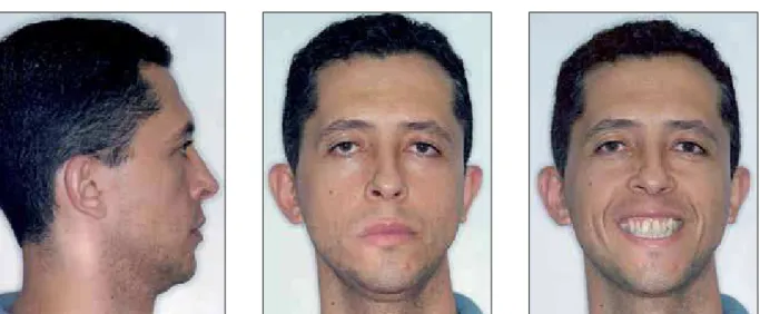
After a careful study of the sample, it was observed that all individuals rated as Long Face Pattern by the raters were dolichofacial. Note-worthy, however, were the presence of lip seal competence and the absence of excessive expo-sure of the upper incisors at rest, or of the gingiva when smiling, essential features for diagnosing the Long Face Pattern. This means that even in individuals with excessive vertical growth whose facial esthetics was compromised, normality may be present indicating that there is sagittal balance between the maxilla and mandible, lip seal com-petence and no excessive vertical growth of the maxilla (Figs 1 and 2). In dolichofacial Pattern I patients, only orthodontic treatment is indicated (Fig 3). For individuals with Long Face Pattern, on the other hand, orthodontic treatment can balance the occlusal relationships but it does not correct the skeletal discrepancy, nor does it ben-efit facial esthetics, which could only be achieved through a combination of orthognathic surgery and orthodontics.
In Pattern I patients who were classified as Pat-tern III, an expressive mandible can be observed in front and side views, although in balance with the maxilla. Among those rated as Pattern II there was again the influence of an increased vertical dimension, which enhances facial convexity. Once again it should be stressed that in Pattern I patients, maxillomandibular relationships are balanced and, therefore, no sagittal or vertical changes can be made.
Among the patients in the sample, 37 were classified as Pattern II according to the Gold Standard. This number is not surprising since Pattern I II III Long Face Short Face Total
I 69.8% 6.2% 11.1% 11.3% 1.6% 100%
II 30.5% 56.5% 0.5% 6.4% 6.1% 100%
III 30.5% 0.7% 53% 11.8% 4.0% 100%
Long Face 10.7% 26.8% 3.1% 59.4% 0% 100%
Short Face 23.7% 11.8% 17.5% 0% 47% 100%
FIGURE 1 - Pattern I, dolichofacial patient diagnosed as Long Face Pattern by 31.25% of raters. Lip seal competence and absence of vertical maxillary excess exclude Long Face diagnosis.
FIGURE 2 - Morphological evaluation of lateral cephalogram confirms basal bone balance.
the sample was collected in orthodontic offices and Class II malocclusion is very common, affect-ing 42% of Brazilians adults,13 75% of whom also
exhibit skeletal discrepancies.12 Pattern II is
charac-terized by increased facial convexity without, how-ever, the presence of vertical discrepancy. Most of these patients present with mandibular deficiency in association (or not) with maxillary excess. Maxil-lary excess seldom occurs in isolation. Mandibular deficiency is characterized by a short chin-neck line,
deficiency in chin protrusion and lower lip ever-sion. Upper lip posture depends on the uprighting or protrusion of the upper incisors.
The major manifestations of Pattern II in fron-tal view are reduced height of the lower lip and chin, and lower lip eversion. Pattern II patients do not present with vertical maxillary excess or any deficiency in dental exposure when smiling. These are the key elements for differential diag-nosis of the Long Face and Short Face patterns, respectively. Such diagnosis may be elusive at times, mainly because most patients with these patterns display a Class II malocclusion.
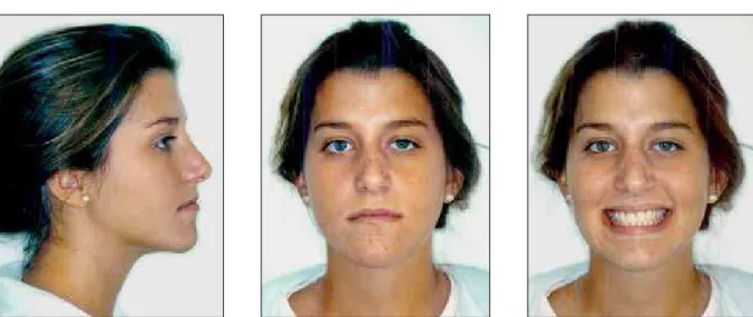
Among sample subjects with Pattern II, 56.5% were correctly classified by the evaluators (Kappa = 0.46, moderate agreement). The most frequent error was classifying individuals with mild Pattern II discrepancies as Pattern I, with raters tending to disregard the minor mandibular deficiencies (Figs 4 and 5). Thirty and half percent of Pattern II pa-tients were rated as Pattern I (Table 4).
Even mild discrepancies between the maxilla and mandible should be identified as treatment differs from that of Pattern I patients. Patients with mild Pattern II should be treated by means
of primary compensatory orthodontic treatment.6
The upper and lower incisors should normally FIGURE 4 - Pattern II patient rated by 75% of raters as Pattern I. Raters overlooked discrepancy given its small size.
preserve any compensations that might be
pres-ent, such as:Reduced buccal inclination of upper
incisors, absence of incisor and canine tip, and buccal inclination of lower incisors7 (Fig 5).
De-pending on Class II size, compensatory atresia is common in the maxilla and should be preserved. Non-identification of Pattern II may affect the appropriate choice of brackets and bonding re-sources and the preservation and acquisition of adequate compensation.
In this study raters only evaluated facial pho-tographs, which may have favored the error com-ponent. Complementary examination of lateral cephalograms assists in the identification of the skeletal discrepancies and dental compensations that should be preserved.
Six percent of Pattern II patients were classified as Long Face Pattern and another 6.5% as Short Face Pattern. Those rated as Long Face Pattern are the Pattern II dolichofacials, and as Short Face the Pattern II brachyfacials. Differential diagnosis must be made by identifying the treatment needs of each patient. When in doubt between Pattern II and Long Face Pattern, professionals must ask themselves if the patient requires upper replace-ment of the maxilla. If the answer is affirmative, the patient is Long Face, if negative he/she is tern II. For a differential diagnosis between Pat-tern II and Short Face the question is whether the maxilla needs to be repositioned inferiorly, and the patient is Short Face if the answer is positive.
Pattern III, in turn, is characterized by reduced facial convexity due to maxillary deficiency, man-dibular prognathism or a combination of both. In patients with maxillary deficiency, infraorbital depression and zygomatic prominence are miss-ing. The middle third is poor. The chin-neck line is long, less pronounced in prognathic dolichofacial patients. The differential diagnosis of these patients relative to the Long Face Pattern is determined by gingival exposure when smiling, which is normal in prognathic patients, who do not require upper replacement of the maxilla in orthognathic surgery.
Individuals with Short Face Pattern may also experience a reduction in facial convexity, which confuses them with Pattern III patients. Differ-ential diagnosis is once again performed accord-ing to the amount of exposure of upper incisors when smiling. A reduced exposure, indicative of the need for lower repositioning of the max-illa, determines the diagnosis of individuals with Short Face Pattern.
In frontal view, Pattern III patients are mainly characterized by excessive lower lip and chin height, and deficiency in the middle third of the face, which is expressionless. Mi-nor discrepancies are not perceived when eval-uated in frontal view, improving the prognosis of nonsurgical treatment.
Seventeen patients were classified as Pattern III according to the Gold Standard. The percent-age of correct classifications was 53%, with Kappa = 0.41 (moderate agreement). Once again, the worst confusion occurred between mild Pattern III and Pattern I individuals (Fig 6). Thirty and a half percent of Pattern III individuals were clas-sified as Pattern I. Lateral cephalograms should be evaluated in order to facilitate differential di-agnosis. Lateral cephalograms allow the identifi-cation of lower incisor uprighting, upper incisor protrusion and skeletal discrepancy (Fig 7). These characteristics should be preserved in the com-pensatory treatment of these patients, thereby influencing the choice of brackets.
Twelve percent of Pattern III patients were rated as Long Face Pattern. These patients, how-ever, are Pattern III dolichofacials and show no vertical maxillary excess.
correct classification for this Pattern was 59.4% (Kappa = 0.49, moderate agreement). Their fa-cial convexity is increased due to mandibular rotation downwards and backwards. Therefore, 26.8% of the patients with Long Face Pattern were classified as Pattern II (Table 3 and Fig. 8). In defining the differential diagnosis one must check the sealing of perioral muscles typically present in Pattern II individuals and
the amount of gingival exposure when smiling, which is normal in Pattern II patients.
In evaluating the profile of Long Face patients one should check gonial angle opening, long and narrow symphysis, increased distance between molar apex and palatal plane, and excessive ex-posure of upper incisor at rest (Fig 9).
When lip seal is present in the patients, it is forced and forms a double chin.
These patients are usually esthetically un-pleasant and require orthognathic surgery as-sociated with orthodontic treatment to restore facial balance and allow proper sealing of the perioral musculature. Surgical planning of these patients includes maxillary replacement and expansion, upward and forward rotation, mandibular advancement or setback, and verti-cal chin reduction.
In 11% of Long Face Pattern evaluations these patients were classified as Pattern I and 3% as Pat-tern III (Table 4).
When Long Face patients fail to be diagnosed as such, the use of unsuitable orthodontic me-chanics may lead to an increase in vertical dimen-sion and the worsening of this pattern, compro-mising facial esthetics and occlusal function (an-terior guidances) by reducing overbite.
FIGURE 6 - Pattern III patients diagnosed as Pattern I by 68.75% of raters. Height of lower lip and chin increased relative to upper lip height allows discrep-ancy identification even in front view photograph.
FIGURE 8 - Patient with Long Face Pattern diagnosed as Pattern II by 43.75% of raters. Vertical excess results in lip seal incompetence and maxillary and mandibular retrusion.
FIGURE 9 - Lateral cephalogram of patient in Figure 8. Opening of gonial angle, long and narrow symphysis, increased distance between molar apex and palatal plane, and excessive exposure of upper incisor at rest.
Only 47% of the evaluations of patients with this pattern were correct. Kappa was 0.33, indicating fair agreement between evaluations. Twenty-four percent of patients with this pattern were con-fused with Pattern I, 11.8% with Pattern II and 17.5% with Pattern III (Table 4). Individuals with Short Face Pattern may have slightly convex pro-file similarly to Pattern I, or a markedly convex profile, typical of Pattern II, or even reduced con-vexity, like Pattern III. This explains why there was such confusion in the evaluation of patients with this pattern (Figs 10, 11 and 12). Differential diagnosis is performed by studying photographs of smiles in which these patients expose very little of their upper incisors since childhood. Another important aspect is soft tissue excess, resulting in compressed lips and facial creases since youth. The impact of aging on this pattern is greater than on any other pattern. Ideally, the orthodon-tic treatment must be associated with surgery for lower replacement of the maxilla, downward and backward rotation with mandibular advancement or setback, and vertical increase of the chin.
When a patient opts for compensatory treat-ment, one should avoid procedures that reduce oral volume and vertical dimension, such as den-tal extractions. Orthodontic treatment of these Patients with Short Face Pattern exhibit small
vertical growth associated with upward and for-ward mandibular rotation. Their chin is usually prominent. There is little upper dental exposure when smiling even in young patients, and exces-sive lip compression at rest, indicating lower re-positioning of the maxilla as a mandatory proce-dure in orthognathic surgery for these patients.
FIGURE 10 - Patient with Short Face Pattern diagnosed as Pattern III by 56.25% of orthodontists. Reduced facial convexity is due to reduced vertical height of maxilla and forward and upward mandibular rotation.
FIGURE 12 - Facial photographs of patient in Figures 10 and 11 after increase in vertical dimension by Surgically Assisted Rapid Maxillary Expansion and provisional prosthetic rehabilitation. Short Face Pattern is still present as it can only be corrected with orthognathic surgery, but significant improvement was achieved in smile esthetics.
patients is usually limited to improving esthetics and establishing satisfactory occlusion.
Evaluation of inter-rater agreement showed a higher percentage of accurate classifications for the entire sample and for all patterns compared with the percentage between raters and the Gold Standard.
Agreement was substantial for the total sam-ple (Kappa = 0.65) and for Patterns I (Kappa = 0.71), II (Kappa = 0.7), III (Kappa = 0.61) and Long Face (Kappa = 0.64), and moderate for the Short Face Pattern (Kappa = 0.51). These results show that rater error tends to be in the same di-rection when classifying a patient in a pattern dif-ferent from the Gold Standard.
COnClusiOns
Identifying points of problem in the diagnosis of facial patterns makes it easier to correct po-tential errors that might result in inappropriate treatment plans and unrealistic prognoses.
An almost perfect agreement was noted be-tween the first and second evaluation by the same rater, which demonstrates that the criteria for di-agnosis of facial pattern are well established.
Agreement between rater evaluation and the Gold Standard was moderate. A tendency was detected whereby raters disregarded minor mandibular deficiencies in Pattern II, and mild
discrepancies in Pattern III. Individuals with Long Face Pattern were often diagnosed as Pattern II due to increased facial convexity resulting from downward and backward mandibular rotation.
Short Face Pattern showed less agreement and these individuals were classified as Pattern I, II or III.
Agreement between raters in evaluating pat-terns was substantial, showing that raters tended to classify individuals under the same pattern, which was different from the Gold Standard. In other words, errors tend to be committed in the same patients and in the same direction.
ACKnOwledgeMents
Contact address Sílvia Augusta Braga Reis Rua Timbiras, 1560, conj. 308
CEP: 30.140-061 – Belo Horizonte / MG, Brazil E-mail: silviabreis@hotmail.com
RefeRenCes
Submitted: March 06, 2008
Revised and accepted: February 25, 2009 1. Ackerman JL, Profit WR. The characteristics of malocclusion:
a modern approach to classiication and diagnosis. Am J
Orthod. 1969;56:443-54.
2. Andrews LF. Straight wire: o conceito e o aparelho. San
Diego: L. A. Wells; 1989. 407 p.
3. Angle EH. Classiication of malocclusion. Dent Cosmos.
1899;41(2):248-357.
4. Atchison KA, Luke LS, White SC. Contribution of
pretreatment radiographs to orthodontists´ decision making. Oral Surg Oral Med Oral Pathol. 1991;71(2):238-45. 5. Borman H, Ozgur F, Gursu G. Evaluation of soft: tissue
morphology of the face in 1,050 young adults. Ann Plast Surg. 1999;42(3):280-8.
6. Capelozza Filho L. Diagnóstico em Ortodontia. Maringá: Dental Press; 2004.
7. Capelozza Filho L, Silva Filho OG, Ozawa TO, Cavassan AO.
Individualização de braquetes na técnica de Straight Wire:
revisão de conceitos e sugestão de indicações para uso. Rev Dental Press Ortod Ortop Facial. 1999;4(4):87-106. 8. Landis JR, Koch GG. The measurement of observer agreement
for categorical data. Biometrics. 1977;33(1):159-74. 9. Lee R, Macfarlane T, O´Brien K. Consistency of orthodontic
treatment planning decisions. Clin Orthod Res. 1999;2:79-84.
10. Luke LS, Atchison KA, White SC. Consistency of patient classiication in orthodontic diagnosis and treatment
planning. Angle Orthod. 1998;68(6):513-20.
11. Michiels G, Sather AH. Validity and reliability of facial proile
evaluation in vertical and horizontal dimensions from lateral cephalograms and lateral photographs. Int J Adult Orthodon Orthognath Surg. 1994;9(1):43-54.
12. Milacic M, Markovic M. A comparative occlusal and cephalometric study of dental and skeletal anteroposterior relationships. Br J Orthod. 1983;10(1):53-4.
13. Reis SAB, Capelozza Filho L, Mandetta S. Prevalência de oclusão normal e má oclusão em brasileiros, adultos,
leucodermas, caracterizados pela normalidade do peril
facial. Rev Dental Press Ortod Ortop Facial. 2002;7(5):17-25. 14. Reis SAB, Abrão J, Capelozza Filho L, Claro CAA. Análise
facial numérica do peril de brasileiros Padrão I. Rev Dental
Press Ortod Ortop Facial. 2006;11(6):24-34.