An interview with
How to cite this section: Tanimoto K. An interview with Kotaro Tanimoto. Dental Press J Orthod. 2016 July-Aug;21(4):24-33. DOI: http://dx.doi.org/10.1590/2177-6709.21.4.024-033.int
Submitted: March 07, 2016 - Revised and accepted: April 22, 2016 E-mail: [email protected]
If I had to describe Tanimoto Sensei using a single word I would say kindness. When I was attending the PhD course in Hiroshima University, I frequently looked at him and thought: “If I have the chance to be an educa-tor, I would like to be like that man.” The generosity, respect and friendship he employed to treat the students is fascinating. By that time, Professor Tanimoto had just come back from his post-doc in the USA full of ideas and dreams. Since that, many of the ideas had become projects and many of the projects had become a real beneit for the patients. Tanimoto Sensei has an extensive and relevant scientiic production and some of them are described in the next pages. I believe this interview is especially interesting because it is enriching to know how professors from the other side of the globe conduct orthodontic education and science. Finally, I would like to express my sincere gratitude to the other professors that assisted in this article and to Dental Press Journal of Orthodontics for the op-portunity to conduct the present interview with Professor Tanimoto, a great friend of mine.
Emanuel Braga – interview coordinator » DDS, Hiroshima University, Japan.
» PhD in Orthodontics, Hiroshima University, Japan.
» Head of Department, Department of Orthodontics, Hiroshima University Institute of Biomedical & Health Sciences, Japan.
DOI: http://dx.doi.org/10.1590/2177-6709.21.4.024-033.int
Kotaro Tanimoto
Se eu tivesse que descrever Tanimoto Sensei em apenas uma palavra, eu diria “bondade”. Na época em que eu estava cursando o doutorado na Hiroshima University, com frequência eu olhava para ele e pensava: “Se eu tiver a opor-tunidade de me tornar um educador, eu gostaria de ser como aquele homem”. A generosidade, o respeito e a amizade com os quais ele tratava seus alunos eram fascinantes. Naquela época, o professor Tanimoto tinha acabado de voltar de seu pós-doutorado nos EUA, cheio de ideias e sonhos. Desde então, muitas de suas ideias se transformaram em projetos e muitos de seus projetos se tornaram benefícios para os pacientes. Tanimoto Sensei tem uma produção cientíica extensa e relevante, e alguns de seus trabalhos estão descritos nas próximas páginas. Acredito que esta entrevista seja muito interessante, pois é enriquecedor saber como professores do outro lado do mundo conduzem a formação e ciência ortodônticas. Por im, eu gostaria de expressar a minha sincera gratidão aos outros professores que contri-buíram para este texto e ao Dental Press Journal of Orthodontics, pela oportunidade de coordenar a presente entrevista com o professor Tanimoto, um grande amigo meu.
Taking Brazil location as a reference, Japan is placed exactly on the other side of the globe. I usually joke that if you are lying from Brazil to Japan and you miss the landing place, you are au-tomatically getting closer to Brazil again. Japan and Brazil are separated by a half planet and many diferences are undoubtedly expected. Regarding education, Japan has a brilliant experience to share and I think many Brazilians are interested in know-ing how dental education is performed there. Dear Tanimoto Sensei, could you please make a brief description about how orthodontic undergraduate education works in Hiroshima University?
Emanuel Braga
In Japan, undergraduate dental education is usually designed as a 6-year program. The program in Hiro-shima University School of Dentistry is characterized by several unique systems, such as the Two-Course Sys-tem, BioDental Education, and Dual-Linguistic Edu-cation. Currently our school has a 2-semester system, and the Orthodontics lecture is held for one year from the second semester of the 3rd grade to the irst semester
of the 4th grade (22 times total). In addition,
Stomato-gnathic Function and Periodontology regarding Ortho-dontics are also lectured. Thereater basic practice of Orthodontics is held in the second semester of 4th grade
(15 times total).
In Hiroshima University School of Dentistry, the Two-Course system, a quite unique system in Japanese dental schools, is given from the second se-mester in the 3rd grade to the first semester in the 5th
grade. Undergraduate students in the 3rd grade can
select “Course for Frontier Science” or “Course for Clinical Dental Science.” In the “Course for Fron-tier Science,” students learn basic science through investigation according to a topic given to each stu-dent. On the other hand, the “Course for Clinical Dental Science” has advanced clinical lectures and practices, such as Typodont practice in Orthodon-tics. Finally, 1-year clinical practice starts from the second semester of the 5th grade.
The undergraduate dental program in Hiroshima University has been continuously reorganized under the concept of BioDental Education and Research since 2010. BioDental Education is designed as a novel dental curriculum to train professionals with scientiic background capable of innovating the next generation
dental medicine through speciic educational pro-grams. The programs are accomplished according to attainment target type education, problem-based learning and research methodology. Scientiic knowl-edge is quite important to every dentist, and training of dental educators with high research background has been an urgent issue in many countries. To date, the nurturing of dental research has been performed mainly in graduate programs in Japan; however, the number of dental students willing to attend graduate courses is not high enough. In addition, few gradu-ates continue research ater completion of the graduate course. Due to this situation, we consider that the up-bringing of scientiic motivation needs to be integrated with undergraduate dental education.
Since another aspect of BioDental Education is to correspond to globalization, Hiroshima University School of Dentistry established the International Den-tal Course (IDC) in 2011. IDC accepts undergraduate dental students from partner universities (Airlangga University in Indonesia, University of Medicine and Pharmacy in Ho Chi Minh City in Vietnam, and Uni-versity of Health Sciences Cambodia in Cambodia in 2016). IDC students join our undergraduate programs from the 2nd grade to the irst semester of 5th grade
ater completion of their irst year education in their home countries. All lectures and practices during this period are given in both English and Japanese (Dual-Linguistic Education). The purpose of IDC is not only to provide English education, but also stimulate learn-ing motivation and speed up future communication among various countries.
During my PhD in Hiroshima University, I was amazed by how well balanced clinical training education and research work in the graduate pro-gram were. In my opinion, all students had the chance to become well-trained orthodontists and brilliant scientists. Could you please talk about the orthodontic PhD course in Hiroshima University? Emanuel Braga
lectures/practices and research practice. Graduate students have to complete all of them. The graduate orthodontic education is designed to complete the at-tainment targets for orthodontic specialist certiica-tion managed by the Japanese Orthodontic Society (JOS). The JOS guidelines for orthodontic specialist certiication require at least a 5-year orthodontic train-ing, consisting of a 2-year or 3-year basic training in a certiied training facility. At present, in Japan, there are 31 training facilities in 29 universities including our department. The orthodontic training program in each facility follows the JOS guidelines, and Bio-logical Science is included in the attainment targets. Therefore, the graduate course in our department pro-vides both orthodontic clinical and research training programs. The orthodontic clinical training consists of Typodont practice, cephalometric analysis, diagno-sis of temporomandibular disorders (TMD) by means of MRI, CT-based tridimensional analysis, caries risk examination, periodontal care, stomatognathic exami-nation, electromyogram examiexami-nation, diagnosis of re-spiratory and speech function, an so on. Applicants are required to contribute with more than 150 cases and inish at least 30 of them.
Furthermore, PhD students receive research training, such as research ethics, research planning, ex-periment techniques, grant application, management of research equipment and material, presentation, and writing. An individual research theme is given to each PhD student, and they have to complete their research to obtain a doctor’s degree. Current main research proj-ects in our department are (1) elucidation of pathogenic mechanism in malocclusion and management of oral maxillofacial skeletal growth; (2) optimization of tooth movement by orthodontic forces; (3) establishment of a new orthodontic treatment based on the image and biochemical diagnosis of temporomandibular disorders (TMD); (4) bone regeneration by use of mesenchymal stem cells and the development of regeneration medi-cine for closure of jaw clet; (5) elucidation of pathogen-esis of amelogenpathogen-esis imperfecta and the restoration of tooth enamel by application of biomineralization pro-cesses; (6) evaluation of the contribution of various oral functions to general itness; and (7) development of new instruments and material for orthodontic treatment.
Tight collaboration with other departments, such as Oral Pathology, Oral Biochemistry, Oral Anatomy, Biomaterial
Technology, and Bacteriology, enables us to correspond to various research ields from basic to clinical research.
In Hiroshima University, irst-year students have to come one hour earlier in order to clean and prepare the rooms for the other members of the Department. In the beginning, I found this task strange and exhausting, especially because PhD students in Brazil usually do not do that. When I became a second-year student, I realized that I was completely unable to throw any piece of garbage in the wrong place. I will never forget this experi-ence. What do you have to say for societies which do not have this sense of community as strong as in
Japan? Emanuel Braga
Since graduate students are qualiied professional dentists as well as students, they need to have common sense and manners as members of society. There is a Japanese proverb that says: “Start with a bow and end with a bow.” Although there are professional sweepers in our university, new members of the department have to clean the department rooms by themselves. As you have mentioned, it may be very diicult to understand sweepers’ work and have sincere gratitude for them un-less we experience the exact same work. In Japan, hav-ing common sense and manners as a member of society are generally reinforced in companies ater employ-ment; whereas there is no such opportunity for most dentists, especially for graduate students. Therefore, we intentionally give them such duties just for one year. This way may be the Japanese traditional style, but al-though there are various senses of values in our society, they are not understood everywhere in our country. Since there are various social systems with diferent manners and rules in diferent countries, I think there can be many ways of education.
Dear Professor Tanimoto, I am very interested in your studies about bone regeneration in cleft sites by means of mesenchymal stem cells and carbonated hydroxyapatite particles. What is the current status of this research and what is the protocol for bone grafting in cleft patients in Hi-roshima University?
Fernando Habib
clet palate. Secondary bone grat for patients with clet palate is usually performed around 8 to 10 years of age, just before canine eruption. Currently, nearly all patients with clet palate receive iliac bone grat. However, iliac bone grat, especially its classic large-incision, open techniques for bone grat harvesting from anterior and posterior parts of the iliac crest, is surgically invasive, leading to a large encumbrance for patients. Common donor site complications for the anterior ilium are pain, sensory loss, gait disturbance, infection, and hematoma formation. In addition, it is occasionally diicult to obtain enough volume for transplantation in young patients.
Tissue engineering with transplantation of mes-enchymal stem cells (MSCs) derived from bone mar-row is a possible candidate to achieve osteogenesis in bone defects and reduce the surgical invasiveness in-cident associated with the extraction of transplant. The bone marrow can be obtained by aspiration us-ing a bone marrow puncture needle with minimum surgical invasion compared with the separation of iliac bone. Although MSCs constitute only 0.001-0.01% of the subcellular components in bone marrow, cultured bone marrow-derived MSCs can proliferate with-out losing the potential to differentiate into multiple mesenchyme lineages, such as chondrocytes, adipo-cytes, and osteoblasts, with corresponding biological stimuli. Therefore, we started the study for regenera-tion medicine using bone marrow-derived MSCs for treatment of cleft palate.
Among many factors believed to be crucial for tissue regeneration, the scafold for alveolar bone regeneration in clet palate patients must possess unique properties. Under the general environment in the oral cavity, vari-ous mechanical forces, such as tongue pressure and biting force, are always loaded onto the transplanted area, lead-ing to deformation or leakage of transplants. Therefore, it is necessary for the scafold to have suicient strength against external forces without any coverage with hard material over the sot scafolds, such as collagen gel. For the treatment of bone defect of clet palate, it is es-sential to guide or move tooth into the regenerated bone area. It is thus required that the alveolar clet area ater regeneration is illed with new bone with normal me-tabolism crucial for tooth movement and subsequent maintenance of tooth. Based on these considerations, the scafolds for bone regeneration in clet palate patients
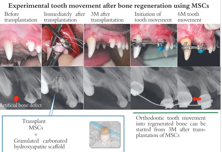
should be digested quickly. Carbonated-hydroxyapatite (CAP) is originally a major mineral constituent of bone and teeth. It is highly substituted with 3-5% carbonate ions instead of phosphate ions compared with the lattice of hydroxyapatite, leading to an unstable crystal structure with high solubility compared with that of hydroxyapa-tite. We examined bone regeneration with bone marrow-derived MSCs and CAP scafold for artiicially created alveolar clet in beagle dogs (Fig 1). The transplantation of MSCs with granulated CAP scafold induced bone re-generation in artiicially created alveolar clet and was fol-lowed by tooth movement to the regenerated bone with a consistent rate of tooth movement.
So far, the above results were obtained using an animal model. Currently, we are going to start clini-cal trials as the next step. The Translational Research Medical Center was established in the newly built Hi-roshima University Hospital outpatient clinic in 2013. The center has cell processing rooms and equipment for regeneration medicine. Continuous basic and clini-cal research is still indispensable to carry out bone re-generation medicine for clet palate in the future.
Professor, Hiroshima University has played a rele-vant role regarding basic research for regeneration medicine over the years. In this context, I would like to highlight your research using amelogenin, an enamel protein, which has proved efective for bone regeneration in several circumstances. How is this topic evolved? Is there any clinical use
al-ready established? Fernando Habib
Amelogenins are enamel matrix proteins that mainly contribute to tooth enamel formation. Such hard tissue formation by speciic proteins in organisms is generally called biomineralization. I conducted a study on biomineralization for enamel formation when I studied abroad at UCSF.
hydrophobic protein with high molecular weight. Con-sequently, it easily precipitates in solutions with neutral pH. That means that full-length amelogenin might not always exert intended biological activities in a vital body.
For this reason, we started to research critical ami-no acid sequences with physiological activity of full-length amelogenin. Since amelogenin is cleaved into small fragments by speciic proteases ater secretion from ameloblasts into enamel matrix, we speculated that some of these fragments with active domain re-main with biological functions. We examined the physiological activity of synthesized potential amelo-genin fragments and speciied a critical amino acid se-quence. The peptide consists of 11 amino acids derived from full-length amelogenin (AMG-peptide) which
exerts an activity almost equal to osteogenic diferen-tiation of MSCs, as compared to full-length amelo-genin. AMG-peptide can be industrially synthesized in bulk and easily dissolved in water due to high hy-drophilic properties. Meanwhile, we found a problem: AMG-peptide is quite easy to difuse in vital tissues ater administration, which suggests that it is diicult to exert its physiological activity sustainably. To solve this problem, we ixed AMG-peptide onto a solid sur-face to prevent difusion. We examined whether or not AMG-peptide activity still remains even ater ixation to a solid surface. AMG-peptide stably kept its activity. For clinical application, AMG-peptide can be ixed onto a scafold surface to enhance bone forma-tion, or support guided bone regeneration using an
Figure 1 -Experimental tooth movement after bone regeneration using MSCs (Source: Tanimoto et al,1 2015).
Experimental tooth movement ater bone regeneration using MSCs
Before
transplantation
Granulated carbonated
hydroxyapatite scafold
Orthodontic tooth movement
into regenerated bone can be
started from 3M ater
trans-plantation of MSCs
Transplant
MSCs
+
Artiicial bone defect
Immediately ater
transplantation
3M ater
transplantation
Initiation of
tooth movement
AMG-peptide-ixed barrier membrane. Although clinical application is currently not attained, AMG-peptide is expected as one of the potential candidates for safe and efective bone regeneration.
An interesting topic investigated at Hiroshima Uni-versity is cryopreservation. Professor, could you talk about cryopreservation and Orthodontics, especially about the beneits of cryopreservation
in surgical cases? Fernando Habib
We have studied about long-term cryopreserva-tion of teeth. This has already been made practical, and the teeth cryopreservation service has been pro-vided to patients. During the freezing process of vital tissue, ice crystals generated in the cell body fatally damage cells. Therefore, it is quite important to sup-press ice crystal formation and its growth, so as to minimize cell damage as much as possible. For this purpose, both temperature management to quickly pass the freezing point of water during the freezing process and the component of stock solution are criti-cal. Right now the optimal condition for tooth cryo-preservation is controlled by means of a specially pro-grammed freezer and stock solution. Cryopreserved teeth can be used for tooth transplantation in case the patient accidentally loses their teeth.
In addition, cryopreserved stem cells can be used for tissue regeneration. Currently, we are investigat-ing bone regeneration usinvestigat-ing dental pulp tissue-derived stem cells (DPSC) from permanent or primary teeth. DPSC can be easily obtained from extracted teeth for orthodontic treatment or by shedding primary teeth, and cryopreserved DPSC can be used for vari-ous purposes in the future. DPSC cryopreservation is expected to become a new medical service by conso-ciation with dental clinic.
Previous studies with the inite element method indicate that disc displacement could afect stress distribution on the condylar articular surface dur-ing prolonged clenchdur-ing. In adult patients with clenching and disc displacement, can occlusal plane splints be helpful? Is there any advantage
to use the splint at night only or all day long?
Márcio Lisboa
I greatly appreciate you for your question regard-ing our research and clinical procedure for
temporo-mandibular disorders (TMD). At our department, we occasionally use the stabilization-type splint for TMD patients with unstable occlusion due to malocclusion, so as to diagnose the presence of bruxism and its fre-quency and time lapse.
For manufacture and application of any type of occlusal splint, we follow the guidelines established by the Japanese Society for the Temporomandibular Joint. The stabilization-type splint is classified in up-per dentition type and lower dentition type, and we usually use the former. The stabilization-type splint can be used for equation of mechanical stress applied to the temporomandibular joint (TMJ) between the right and left side. In such cases, it is important to use the splint at the time when patients are clenching their teeth. If timing is unclear, patients are encour-aged to start splint therapy at night. The dentist in charge carefully checks for changes in clinical symp-toms and abrasion of splint surface; if no change can be observed, patients start using the splint during the day. Either way we prohibit patients to use the splint continuously for more than 15 hours, so as to prevent irreversible occlusal changes due to mandibular shift and/or tooth movement.
The articular disc appears to be crucial in the regeneration of a damaged condyle, suggest-ing that defects or damage to the articular disc may influence mandibular growth and regen-eration or repair of the condyle. In this con-text, teenagers with disc displacement with and without reduction may have problems with the TMJ and occlusion in adulthood? Márcio Lisboa
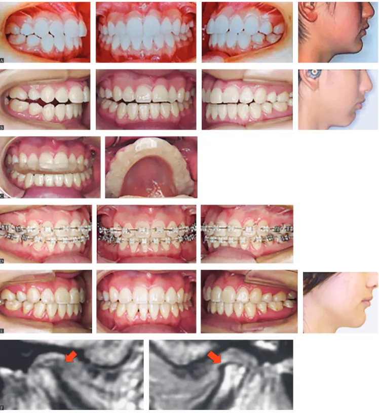
Disc displacement does not necessarily cause seri-ous adverse effects on mandibular condylar growth. However, TMJ osteoarthritis (TMJ -OA) in young patients occasionally causes severe changes in occlu-sion and facial configuration, such as skeletal open bite, mandibular shift, and mandibular retrusion. Figure 2shows a treatment case of open bite caused by TMJ-OA. Surgical orthodontic treatment is also frequently selected to treat these severe malocclu-sions. A sufficient observation period is indispens-able before initiation of orthodontic treatment, and close attention must be paid to pathologic status of TMD during treatment. Furthermore, a long-term retention period is desirable after orthodontic treat-ment. In our clinic, all orthodontic patients with TMD undergo TMJ-MRI examination. Since near-ly all patients with mandibular condylar resorption are accompanied by disc displacement, these find-ings seem to have some relation. Disc displacement might be no more than a symptom of degenerative TMJ disease, such as OA.
The Gnathology Society showed the role of occlusion in Dentistry. Nowadays, what is the importance of occlusion to TMD?
Márcio Lisboa
Occlusion is no more than one factor among var-ious causes of TMD, and its contribution to TMD is not so strong as previously believed. There are
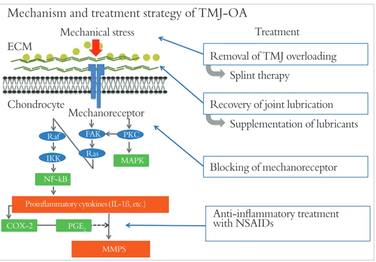
many severe malocclusion patients without TMD. So far, the prevalence of TMD in orthodontic pa-tients, at least in our clinic, is not very different from that of the general population. Major causes of OA and internal derangement are believed to be exces-sive forces applied to TMJ, whereas internal causes, such as changes in the property of synovial fluid, are also suggested.
Our previous study indicated that intra-articular inflammation induces degradation and decreased synthesis of lubricants in joint fluid, such as hyaluro-nan (HA) and superficial zone protein (SZP). Due to this process, homeostasis of synovial fluid com-ponents cannot be maintained, thereby resulting in deterioration of lubricating and buffering functions in the joint. Disc displacement can be induced un-der these pathological changes of intra-articular en-vironment. If this condition is kept in the long term, severe degeneration or resorption of articular carti-lage and subchondral bone are likely to be caused.
Figure 2 - A treatment case with open bite accompanied by osteoarthritis of the temporomandibular joint. A) Initiation of retention at a previous hospital. B ) Ini-tial arrival at our hospital. C) Initiation of splint therapy. D) Orthodontic treatment with multi-bracket appliances. E) Removal of all appliances. F) MRI indicated TMJ-osteoarthritis on both sides at initial arrival (Source: Tanimoto2, 2011).
A
C
E B
D
It is known that good orthodontic treatment outcomes lead to TMJ health. In your opinion, is it appropriate to treat orthodontically pa-tients with TMD muscle or articular
symptom-atology?Márcio Lisboa
The important thing for establishing good TMD treatment goals is not occlusion itself, but to man-age present disharmony of masticatory muscles and overload onto the TMJ. Outer forces enough to have a bad influence over TMJ hard-tissue re-modeling as well as internal causes would become a trigger of pathogenesis. Therefore, we attempt to carefully examine TMJ pathology and dysfunction before starting orthodontic treatment, but never plan it for TMD treatment as a main purpose. If any
symptom of TMD is observed in orthodontic pa-tients, differential diagnosis for TMD is performed after careful examination. According to the Japanese Society for the Temporomandibular Joint, TMD is roughly classified into four types: disorders by mas-ticatory muscle pain, disorders by TMJ pain, disor-ders by disc displacement, and OA. Management of TMD is determined according to each classification. Orthodontic treatment is never initiated if patients have acute symptoms or progressive pathology of TMJ. Adequate initial TMD treatment followed by a sufficient observation period is usually considered in advance. Particularly, if TMD patients have oral habits, such as bruxism, it would also affect the sta-bility of occlusion after orthodontic treatment.
Mechanism and treatment strategy of TMJ-OA
Mechanical stress
Treatment
ECM
Chondrocyte
Raf
IKK
COX-2
NF-kB
Proinlammatory cytokines (IL-1ß, etc.) FAK
Ras
PGE2
MMPS PKC
MAPK
Mechanoreceptor
Splint therapy
Removal of TMJ overloading
Recovery of joint lubrication
Supplementation of lubricants
Blocking of mechanoreceptor
Anti-inlammatory treatment
with NSAIDs
Emanuel Braga
» Professor of Orthodontics and Pediatric Dentistry, Universidade Federal da Bahia (UFBA), Salvador, Bahia, Brazil.
» PhD in Orthodontics, Hiroshima University, Japan. » Vice-president, Association of Former Research
Fellows Brazil/Japan (ABRAEX), Brasília, Distrito Federal, Brazil.
Fernando Habib
» Professor of Orthodontics, Universidade Federal da Bahia (UFBA), Salvador, Bahia, Brazil.
» PhD in Dentistry (Laser Therapy), Universidade Federal da Bahia (UFBA) and Universidade Federal da Paraíba (UFPB).
» Specialist in Orthodontics, Universidade Federal do Rio de Janeiro (UFRJ), Rio de Janeiro, Rio de Janeiro, Brazil.
» Coordinator, Specialization course in Orthodontics, Universidade Federal da Bahia (UFBA), Salvador, Bahia, Brazil.
1. Tanimoto K, Sumi K, Yoshioka M, Oki N, Tanne Y, Awada T, et al. Experimental tooth movement into new bone area regenerated by use of bone marrow-derived mesenchymal stem cells. Cleft Palate Craniofac J.
2015 July;52(4):386-94.
2. Tanimoto K. An Angle Class II open bite case with temporomandibular joint osteoarthritis. In: Ito G et al, eds. Quintessence YEAR BOOK 2011. Orthodontics for clinicians. Tokyo: Quintessence; 2011. p. 171-8.
REFERENCES
Márcio Lisboa
» Professor of Prosthesis and Occlusion, Universidade Federal da Bahia (UFBA), Salvador, Bahia, Brazil. » PhD in Dentistry (Laser Therapy), Universidade
Federal da Bahia (UFBA) and Universidade Federal da Paraíba (UFPB).