Dementia & Neuropsychologia 2008 June;2(2):151-154
Rodrigues MA, et al. Cognitive deficits associated with optic aphasia 151
Cognitive deficits associated with optic aphasia
Neuropsychological contribution to a differential diagnosis
Melissa de Almeida Rodrigues
1, Carla Cristina Adda
2,
Mara Cristina de Souza Lucia
3, Milberto Scaff
4, Eliane Correa Miotto
3Abstract – Optic aphasia is characterized by a deficit in naming objects presented visually, as a result of left occipito-temporal lesion. It differs from other neuropsychological disorders due to the nature of the deficits and impairment of cognitive function. A 52 year-old patient, admitted after an episode of sub-acute infarction in the territory of the left posterior cerebral artery involving the temporo-occipital region, was submitted to neuropsychological evaluation as part of a diagnostic investigation and presented specific characteristics of this disorder, as well as impairment to episodic memory. The relevance of the present case is justified not only due to the rarity of the disorder, but also because it highlights the importance of differential diagnosis in the treat-ment of patients.
Key words: optic aphasia, neuropsychological assessment, differential diagnosis.
Déficits cognitivos associados à afasia óptica: contribuição neuropsicológica para o diagnóstico diferencial.
Resumo – Afasia óptica consiste num déficit de nomear objetos apresentados visualmente como resultado de lesão occipito-temporal esquerda. Difere de outras desordens neuropsicológicas devido à natureza do déficit e comprometimento de funções cognitivas. Um paciente de 52 anos, internado após episódio de infarto subagudo em território de artéria cerebral posterior esquerda com acometimento da região têmporo-occipital, foi subme-tido à avaliação neuropsicológica como parte de investigação diagnóstica e apresentou características específicas desta desordem, bem como comprometimento de memória episódica. A relevância deste estudo é justificada não somente pela raridade com que a afasia óptica se apresenta, mas porque evidencia a importância do diagnóstico diferencial no tratamento dispensado aos pacientes.
Palavras-chave: afasia óptica, avaliação neuropsicológica, diagnóstico diferencial.
1Neuropsychologist of the Psychology Division, Hospital das Clinicas, University of Sao Paulo, Brazil. 2Neuropsychologist of the Psychology Division. 3Director of the Psychology Division. 4Professor of the Neurology Department.
Melissa Rodrigues – Rua Arruda Alvim, 49 / aptº 121 - 05410-020 São Paulo SP - Brazil. E-mail: [email protected] Received March 24, 2008. Accepted in final form May 14, 2008.
Optical aphasia was first described in 1889 by Freud
1,2as a neuropsychological disorder that is chiefly
character-ized by a difficulty in naming objects presented visually.
The dysfunction may be followed by right homonymous
hemianopsia, alexia and disturbances in the visual
identi-fication of faces and colors.
3It is secondary to a lesion in
the temporo-occipital territory and splenium of the
cal-losal corpus, generated by infarct in the territory of the left
posterior brain artery.
1,4,5In an attempt to describe this disorder, several theories
have been put forward (Table 2). The superadditive
im-pairments in vision and naming theory holds that, based
on the fact that the left temporo-occipital region plays an
important role in both visual and semantic processes,
2,5optic aphasia may be a result of lesions to the pathway that
maps visual input to semantics, and the pathway that maps
semantics to name responses, if the effects of theses lesions
were cumulative.
2Thus, a task that requires both pathways
(naming visually presented objects for instance) manifests
a much higher error rate then the sum of errors in tasks
involving one pathway or the other.
Although optic aphasia is often associated with
agno-sia,
6,7it differs in the capacity to copy shapes – a
compro-mised ability in aperceptive agnosia
3,8,9– and in recognition
of the presented object through other sensorial means
10– also compromised in associative agnosia.
3,8,9,11Four
Dement Neuropsychol 2008 June;2(2):151-154
152 Cognitive deficits associated with optic aphasia Rodrigues MA, et al.
are mistakes while naming the objects, however, these are
semantically related to the presented object; 3 – There is
insensitivity concerning the quality of visually presented
stimulus such as drawings or tri-dimensional objects; 4
– There is no compromise in daily life activities.
2,6,12It is also important to discriminate optic aphasia from
anomia, which presents as an incapacity to name objects
based on their definitions, as well as objects that are
pre-sented through tactile and auditory means.
2Despite the differences described, the difficulty in
iden-tifying optical aphasia is common, not only because of its
similarities to other pathologies, but also due to its rarity.
The differential diagnosis is possible through a group of
ex-ams and evaluations performed by a multi-professional team
which has the capacity to recognize the symptoms. Among
these evaluations, the importance of the
neuropsychologi-cal assessment is confirmed because this kind of evaluation
attempts to determine which brain functions are
particu-larly compromised, which are preserved and how the
defi-cits influence behavior patterns displayed by the patient.
13Case report
A.M.B., male, 52 years-old, truck driver and 11 years of
education, was admitted in the emergency room after an
episode of vision darkening and weakness in the inferior
limbs that progressed to difficulty in naming people and
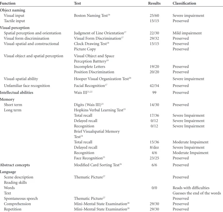
Table 1. Neuropsychological tests and results.
Function Test Results Classification
Object naming
Visual input Tactile input
Boston Naming Test16 25/60
15/15
Severe impairment Preserved
Visual perception
Spatial perception and orientation Visual form discrimination Visual-spatial and constructional
Judgment of Line Orientation17
Visual Form Discrimination17
Clock Drawing Test18
Picture Copy
22/30 29/32 15/15
Mild impairment Preserved Preserved Preserved Visual object and spatial perception Visual Object and Space
Perception Batttery19
Incomplete Letters Position Discrimination
19/20 20/20
Preserved Preserved Visual-spatial ability Hooper Visual Organization Test20 Severe impairment
Unfamiliar face recognition Facial Recognition17 42/54 Preserved
Intellectual abilities Wais III21,22 99 Preserved
Memory
Short term Long term
Digits (Wais III)21
Hopkins Verbal Learning Test23
Total recall Delayed recall Recognition
Brief Visualspatial Memory Test24
Total recall Delayed recall Recognition Face Recognition25
14/30 17/36 0/12 0/12
15/36 8/dez 4/6 23/25
Preserved
Severe Impairment Severe Impairment Severe Impairment
Moderate Impaiment Severe Impairment Moderate Impaiment Preserved
Abstract concepts Modified Card Sorting Test26 6/6 Preserved
Language
Scene description Reading skills Words Text
Spontaneous speech Comprehension Repetition
Thematic Picture27
Thematic Picture27
Mini-Mental State Examination28
Mini-Mental State Examination28
0/0
29/30 29/30
Preserved
Reads with difficulties Guesses the end of the words Preserved
Dement Neuropsychol 2008 June;2(2):151-154
Rodrigues MA, et al. Cognitive deficits associated with optic aphasia 153
objects after 12 hours. A CT scan revealed a hypodensity
area in the left occipito-temporo region and MRI revealed
subacute infarct in the left posterior cerebral artery
terri-tory. This had affected the temporo-occipital region,
in-cluding the left hippocampus.
The preliminary neurological exam detected difficulty
in identifying colors, reading words in spite of the
pre-served capacity of recognizing letters and writing, and right
hemianopsia. Eye ground exams were normal, confirmed
by ophthalmologic exam. According to the evaluation by a
speech-language therapist, the patient displayed difficulty
in naming objects; slow reading performance with
pres-ence of repetitions and prolongation; preserved writing
and naming of pictures of action. The patient was
subse-quently submitted to neuropsychological evaluation
com-prising quantitative and qualitative tests.
During the neuropsychological evaluation, difficulties
in naming visually presented objects, reading and episodic
memory were observed (Table 1).
Discussion
The location of the lesion and neuropsychological
find-ings of this evaluation confirm the clinical suspicion that,
concerning language skills, the patient showed specific
dif-ficulty in naming objects through visual presentation of
stimulus. This confirmed suspected optic aphasia. However,
this condition is similar to other pathologies suspected by
the team during the period of diagnostic investigation. The
first hypothesis, anomia, was refuted because the subject was
able to name objects presented by other sensorial means.
9The difficulty in naming visually presented objects may
be a consequence of perceptual disorder, observed in visual
agnosia,
4,13however, because of the normal performance
in visual-perceptive and visual-spatial tests, and semantic
knowledge about the objects that he was unable to name,
the hypothesis of aperceptive or associative agnosia was
ruled out. The capacity for identifying shapes and faces
through different points of view (object constancy)
sug-gests that the difficulty in naming objects occurs even if
the structural representations are preserved.
5The naming deficit could be attributed to some kind of
aphasia, however, in the case of optic aphasia, the
represen-tations and processes that constitute language are intact as
well as some aspects of language such as spontaneous speech,
Table 2. Models of optic aphasia2.
Models Theories Limitations
Canonical Model A visual processing system feeds its output into a semantic system which in turn feeds its output into a naming system. One cannot name a visually pre-sented object until one first knows what the object is.
One cannot place the lesion in vision, semantics, or the pathway connecting them, because patients can non-verbally demonstrate their recognition of visually presented objects. Neither can one place the lesion in naming or the pathway between seman-tics and naming, because patients are unimpaired in their ability to name objects presented in the tactile or auditory modalities.
Direct Visual Naming Pathway
There is a direct, uninterrupted pathway between vi-sion and naming. Optic aphasia results when the di-rect visual naming pathway becomes disconnected.
There are no documented cases of individuals who can name visual objects without any knowledge of what the objects are.
Modality-Specific Semantic Systems
Each modality has a corresponding semantic system. Optic aphasia arises when there is a disconnection between verbal semantics and visual semantics.
It does not explain the ability of optic aphasics. To sort visually dissimilar items into the same super-ordinate category.
Impaired Access to Semantics from Vision
There is an impairment in accessing a unified seman-tic system from vision. Whereas nonverbal responses may be initiated by activation of isolated semantic features from isolated visual features, naming re-quires access to a complete semantic representation.
Studies indicating poor performance on difficult nonverbal tasks may simply point to the fact that some patients indeed have a greater semantic deficit than others, apart from their inability to name visu-ally presented objects.
Hemisphere-Specific Semantic System
There is an independent semantic system for each hemisphere. Optic aphasia occurs when there is a disconnection between visual input and left hemi-sphere semantics
Dement Neuropsychol 2008 June;2(2):151-154
154 Cognitive deficits associated with optic aphasia Rodrigues MA, et al.
repetition and comprehension. The impairment is found in
access to it,
5which also occurs in pure alexia, reported by
various authors as a comorbidity to optic aphasia.
15In both
cases the difficulty in reading words and texts is associated
to a disconnection between occipital areas and language
ar-eas within the left hemisphere. The fact that the majority of
optic aphasia cases are associated to a deficit in the right
vi-sual field
9explains the difficulty described by the patient in
seeing the second half of the words. This difficulty was not
specific to reading (the patient found watching TV difficult
for the same reason), but it did not compromise his
per-formance in copying pictures as well as in perception tests.
In relation to the affected area in the brain, although
the patient did not show compromise in the splenium of
the callosal corpus on imaging exams, we found reports in
the literature with the same characteristics,
4which taken
together with the clinical evidence and results of
neuropsy-chological assessment, constitute a fundamental triad for
the diagnosis of neuropsychological impairment.
Although it would be valuable to analyze a larger
amount of data collected from other patients with this
same disorder in order to obtain consistent results,
includ-ing a more specific language evaluation, the relevance of
this present case study is justified not only due to its rarity
but also because it highlights the importance of differential
diagnosis concerning patient treatment.
References
1. Gainotti G. Anatomical functional and cognitive determi-nants of semantic memory disorders. Neurosci Biobehav Rev 2006;30:577-594.
2. Sitton M, Mozer M, Farah M. Superadditive effects of multiple lesions in a connectionist architecture: implications for the neu-ropsychology of optic aphasia. Psychol Rev 2000;107:709-734. 3. Hodges JR. Localized cognitive Functions in cognitive assessment for clinicians. 3rd ed. New York : Oxford University Press; 1994.
4. Ferreira CT, Giusiano B, Ceccaldi M, Poncet M. Optic aphasia: evidence of the contribution of different neural systems to object and action naming. Cortex 1997;33:499-513. 5. Hillis AE. Aphasia: progress in the last quarter of a century.
Neurology 2007;69:200-213.
6. De Renzi E, Zambolin A, Crisi G. The Pattern of neuropsy-chological impairment associated with left posterior cerebral artery infarcts. Brain 1987;110:1099-1116.
7. Riddoch MJ, Humphreys, GW. Visual object processing in optic aphasia: a case of semantic access agnosia. Cogn Neu-ropsychol 1987;4:131-185.
8. De Renzi E. Disorders of visual recognition. Semin Neurol 2000;20:479-486.
9. Gil R. Neuropsicologia. 2ª ed. São Paulo: Livraria Santos Edi-tora Ltda; 2002.
10. Mesulan MM. Disorders of complex visual processing in prin-ciples of behavioral and cognitive neurology. 2ª ed. New York: Oxford University Press; 2000.
11. Gazzaniga M, Ivry R, Mangun G. Funções perceptivas supe-riores in neurociência cognitiva: a biologia da mente. 2ª ed. Porto Alegre: Artmed; 2006.
12. Farah MJ. Visual agnosia: disorders of object recognition and what they tell us about normal vision. Cambridge: MIT Press; 1990. 13. Lezak MD. Neuropsychological Assessment. 3ª ed. New York:
Oxford University Press; 1995.
14. Frota N, Pinto L, Porto C, Águia P, Castro L, Caramelli P. Visual agnosia and prosopagnosia secondary to melanoma metástases. Dement Neuropsychol 2007;1:104-107. 15. Chanoine V, Ferreira CT, Demonet JL, Nespoulous, Pocet M.
Optic aphasia with pure alexia: a mild form of associative agnosia? A case study. Cortex 1998;34:437-448.
16. Kaplan E, Goodglass H, Weintraub S. The Boston Naming Test. Philadelphia: Lea & Febiger; 1983.
17. Benton AL, Sivan AB, Haqmsher KS, Vaeney NR, Spreen O. Contributions to neuropsychological assessment. New York: Oxford University Press; 1994.
18. Strauss E, Sherman EMS, Spreen O. A Compendium of neu-ropsychological test: administration, norms, and commen-tary. 3ª ed. New York: Oxford University Press; 2006. 19. Warrington EK, James M. The visual object and space
percep-tion battery. Suffolk: Thames Valley Test Company; 1991. 20. Hooper HE. The Hooper visual organization test: Manual.
Beverly Hills, California: Western Phychological Services; 1958. 21. Nascimento EN. Adaptação e validação do teste WAIS III para
o contexto brasileiro.Tese. Brasília: Instituto de Psicologia da Universidade de Brasília; 2000.
22. Ringe WK, Saine KC, Lacritz LH, Hynan LS, Cullum CM. Dyatic short forms of the Wechsler Adult Intelligence Scale-III. Assessment 2002;9:254-260.
23. Brandt J, Benedict RHB. Hopkins verbal learning test- revised. Odessa: Psychological assessment resource; 2001.
24. Benedict RHB. Brief visualspatial memory test-revised. Odes-sa: Psychological Assessment Resource; 1997.
25. Warrington EK. The Camden memory tests. Hove (UK): Psy-chology Press; 1996.
26. Nelson HE. A modified card sorting test sensitive to frontal lobe defects. Cortex. 1976;12:313-24.
27. Terman LM, Merril MA. The Stanford-Binet Intelligence Scale, Form L-M. Boston: Houghton-Mifflin Co; 1973 28. Folstein MF, Folstein SE, McHugh PR. Mini-mental state: a