Radiol Bras. 2015 Mai/Jun;48(3):181–191 181
Abdominal tuberculosis: a radiological review with emphasis
on computed tomography and magnetic resonance imaging
findings
*
Tuberculose abdominal: uma revisão radiológica com ênfase em achados de tomografia computadorizada e ressonância magnética
Rocha EL, Pedrassa BC, Bormann RL, Kierszenbaum ML, Torres LR, D’Ippolito G. Abdominal tuberculosis: a radiological review with emphasis on computed tomography and magnetic resonance imaging findings. Radiol Bras. 2015 Mai/Jun;48(3):181–191.
Abstract
R e s u m o
Tuberculosis is a disease whose incidence has increased principally as a consequence of HIV infection and use of immunosuppressive drugs. The abdomen is the most common site of extrapulmonary tuberculosis. It may be confused with several different conditions such as inflammatory bowel disease, cancer and other infectious diseases. Delay in the diagnosis may result in significantly increased morbidity, and therefore an early recognition of the condition is essential for proper treatment. In the present essay, cases with confirmed diagnosis of abdominal tuberculosis were assessed by means of computed tomography and magnetic resonance imaging, demonstrating the involvement of different organs and systems, and presentations which frequently lead radiologists to a diagnostic dilemma. A brief literature review was focused on imaging findings and their respective prevalence.
Keywords: Tuberculosis; Abdomen; Computed tomography; Magnetic resonance imaging.
A tuberculose é uma doença que vem crescendo, principalmente em consequência da infecção por HIV e do uso de drogas imunossu-pressoras. O abdome é o foco extrapulmonar mais comum de tuberculose. Pode ser confundida com diversas condições, como doença intestinal inflamatória, neoplasias e outras doenças infecciosas. O retardo no diagnóstico pode resultar em um aumento significativo da morbidade, sendo essencial o seu reconhecimento precoce para o tratamento adequado. Neste estudo avaliamos, por meio de tomo-grafia computadorizada e ressonância magnética, casos com diagnóstico confirmado de tuberculose abdominal, demonstrando o aco-metimento de diversos órgãos e sistemas, e apresentações que podem levar o radiologista a um dilema diagnóstico. Uma breve revisão da literatura foi realizada enfocando os achados já descritos e a sua prevalência.
Unitermos: Tuberculose; Abdome; Tomografia computadorizada; Ressonância magnética.
* Study developed at Department of Imaging Diagnosis – Escola Paulista de Medicina da Universidade Federal de São Paulo (EPM-Unifesp), São Paulo, SP, Brazil. 1. MDs, Radiologists at Unit of Abdomen, Department of Imaging Diagnosis – Escola Paulista de Medicina da Universidade Federal de São Paulo (EPM-Unifesp), São Paulo, SP, Brazil.
2. Fellow, Department of Imaging Diagnosis – Escola Paulista de Medicina da Universidade Federal de São Paulo (EPM-Unifesp), São Paulo, SP, Brazil.
3. Professor at Department of Imaging Diagnosis – Escola Paulista de Medicina da Universidade Federal de São Paulo (EPM-Unifesp), São Paulo, SP, Brazil.
Mailing Address: Dr. Giuseppe D’Ippolito. Departamento de Diagnóstico por Ima-gem – EPM-Unifesp. Rua Napoleão de Barros, 800, Vila Clementino. São Paulo, SP, Brazil, 04024-012. E-mail: [email protected].
Received May 16, 2013. Accepted after revision April 25, 2014.
data available at Datasus (www.datasus.gov.br), the tuber-culosis incidence rate, including all the disease presentations, in 2010, corresponded to 37.57:100,000 inhabitants, while the incidence rate for the extrapulmonary presentation of the disease was 5.28. In 2010, 71,658 new cases of tuberculosis were recorded in Brazil, as all the disease presentations are considered; and among those cases, 10,071 were extrapulmo-nary tuberculosis.
The abdominal presentation may involve different struc-tures such as gastrointestinal tract, genitourinary tract, solid organs (liver, spleen, pancreas), gallbladder, aorta and its branches, peritoneum and lymph nodes, frequently with concomitant involvement of those organs(6,7).
The disease may mimic several other conditions such as lymphoma, Crohn’s disease, amebiasis, adenocarcinoma, among others. Imaging findings are not pathognomonic, but may be highly suggestive of the disease as considered in conjunction with clinical findings, immunological conditions and the demographic origin of the patient(7).
Abdominal tuberculosis may affect practically any int-racavitary organ, presenting quite nonspecific symptoms. In
Eduardo Lima da Rocha1, Bruno Cheregati Pedrassa1, Renata Lilian Bormann1, Marcelo Longo Kierszenbaum1, Lucas Rios Torres2, Giuseppe D’Ippolito3
INTRODUCTION
Tuberculosis is responsible for about 1.7 million deaths annually worldwide, and the number of new cases (more than 9 million) is the greatest in history(1). Tuberculosis is known to be associated with poverty, deprivation and immunodefi-ciency(2).
a series with 49 patients with abdominal tuberculosis, Sinan et al.(8) stratified in detail the main symptoms and imaging findings. Fever (75%), abdominal pain (65%) and weight loss (36%) had higher prevalence than other signs and symp-toms. Peritonitis (38%) was the main tomographic finding, followed by lymph node disease (23%), involvement of the gastrointestinal tract (19%) and solid organs (10%). Diffuse pattern of lymph node commitment was most commonly observed (48%). In the gastrointestinal tract, the terminal ileum and the ileocecal region were most remarkably affected (50%). Among the solid organs, liver and spleen presented greater involvement (70%).
The present essay is aimed at reviewing and illustrating the main presentations of abdominal tuberculosis by means of images acquired by computed tomography (CT) and mag-netic resonance imaging (MRI).
PERITONEUM
Peritoneal tuberculosis is the most common presenta-tion of abdominal tuberculosis(7,8) and includes the involve-ment of the peritoneal cavity, mesenterium and oinvolve-mentum(7). It is believed that its origin is hematogenous, but it may be secondary to lymph node rupture, gastrointestinal dissemi-nation or tubal involvement(9). Most probably, it results from rupture of mesenteric lymph nodes compromised by he-matogenous dissemination of a distant primary focus (usu-ally located in the lungs). Other accepted dissemination pathways include direct extension and the lymphatic chain. It rarely results from genitourinary tract infection(10).
Despite a certain difficulty in differentiating between the several abdominal tuberculosis presentations, besides a con-siderable superimposition of presentation patterns, perito-neal tuberculosis is classically classified into three types ac-cording to its macroscopic aspects, namely: dry, wet and fibrous types(7,9,11–13). The wet type (Figure 1) presents pri-marily either as free or loculated ascites, associated or not with diffuse and smooth peritoneal thickening; in the dry type, there is a predominance of peritoneal and mesenteric thick-ening with caseous nodules, lymph nodes enlargement and fibrinous adhesions; on its turn, the fibrous type (Figure 2) is characterized by remarkable omental thickening and en-tanglement of bowel loops clinically resembling a mass, occasionally with loculated ascites and that may be similar to peritoneal carcinomatosis(7,14).
Free or loculated ascites may be present in 30–100% of cases, and the tomographic density is variable (20–45 HU), depending on the phase of the disease. Only 3% of patients present with the dry-type tuberculous peritonitis. The pres-ence of fat-fluid level in association with necrotic lymph nodes is highly specific for tuberculous ascites(6).
The omentum may be altered in up to 80% of cases, ap-pearing as diffuse infiltration, nodule, and omental cake. Dif-fuse densification is most commonly found (Figure 2), while the omental cake pattern, characterized by omental fat thick-ening and densification, is less frequently seen, occurring in
< 20% of cases, but it is most typically found in peritoneal carcinomatosis, in up to 40% of cases(6,14).
Mesenteric disease is a common abnormality that may be observed at CT as early as at the initial stages of perito-neal tuberculosis, in up to 98% of cases(14), ranging from a
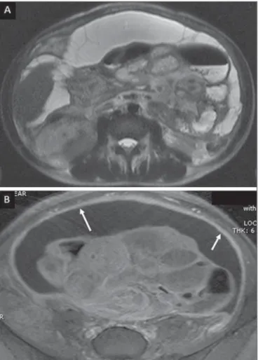
Figure 1. Wet peritonitis in a 27-year-old, female patient. Axial MRI T2-weighted sequence (A) demonstrates large ascites with multiple fine septa. Gadolinium-enhanced MRI T1-weighted image (B) shows diffuse, smooth and regular perito-neal thickening (arrows).
Figure 2. Fibrinous peritonitis. MRI T2-weighted image with fat-suppression showing omental (thin arrow) and diffuse peritoneal (thick arrow) thickening asso-ciated with bowel loops conglomerate occupying the pelvic cavity (arrowhead).
mild involvement (linear striations, vascular engorgement, star-shaped appearance, fat densification) to a more exten-sive involvement (diffuse infiltration of the mesenteric leaves). Mesenteric abscesses probably result from extension of a caseous process of large lymph node masses(13). Thick stria-tions with vascular engorgement constitute the most com-mon finding (65% of cases), followed by the nodular pat-tern (29%)(14).
The diagnosis of peritoneal tuberculosis remains as a challenge due to the wide range of clinical presentations, non-specific laboratory findings and superimposition of imaging findings with other diseases, particularly with peritoneal carcinomatosis, whose treatment and prognosis are com-pletely different. Thus, the most common CT findings in peritoneal tuberculosis include: a) ascites (70–90% of cases); b) smooth peritoneal thickening with marked enhancement after intravenous contrast injection; c) densification of the mesenteric root fat planes, which may occur in up 70% of cases; d) lymph node enlargement with areas of central ne-crosis or calcification(6,14,15).
On the other hand, the most frequent findings in perito-neal carcinomatosis include: a) multinodular and irregular peritoneal thickening; b) homogeneous retroperitoneal lymph nodes enlargement; c) omental cake, as above men-tioned(6,15).
The most useful tomographic sign to differentiate be-tween peritoneal tuberculosis and peritoneal carcinomato-sis is peritoneal thickening that, in the first condition, is smooth and regular, and in the second, is nodular and ir-regular(6).
Nontuberculous peritonitis (bacterial peritonitis, for ex-ample), peritoneal pseudomyxoma and mesothelioma con-stitute the spectrum of the main differential diagnoses of peritoneal tuberculosis(9,11,12,15).
LYMPH NODES
Lymph node involvement is usually associated with gas-trointestinal tuberculosis and less commonly with the peri-toneal and solid organs presentations, but it may be the only sign of disease, particularly in the periportal region(16). The most common involvement of lymph node chains (mesen-teric, celiac, porta hepatis, and peripancreatic lymph nodes) may be explained by the lymphatic drainage of the ileoce-cal, jejunal, ileal and right colonic regions after ingestion of infected material(16).
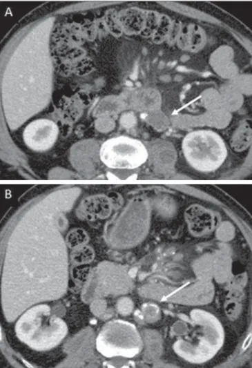
The lymph node disease pattern is variable at CT, most frequently demonstrating lymph node enlargement (40–60%) with hypoattenuation in the center and hyperattenuation in the periphery, after intravenous contrast injection, which is typical but not pathognomonic of caseous necrosis (Figure 3)(9). Lymphoma, metastasis, pyogenic infection and Whipple’s disease are the main differential diagnoses(11,16). Other lymph node involvement patterns include increase in the number but not in volume of lymph nodes, large, local-ized lymph nodes clusters and conglomerates (Figure 4)(16).
GASTROINTESTINAL TRACT
The intestinal presentation of abdominal tuberculosis is not uncommon(16). Some mechanisms may lead to bowel in-volvement by the disease: ingestion of infected material in
Figure 3. Tuberculous lymph node disease in a 54-year-old, male patient. Pres-ence of multiple enlarged lymph nodes in the mesenteric root and periaortic re-gion. Enlarged lymph node with central necrosis (arrow on A). Also, gross lymph node calcifications are observed (arrow on B).
active pulmonary tuberculosis; reactivation of a quiescent intestinal focus resulting from hematogenous dissemination in the childhood, hematogenous dissemination of active tu-berculosis, or direct spread from other organs(17).
Tuberculosis may involve any gastrointestinal tract seg-ment, but there is a preponderance in the ileocecal valve, terminal ileum and cecum(7,12,16,18), which occurs in up to 90% of intestinal tuberculosis cases (Figure 5A)(9,12,16).
Main imaging findings of intestinal tuberculosis include symmetrical or asymmetrical parietal thickening, extrinsic compression by enlarged lymph nodes which, on their turn may represent heterogeneous masses as associated with ad-herent loops and mesenteric thickening(16).
The clinical presentation of rectal tuberculosis is rarer and different from tuberculosis in proximal segments. Hematochezia (88%) and constipation (37%) are the most common symptoms. Luminal narrowing is usually signifi-cant, with variable length, areas of deep ulceration, most commonly located about 10 cm from the anal border (Fig-ure 5B). Prominent fibrosis associated with rectal inflam-mation may increase the presacral space(19). A study published by Nagi et al.(18) revealed an incidence of 10.8% of colorectal
involvement in a series of 684 patients affected by gastrointes-tinal tuberculosis along 10 years, in spite of large, previously published series revealing an incidence between 3% and 9%, with scarce reports about cases of single rectal involve-ment(18).
Perforation (Figure 6) and fistulas are the most frequent gastrointestinal complications of tuberculosis, with inci-dence of 7.6%; the small bowel and the colon are the most common sites(20). Other complications include vascular com-plications, intussusception and obstruction of the small bowel(7).
The differential diagnosis for intestinal tuberculosis varies with the involvement degree and pattern, and includes Crohn’s disease, lymphoma, amebiasis and adenocarcinoma. The presence of pulmonary compromise at chest radiogra-phy may help in the diagnostic rationale in spite of the fact that it is absent in up 50–60% of cases(7,16).
Figure 5.A: Tuberculosis of the cecum and ascending colon. Note the marked, irregular parietal thickening, decreased luminal diameter and densification of ad-jacent fat planes at the level of the cecum, segment most frequently affected by tuberculosis, adjacent to the region of the ileocecal valve and ascending colon. B:
Rectal tuberculosis in another patient. CT image demonstrates irregular, focal thickening, decreased luminal diameter and densification of adjacent fat planes at the level of the rectum, a segment that is rarely affected by tuberculosis. This lesion resembles neoplastic involvement.
Figure 6. Female, 25-year-old patient presenting intestinal perforation secondary to intestinal tuberculosis, producing a large fluid collection (arrow).
LIVER AND BILIARY TRACT
Isolated hepatic tuberculosis is a rare condition and is usually associated with concomitant involvement of other or-gans(21). On the other hand, the prevalence of hepatic com-mitment in autopsies of patients with disseminated tubercu-losis is of 80–100%(16,21).
Manifestations of hepatic tuberculosis may be divided into two types, namely, miliary and macronodular. The miliary form (Figure 7) is associated with hematogenous dis-semination, and hence the diffuse involvement of the liver(4,21). There is a diffuse enlargement of the liver and, despite the increase in hepatic enzyme levels, biliary dilata-tion may not be noticeable due to the predominance of small-caliber ducts involvement(21). Most commonly it is related to miliary pulmonary tuberculosis(4,21).
form (Figure 9B) is extremely rare and is seen either as multiple or solitary, rounded or ovoid nodules with variable appearance both at CT and MRI, which may represent dif-ferent disease stages. At contrast-enhanced T1-weighted se-quences, one can observe peripheral enhancement or, less commonly, gradual and progressive enhancement(24).
The differential diagnosis for multiple focal splenic le-sions includes lymphoma, Kaposi’s sarcoma, metastasis, sar-coidosis, bacillary angiomatosis, pyogenic/fungal abscesses, histoplasmosis and disseminated Mycobaterium avium-intracellulare and Pneumocystis carini infection(9,16,24).
PANCREAS
In spite of the fact that pancreatic compromise by tu-berculosis is rarely identified at imaging studies, at least one series has reported a prevalence of 8.3% of pancreatic in-volvement in 384 patients with diagnosis of abdominal tu-berculosis. The described alterations include increased pan-creas dimensions (Figure 10), hypodense intrapancreatic col-lections or complex masses, besides peripancreatic lymphad-enopathy(25).
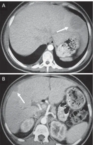
Figure 8. Macronodular hepatic tuberculosis. Contrast-enhanced abdominal CT demonstrating the presence of hypovascular nodules in the left (A) and right (B) lobes.
enlarged liver. At MRI, the lesions present low signal inten-sity and minimal peripheral enhancement, with a honeycomb-ing pattern in the miliary form at T1-weighted sequences (Figure 7). At T2-weighted sequences, the lesions are hypoin-tense, with a less hypointense halo in relation to the surround-ing liver parenchyma(12).
The differential diagnosis for the micronodular form of hepatic tuberculosis includes metastasis, fungal infection, sar-coidosis and lymphoma(12); and in the macronodular form, it is made primarily with abscess and metastasis(12,16,21).
The involvement of the biliary tree by tuberculosis is even more rarely observed and its annual incidence is esti-mated to be 0.1%(22). The involvement may be either pri-mary, with small ducts stenosis, or secondary to compres-sion by hepatic granulomas, many times making it more difficult to differentiate with primary sclerosing cholangitis and cholangiocarcinoma(21).
The gallbladder is very rarely involved. Mural thicken-ing, irregular septa, and lymphadenopathy may be found. There is no typical presentation, and it may be quite vari-able(23). The diagnosis is usually made on the basis of histo-pathology(7).
SPLEEN
Splenic tuberculosis is usually associated with the dis-seminated form of miliary tuberculosis and, in spite of be-ing reported in up to 80–100% of autopsies of patients with disseminated tuberculosis, it is much less frequently identi-fied by imaging methods(24). However, a recent series re-ported a rate of splenic involvement diagnosed by imaging methods (ultrasonography, CT or MRI) of 45.8% in cases of disseminated tuberculosis(24).
As well as hepatic tuberculosis, there are two types of presentations of splenic tuberculosis, namely, miliary and macronodular. The first and most common type (Figure 9A) usually manifests as moderate splenomegaly, but minute hypodense lesions may be seen at CT. The macronodular
SUPRARENAL GLANDS
Suprarenal glands are not a rare site of involvement by tuberculosis(26), and this is the main cause of suprarenal gland failure (Addison’s disease)(26).
Suprarenal tuberculosis manifests with increase in gland dimensions (Figure 11A), central necrosis and either uni-lateral or biuni-lateral calcifications (Figure 11B)(11,12,26). Su-prarenal glands enlargement is also observed in patients with renal tuberculosis(11). Atrophy and calcifications may be seen in previously treated patients(11,26).
The radiological differential diagnosis includes metasta-sis, lymphoma, primary neoplasm, blastomycosis(27) and hemorrhage(26).
KIDNEYS
In spite of the fact that renal tuberculosis is usually a result of hematogenous dissemination originating from the lungs, less than 50% of patients present with radiological evi-dences of pulmonary tuberculosis and only 10% present with active disease(28). Renal tuberculosis is usually a consequence
Figure 9.A: Miliary splenic tuberculosis. Contrast-enhanced CT showing minute hypoattenuating, nodules randomly distributed in the spleen. B: Macronodular splenic tuberculosis. Contrast-enhanced CT reveals a single, peripheral nodule in the spleen.
Figure 10. Pancreatic tuberculosis. CT identifies a slight increase in the dimen-sions of the organ and loss of the habitual, lobulated contours of the pancreas.
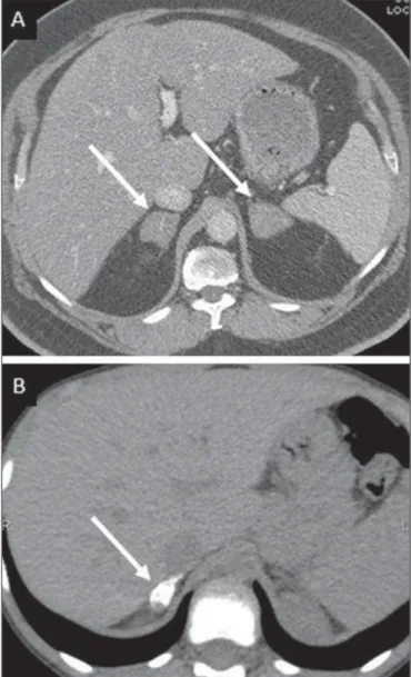
Figure 11. Adrenal tuberculosis. A: CT shows hyperplasia of both adrenal glands.
from a primary pulmonary infection that might have occurred several years before. Tubercle bacilli lodge at the corticomed-ullary junction, forming granulomas in the papilla which remain stable for many years; if reactivation occurs, the or-ganisms spread into the medulla, causing papillitis(28).
Focal tissue edema and vasoconstriction caused by ac-tive inflammation result in local hypoperfusion identifiable at CT and MRI(26). Calyceal deformity is the initial find-ing(28). Multiple or solitary parenchymal nodules (Figure 12) with no urinary tract involvement are rare manifestations and may mimic neoplasms(26).
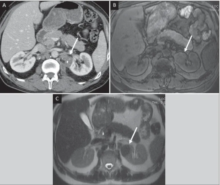
The most characteristic imaging finding is caliectasis (Figures 13 and 14)(11,26).
The disease progression might result in extensive papil-lary necrosis, cavitation and later cortical scars, pyeloinfun-dibular strictures and hydronephrosis. At a terminal stage of the disease, lost of renal function and calcifications are ob-served(28). Calcifications may present with several patterns
such as amorphous, granular, lobar and diffuse (named autonephrectomy)(28).
URETERS
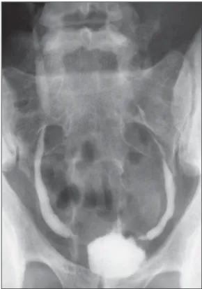
Dilatation and irregular thickening of the urothelium (Figure 15) represent the first signs of ureteral tuberculosis. The dilatation results primarily from ureterovesical junction stricture secondary to cystitis and tuberculous urethritis.
At advanced stages of the disease, ureteral stenosis, short-ening, filling defects and ureteral calcifications may be seen (Figure 16)(12).
BLADDER
Initially, tuberculous cystitis produces mucosal ulcer-ation and edema. The disease extension towards the muscle layer leads to fibrosis and, consequently, to mural thickening and decrease in contractility. For this reason, the main sign of tuberculous cystitis is a thickened bladder and reduced
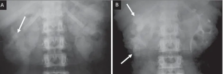
Figure 13. Renal tuberculosis at right. Plain radiography demonstrates spotted calcifications (arrow on A). Excretory urography shows deformity of the pyelocalyceal system, decreased thickness of the renal parenchyma and cavitations filled with contrast agent in contiguity with the renal infundibulum (arrows on B).
Figure 14. Left renal tuberculosis evolving with calicectasis. Axial CT images (A) and coronal reconstruction (B) demonstrate calicectasis in middle calyceal group at excretory phase (arrows on A and B).
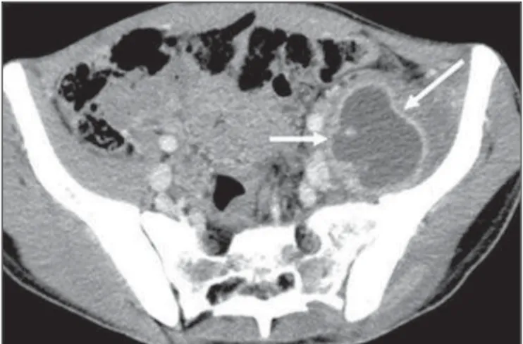
bladder capacity (Figures 16 and 17). Ureteral reflux may be observed. Calcified tuberculous cystitis is rare and should be differentiated from other conditions such as schistosomia-sis, amyloidoschistosomia-sis, cyclophosphamide-induced actinic cystitis, carcinoma and foreign bodies(28).
FEMALE GENITAL ORGANS
Tuberculous infection of the female genital system may cause menstrual disorders, gestational complications, neo-natal tuberculosis, antituberculosis drugs side effects during pregnancy, increased drug-resistance and infertility(29). Chavhan et al. have published a study showing 7.5% incidence of female genital tuberculosis in 492 patients undergoing
investigation for infertility and submitted to hysterosalpin-gography(30).
Most women with genital tuberculosis present with in-fertility secondary to tubal involvement (Figure 18A), which occurs in up to 94% of such patients. Typically, it is bilat-eral and causes multifocal stricture and calcifications(12). Tubo-ovarian abscess extending through the peritoneum and extraperitoneal compartment is also suggestive of tubercu-losis(12).
Endometrial tuberculosis occurs in up to 50% os patients with tubal tuberculosis(26). Tuberculous endometritis may mimic relevant uterine adhesions and endometrial carcinoma (Figure 18B)(11).
MALE GENITAL ORGANS
Tuberculosis may affect the whole male genital tract, with lesions in the prostate, seminal vesicles, deferent ducts, epididymis, penis and testes. Genital tuberculosis occurs by hematogenous dissemination to the prostate and epid-idymides or through the urinary system to the prostate with
Figure 17. Vesical tuberculosis. CT identifies diffuse and asymmetrical thicken-ing of the vesical wall (arrows).
Figure 18.A: Adnexal tuberculosis. MRI shows diffuse parietal thickening with marked contrast uptake and dilatation of both uterine tubes at T1-weighted se-quence with fat-suppression after intravenous contrast injection. B: Uterine tuber-culosis. Axial MRI T2-weighted image demonstrates diffuse thickening of the endometrium and junctional zone (arrows), besides heterogeneous endometrial content.
canalicular dissemination to the seminal vesicles, deferent ducts and epididymides. It may be either associated with renal lesions or present as an isolated condition(31).
Epididymides are involved in 10–55% men with uro-genital tuberculosis(31). Generally, it starts at the epididy-mal tail and later may propagate to the entire structure(26). Sonographic findings include edema and heterogeneous echotexture of the involved segment. At MRI, increased vol-ume and low signal intensity are observed at T2-weighted sequences, suggesting chronic inflammation and fibrosis(11,26). The seminal vesicles and deferent ducts may present wall thickening, stricture, and parietal or intraluminal calcifica-tions at sectional images(26).
Tuberculous prostatitis is characterized by decreased echogenicity and increased vascularization at Doppler ultra-sonography, similarly to prostate cancer(12). In patients with prostatic abscess (Figure 19), CT scans and MRI reveal cystic lesion with peripheral enhancement, indistinguishable from other causes of abscess. Dystrophic and nonspecific calcifica-tions may be observed in the chronic phase of the disease(26).
radiologist recognizes the imaging findings, allowing for the establishment of a more effective strategy to confirm the diagnosis and to institute the appropriate treatment as soon as possible.
REFERENCES
1. Lawn SD, Zumla AI. Tuberculosis. Lancet. 2011;378:57–72. 2. Aston NO. Abdominal tuberculosis. World J Surg. 1997;21:492–9. 3. Kapoor VK. Abdominal tuberculosis. Postgrad Med J. 1998;74:459–
67.
4. Saluja SS, Ray S, Pal S, et al. Hepatobiliary and pancreatic tuber-culosis: a two decade experience. BMC Surg. 2007;7:10. 5. Xu XF, Yu RS, Qiu LL, et al. Gallbladder tuberculosis: CT findings
with histopathologic correlation. Korean J Radiol. 2011;12:196– 202.
6. Peixoto Filho AAA, Peixoto M, D’Ippolito G. Tuberculose perito-neal: como diagnosticar? Rev Imagem. 2007;29:47–52.
7. Gulati MS, Sarma D, Paul SB. CT appearances in abdominal tu-berculosis. A pictorial essay. Clin Imaging. 1999;23:51–9. 8. Sinan T, Sheikh M, Ramadan S, et al. CT features in abdominal
tuberculosis: 20 years experience. BMC Med Imaging. 2002;2:3. 9. Burrill J, Williams CJ, Bain G, et al. Tuberculosis: a radiologic
re-view. Radiographics. 2007;27:1255–73.
10. Epstein BM, Mann JH. CT of abdominal tuberculosis. AJR Am J Roentgenol. 1982;139:861–6.
11. Harisinghani MG, McLoud TC, Shepard JA, et al. Tuberculosis from head to toe. Radiographics. 2000;20:449–70.
12. Engin G, Acunas B, Acunas G, et al. Imaging of extrapulmonary tuberculosis. Radiographics. 2000;20:471–88.
13. Suri S, Gupta S, Suri R. Computed tomography in abdominal tu-berculosis. Br J Radiol. 1999;72:92–8.
14. Na-ChiangMai W, Pojchamarnwiputh S, Lertprasertsuke N, et al. CT findings of tuberculous peritonitis. Singapore Med J. 2008;49: 488–91.
15. Ha HK, Jung JI, Lee MS, et al. CT differentiation of tuberculous peritonitis and peritoneal carcinomatosis. AJR Am J Roentgenol. 1996;167:743–8.
16. Pereira JM, Madureira AJ, Vieira A, et al. Abdominal tuberculosis: imaging features. Eur J Radiol. 2005;55:173–80.
17. De Backer AI, Mortelé KJ, De Keulenaer BL, et al. CT and MR im-aging of gastrointestinal tuberculosis. JBR-BTR. 2006;89:190–4. 18. Nagi B, Kochhar R, Bhasin DK, et al. Colorectal tuberculosis. Eur
Radiol. 2003;13:1907–12.
19. Sharma MP, Bhatia V. Abdominal tuberculosis. Indian J Med Res. 2004;120:305–15.
OTHERS
Iliopsoas muscle abscess (Figure 20) was a well known complication of vertebral tuberculosis until the implemen-tation of modern chemotherapy schemes(32). It may be clas-sified into primary (30%) or secondary (70%), depending on the presence of an underlying disease, such as, tubercu-losis. In developing countries, vertebral tuberculosis (Pott’s disease) is considered to be the most common cause of psoas muscle abscess; however, few reports are found in the litera-ture about psoas muscle abscess as a primary presentation of tuberculosis(33). The presence of calcification in the ab-scess is virtually pathognomonic of tuberculosis(12).
CONCLUSION
Tuberculosis presents a wide spectrum of clinical and imaging findings, and may affect many different organs in different ways. The diagnosis requires a high degree of sus-picion and, despite the fact of being defined only by means of biopsy and specimens culture, it is important that the
Figure 19. Prostatic tuberculosis. Hypoattenuating nodular lesion with periph-eral enhancement compatible with abscess.
20. Nagi B, Lal A, Kochhar R, et al. Perforations and fistulae in gas-trointestinal tuberculosis. Acta Radiol. 2002;43:501–6.
21. Chong VH, Lim KS. Hepatobiliary tuberculosis. Singapore Med J. 2010;51:744–51.
22. Chong VH. Hepatobiliary tuberculosis: a review of presentations and outcomes. South Med J. 2008;101:356–61.
23. Kumar K, Ayub M, Kumar M, et al. Tuberculosis of the gallblad-der. HPB Surg. 2000;11:401–4.
24. De Backer AI, Vanhoenacker FM, Mortelé KJ, et al. MRI features of focal splenic lesions in patients with disseminated tuberculosis. AJR Am J Roentgenol. 2006;186:1097–102.
25. Nagar AM, Raut AA, Morani AC, et al. Pancreatic tuberculosis: a clinical and imaging review of 32 cases. J Comput Assist Tomogr. 2009;33:136–41.
26. Jung YY, Kim JK, Cho KS. Genitourinary tuberculosis: compre-hensive cross-sectional imaging. AJR Am J Roentgenol. 2005;184: 143–50.
27. Mayo-Smith WW, Boland GW, Noto RB, et al. State-of-the-art adrenal imaging. Radiographics. 2001;21:995–1012.
28. Muttarak M, ChiangMai WN, Lojanapiwat B. Tuberculosis of the genitourinary tract: imaging features with pathological correlation. Singapore Med J. 2005;46:568–74.
29. Ghosh K, Ghosh K, Chowdhury JR. Tuberculosis and female re-productive health. J Postgrad Med. 2011;57:307–13.
30. Chavhan GB, Hira P, Rathod K, et al. Female genital tuberculosis: hysterosalpingographic appearances. Br J Radiol. 2004;77:164–9. 31. Figueiredo AA, Lucon AM. Urogenital tuberculosis: update and review of 8961 cases from the world literature. Rev Urol. 2008;10: 207–17.
32. Mallick IH, Thoufeeq MH, Rajendran TP. Iliopsoas abscesses. Postgrad Med J. 2004;80:459–62.